Infective endocarditis still carries with it a high risk of morbidity and mortality. Early diagnosis and prompt effective treatment are the main determinants of favourable outcomes. Nevertheless, current mortality represents about one-fifth of the infective endocarditis episodes, and more than one-fifth of the cases will require early emergency surgery.Reference Chatzis, Saroglou, Giannopoulos and Sarris1
Infective endocarditis is less common in children, especially in infants than in adults, and is increasingly observed in patients with congenital cardiac disease.Reference Ferrieri, Gewitz and Gerber2 Clinical typical presentation with sepsis and/or cardiac failure easily leads to diagnosis, with Streptococcus and Staphylococcus being the two most common agents responsible.Reference Pellicelli, Pino, Terranova, D’Ambrosio and Soccorsi3, Reference Bashore, Cabell and Fowler4
However, in some cases such as ours, clinical presentation is atypical and/or the microbial organisms causing the infection are rare.
Case report
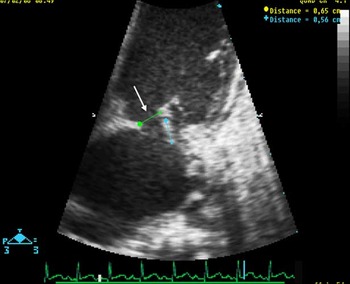
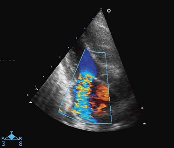
R. is a 9.5-month-old male infant, born at full term. His birth weight was 3.2 kilograms. The infant had no known cardiac murmur. He was referred to the Department of Pediatric Cardiology for symptoms of respiratory distress and cardiomegaly on chest X-ray. The current history goes back to 15 days before, when the infant was treated for acute bronchiolitis with aerosolised salbutamol and physiotherapy: at day 4 after the onset of treatment, he became suddenly hypotonic and pale with shortness of breath while feeding; concomitantly fever peaked at 40°C. The infant was admitted to the hospital and received a dose of antibiotics (cefpodoxime) because of a high suspicion of sepsis. On physical examination he appeared tachypneic, pale and hypotonic, but his neurological examination was normal. A systolic murmur, significant hepatomegaly and mild splenomegaly were present. The child became apyretic, 37°C, within few hours after admission. Pulse oximetric saturation was 93% in room air, respiratory rate was 56 per minute, heart rate was 155 beats per minute, and blood pressure was 110/71 millimetres of mercury. The mother had noticed a transient purpura in the lower limbs 1 week before. Laboratory tests showed normal white blood cell and platelet counts. Haemoglobin was 93 grams per litre and C-reactive protein ranged in the low values – 4.7 milligrams per litre. Repeated blood cultures were performed. Electrocardiogram records were normal; chest X-ray showed cardiomegaly and diffuse pulmonary oedema infiltrates. Cardiac ultrasound showed a mass 6 × 6 millimetres in diameter at the anterior leaflet of the mitral valve (Fig 1) and significant mitral valve regurgitation (Fig 2) due to perforation of the anterior leaflet. Left cardiac cavities were enlarged with the hyperdynamic left ventricle. Clinical symptoms led to the diagnosis of cardiac failure because of severe mitral regurgitation in the context of mitral infective endocarditis with valve perforation. However, there were neither clinical symptoms nor biological criteria of ongoing infection. The clinical symptoms promptly improved with diuretics and anti-converting enzyme inhibitor therapy. Antibiotics were initiated with ceftriaxone (50 milligrams per kilogram per day) associated with vancomycine (40 milligrams per kilogram per day), and amikacine (14 milligrams per kilogram per day). Cerebral tomodensitometry was normal, whereas abdominal tomodensitometry showed several small lesions, highly suspect of septic emboli and located in the right kidney. Blood cultures were all negative. The infant underwent surgical plasty of the mitral valve on day 6 after admission: the vegetation was entirely removed and patch closure of the leaflet perforation was performed. Polymerase chain reaction culture of the specimen was positive for Kingella kingae. Further antibiotic treatment was continued with ceftriaxone alone. Cardiac ultrasound, performed on day 11 after surgery, showed mild mitral regurgitation and no stenosis. The infant was discharged on day 13 post-operatively under anti-converting enzyme inhibitor therapy. He completed a 4-week intravenous antibiotic treatment.
The four-chamber view of the mitral valve shows vegetation and perforation (arrow) on the anterior leaflet of the mitral valve.

Color-Doppler four-chamber view of the mitral valve shows significant mitral regurgitation.

Discussion
Infective endocarditis is an uncommon disease in infants. It commonly occurs in patients with underlying cardiac malformation, either native lesions or congenital cardiac disease that is treated with either residual shunts, conduits, prosthesis, or valvular dysfunction. Lesions with turbulent flow and high-velocity jets are common targets for the growth of vegetation.Reference Bashore, Cabell and Fowler4
The first particular characteristic of our case is the young age of the patient and the absence of known and identified congenital cardiac disease. Infective endocarditis episodes have been reported to occur in normal hearts, mostly in adults, drug abusers, patients with a central venous line, or pacemaker; in these cases, endocarditis is localised in the right heart.Reference Pellicelli, Pino, Terranova, D’Ambrosio and Soccorsi3, Reference Bashore, Cabell and Fowler4 To our knowledge, very scarce literature has reported infants with normal cardiac and mitral valve infective endocarditis.
Second, the clinical presentation of infective endocarditis usually runs in a sub-acute course accompanied by moderate-to-high grade fever, fatigue, poor feeding, weight loss, increased cyanosis, cough, dyspnoea, or systemic embolisation. Subtle infective endocarditis can also occur in patients with congenital cardiac disease;Reference Chatzis, Saroglou, Giannopoulos and Sarris5 our case may range in the latter category, although underlying cardiac disease could not be identified. He presented with symptoms of cardiac failure, but no ongoing sepsis, and promptly improved with diuretics. On the basis of cardiac ultrasound and abdominal tomodensitometry findings a diagnosis was made. Subtle presentation and very few symptoms of sepsis were the second remarkable point in this case.
The third remarkable point is that the causative microbial agent belongs to the HACEK group. Very few cases of K. kingae infective endocarditis have been reported and the majority of these has been diagnosed in patients with either prosthetic cardiac valves, or previously recognised cardiac disease. Normal undamaged cardiac endothelium usually is not the target for bacterial colonisation whenever a bacteremia occurs.Reference Wilson, Taubert and Gewitz6 In our case, no anatomical cardiac abnormality could be identified. K. kingae is a member of HACEK group of Gram-negative bacteria (Haemophilus spp., Actinobacillus actinomycetemcomitans, Capnocytophaga spp., Cardiobacterium hominis, Eikenella corrodens, and K. kingae), which were recently implicated as causative agents for infective endocarditis, sharing the ability to infect the human valve.Reference Bosworth7–Reference Dubnov-Raz, Scheuerman, Chodick, Finkelstein, Samra and Garty9 K. kingae is part of the bacterial flora of the pharynx in young children and rarely causes serious infections.Reference Bosworth7 It is transmitted from child to child. The clinical presentation is often subtle so that making the diagnosis requires a high index of suspicion; arthritis is the most common presentation for systemic infections.Reference Yagupsky8 In our case, the diagnosis was insured by cardiac ultrasound, which showed the vegetation and perforation on the anterior mitral valve. K, kingae is susceptible to a wide array of antibiotics given empirically to young children, like β-lactams, with the notable exception of complications.Reference Dubnov-Raz, Scheuerman, Chodick, Finkelstein, Samra and Garty9
Lastly, this case shows the high ability of polymerase chain reaction to retrieve the causative agent when blood cultures are negative. This technique, when applied peri-operatively, removed valvular infected tissues, and has been shown to enhance microbial diagnosis by 20%.Reference Di Filippo, Delahaye and Semiond10
Conclusion
The present case report is remarkable because of the atypical subtle presentation of K. kingae infective endocarditis in an infant with mitral valve location on a previously normal heart, which was successfully treated with early surgical mitral valve plasty, with the bacterial agent being identified by polymerase chain reaction culture of the valvular tissue, belonged to the HACEK group.