Cardiac catheterisation in the field of CHD has evolved from a diagnostic to predominantly a therapeutic modality, extending its remit to provide non-surgical options for patients with heart failure, pulmonary hypertension, and valvar heart disease. Hybrid approaches continue to complement rather than compete with surgery and will reshape therapy for congenital heart lesions over the next decade. The interventional cardiology programme at the Seventh World Congress of Pediatric Cardiology & Cardiac Surgery (WCPCCS) reflected this shift in approach with interventional topics discussed not only within the main interventional track but also in the surgical, fetal, heart failure/pulmonary hypertension, and adult congenital programmes. The benefits and challenges of registry data for interventional cardiology were also discussed outside of the main track and data were presented on numerous late-breaking clinical trials, reflecting the rapid advancements taking place in the field. Live cases, although sometimes contentious, provide a unique learning experience and were presented from four centres in Europe and North America. A total of 28 high-quality lectures were delivered with four debates, 18 oral abstracts, a nightmare case session, and a productive round table discussion with representatives from the Food and Drug Administration in the United States of America to determine whether streamlined pathways for optimising a global approach to device approval for congenital heart interventions can be achieved. This article will provide a summary of this unique learning experience for congenital interventions at the Seventh WCPCCS.
Live cases
Two live cases were presented from Nationwide Children’s Hospital, Columbus, Ohio on Monday 17th July. These were particularly notable for organising a faultless transmission at 2 AM local time! The first live case highlighted the benefits of three-dimensional rotational angiography overlay in the setting of native coarctation of the aorta. The rotation was performed with the stiff wire in position so as to ensure the overlay matched the anatomy of the aortic isthmus at the time of stent deployment. Minimal contrast was used to position the stent before deployment with an excellent result. Discussion centred on the use of bare metal versus covered stents in this setting. Indeed a recent randomised trial comparing bare versus covered Cheatham-Platinum stents for native coarctation of the aorta did not demonstrate any difference in aortic wall injury between the two groupsReference Sohrabi, Jamshidi and Yaghoubi 1 ; however, one of the panellists suggested this study was under-powered to do so. Two talks were delivered later in the week on the topic of stenting for coarctation of the aorta, underlying its ongoing relevance to the interventional community. Progress in stent development has translated into a greater variety of stent choices and the nuances of choosing the most appropriate stent type to match the varieties of anatomical substrates encountered was discussed. Data were presented from the Congenital Cardiac Interventional Study Consortium (CCISC) coarctation registry, which has now recruited more than 900 patients undergoing surgical and interventional therapy for coarctation of the aorta. There has been a clear reduction in complications in the stent cohort (~650 patients) with growing experience and improvements in stent design.
The second live case from Columbus, performed by Dr Darren Berman, demonstrated the implantation of CardioMEMS system (St Jude Medical, St Paul, Minnesota) in a patient with a total cavopulmonary connection. This system compromises a silicone encapsulated sensor and pressure sensitive capacitor, which acts as a wireless radiofrequency sensor. Two nitinol loops at either end of the device assist in anchoring the device, usually within a branch of the left pulmonary artery. The entire device comes pre-mounted on a delivery catheter and tracks over a 0.018″ wire requiring an 11 Fr introducer. The group from Nationwide has previously published on the potential advantages of this system in monitoring pulmonary artery pressures in patients with a total cavopulmonary circulation.Reference Bradley, Berman and Daniels 2 It remains to be seen whether the data will optimise chronic management of single ventricle patients in a similar fashion to its evolving role in the management of chronic heart failure in adults. The live case was preceded by a full session dedicated to interventions for heart failure. Dr Tom Jones (Seattle, United States of America) presented experience with various heart failure therapies including the use of MitraClip (Abbot, Lake Bluff, Illinois, United States of America) in a child with dilated cardiomyopathy and severe functional mitral regurgitation.Reference Joffe, Jones and Reisman 3 The patient had an appreciable reduction in mitral regurgitation with increased in left ventricular ejection fraction and improvement in symptoms while weaning medications. As transcatheter therapies for mitral valve disease evolve in adult heart disease, it is important that we maintain an awareness of the potential application of these technologies to CHD patients. In fact, in a combined session with Adult Congenital Heart Disease, Professor Horst Sievert (Frankfurt, Germany) spoke about the future application of transcatheter left heart valve repair and replacement in adult CHD patients.
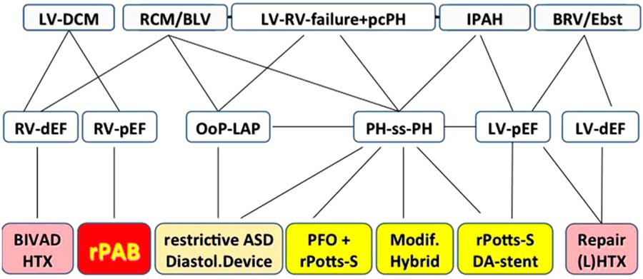
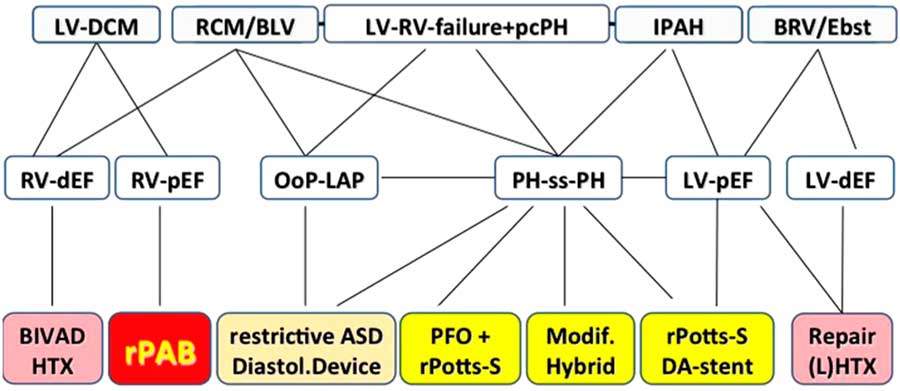
Another notable area where transcatheter interventions are broadening therapeutic options is in the management of pulmonary hypertension. Talks by Professor Kevin Walsh (Dublin, Ireland) and Professor Dietmar Schranz (Giessen, Germany) outlined established (atrial septostomy) and evolving (transcatheter Pott’s shunt) therapies. Atrial septostomy has been shown to improve symptoms and survival; however, spontaneous closure rates of up to 30% have been reported.Reference Sandoval, Gaspar and Pulido 4 Atrial septal stenting may be performed to maintain patency; however, early clinical experience with newer devices including the atrial flow restrictor (Occlutech International AB, Helsingborg, Sweden) are promising. Transcatheter creation of a reverse Pott’s shunt was also discussed with early data demonstrating improvement in symptomsReference Baruteau, Belli and Boudjemline 5 ; however, the timing and technical aspects of the procedure are still evolving. Professor Schranz discussed an algorithmic approach to heart failure and pulmonary hypertension in younger patients including reverse PA banding for a failing left ventricle facilitating a native right ventricular assist (Fig 1)Reference Schranz, Akintuerk and Voelkel 6 and it is clear that interventional cardiology will contribute in an increasing way to these conditions.
Algorithm for the management of patients with heart failure, borderline ventricle, or pulmonary hypertension.Reference Schranz, Akintuerk and Voelkel 6 BIVAD, biventricular assist device; BLV, borderline left ventricle; BRV, borderline right ventricle; DA, ductus arteriosus; DCM, dilated cardiomyopathy; dEF, depressed ejection fraction; diastol., diastolic; Ebst., Ebstein anomaly; HTX, heart transplantation; IPAH, idiopathic pulmonary hypertension; (L)HTX, (lung) heart transplantation; LV-DCM, left ventricular-dilated cardiomyopathy; LV, left ventricle; LVAD, left ventricular assist device; Modif., modified; OoP-LAP, out-of-proportion left atrial pressure; pcPH, precapillary pulmonary hypertension; pEF, preserved ejection fraction; PFO, persistent foramen ovale; PH-ss-PH, pulmonary hypertension to suprasystemic pulmonary hypertension; rASD, restrictive atrial septum defect; RCM, restrictive cardiomyopathy; rPAB, reversible pulmonary artery banding; rPotts-S, reverse Potts shunt; RV, right ventricle.
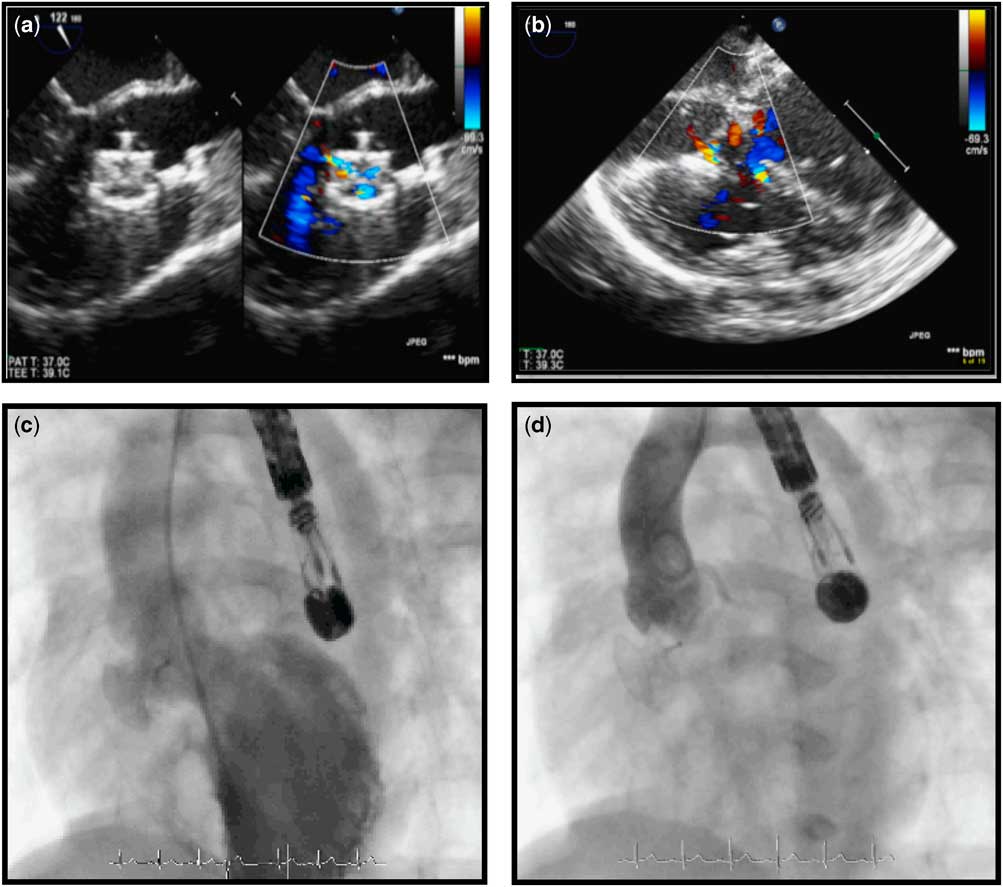
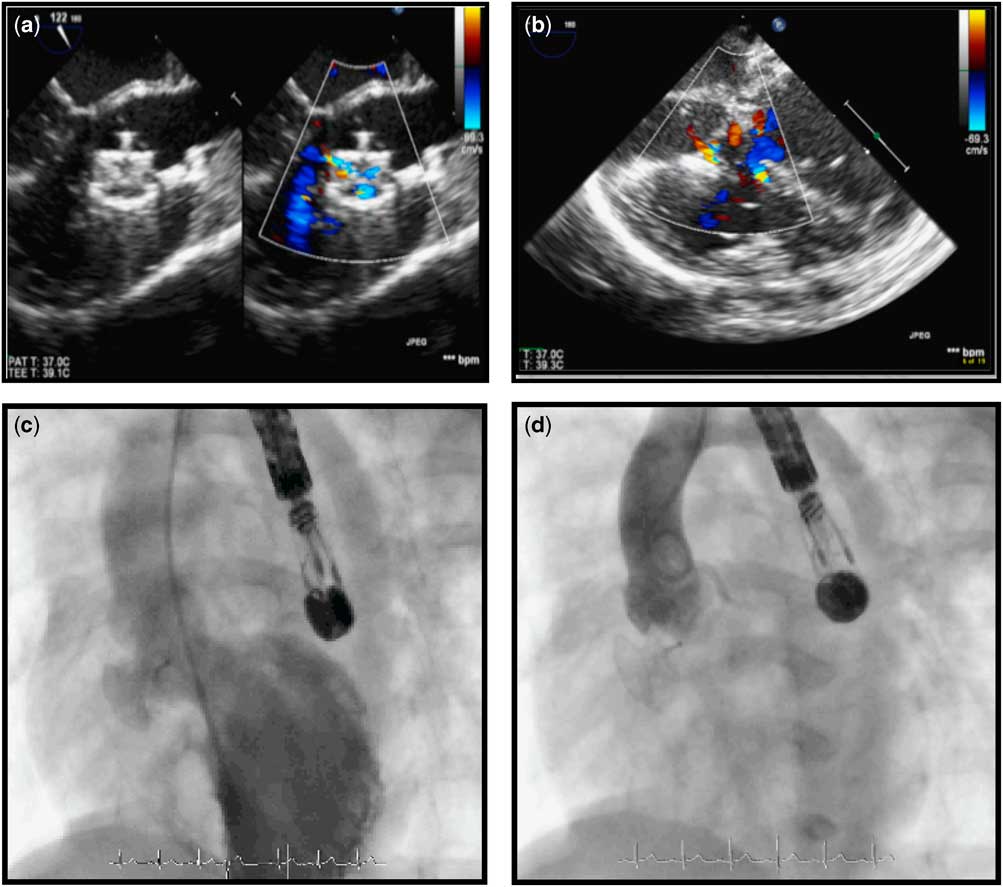
Live cases on Tuesday were presented from Dublin, Ireland with the first case demonstrating transcatheter closure of a clinically significant perimembranous ventricular septal defect in a 7 kg infant via a retrograde approach from a left common carotid cutdown. The approach facilitated easy recrossing of the defect when initial device deployment led to impingement on the tricuspid valve. Crossing the more anterior fenestration brought the right ventricular disc of the device away from the tricuspid valve with no increase in tricuspid regurgitation and good defect closure (Fig 2). It remains to be seen whether this approach coupled with softer device design will compete with surgical closure of perimembranous defects in smaller infants. The second case from Dublin demonstrated transcatheter pulmonary valve replacement in an 11-year-old with previously repaired tetralogy of Fallot and severe native outflow pulmonary regurgitation (right ventricular volume of 192 ml/m2). Previous pre-stenting of the right ventricular outflow tract had been carried out with a 43 mm Andrastent XXL (Andramed GmbH, Reutlingen, Germany) on a 24 mm BiB (NuMed Inc., Hopkinton, New York, United States of America) and this was further dilated with a 26 mm Atlas balloon (Bard PV Inc., Tempe, Arizona, United States of America) with subsequent implantation of the a 26 mm SAPIEN XT valve (Edwards Lifesciences, Irving, California, United States of America) with a good result. Transcatheter pulmonary vale replacement remains a hot topic in the field and multiple presentations were delivered, including impact of 15 years’ experience with tPVR on clinical outcomes (Professor Felix Berger, Berlin, Germany) to newer approaches and valve systems for the native right ventricular outflow tract. The Venus Pulmonary (Venus MedTech, Hangzhou, China) valve is a self-expanding valve system, with a porcine pericardial valve mounted on a nitinol stent suitable for right ventricular outflow tract’s up to 33 mm in diameter. Clinical experience to date in almost 100 patients worldwide was presented by Professor Ziyad Hijazi (Doha, Qatar), with details of a clinical trial in Europe presented in the Late Breaking catheterisation session by Professor Shak Qureshi (London, United Kingdom).
Series of echocardiographic and angiographic images “following” retrograde closure of a perimembranous ventricular septal defect (VSD) with a 10 mm Occlutech membranous VSD occluder. ( a ) Long-axis “transoesophageal” transesophagea echocardiographic (TEE) image demonstrating the device in position with no impingement on the aortic valve. ( b ) TEE image demonstrating minimal tricuspid regurgitation following device release. ( c ) Left ventricular angiography demonstrating the device in position with minimal leak. ( d ) Ascending aortogram confirming no aortic regurgitation following device release.
Live cases on Wednesday were due to be presented from Milan; however, due to technical challenges with the case, a taped case of a transcatheter tricuspid valve-in-valve replacement was presented by Allison Cabelka (Rochester, New York, United States of America). Data from an evolving registry of over 150 patients suggest this is an effective approach leading to good outcomes in a sick cohort of patients.Reference McElhinney, Cabalka and Aboulhosn 7 In the afternoon live case session, Professor Mario Carminati (Milan, Italy) presented the outcome of the challenging morning case. The patient had a short landing zone for a Melody valve with instability of the initial pre-stent. The case highlighted nicely the continuing challenges that exist with this approach due to significant anatomical diversity. Options such as Melody valve shorteningReference Malekzadeh-Milani, Patel and Boudjemline 8 and telescoping a longer stent from one of the branch pulmonary arteries were discussed.Reference Boudjemline, Legendre and Ladouceur 9
Finally on Thursday morning, Dr Iqbal Malik presented two atrial septal closure cases, including a patent foramen ovale closure in a patient with an atrial septal aneurysm. Transcatheter patent foramen ovale closure has re-emerged as a potentially clinically superior therapy to medical therapy for crytogenic stroke in younger patients, particularly those with larger defects, following presentation of data from both the RESPECT and REDUCE trials, which were discussed by Dr Mark Turner (Bristol, United Kingdom) in a session on Tuesday morning dedicated to atrial interventions. Other speakers in this session included Dr Teiji Akagi (Japan) who discussed the benefits of complete registry follow-up data on atrial septum defect closure patients with the Amplatzer Septal Occluder. Complete data sets on complications including erosion are available in Japan with 14 cases of erosion reported from over 8000 atrial septum defect implants (0.18%). The benefits of complete registry data sets are clear; however, challenges exist in relation to accuracy of data recording, funding for personnel, and variances in international practices. Finally, this session was closed with an entertaining but relevant debate between Dr Michel Ilbawi (Chicago, United States of America) and Dr Zahid Amin (Augusta, United States of America) as to the merits and disadvantages of both transcatheter and surgical approaches to atrial septum defect closure.
Highlight lectures
Working closely with our surgical colleagues to provide the least invasive optimal repair or palliation of CHD continues to be the goal of hybrid procedures. A dedicated session on hybrid therapies on Wednesday morning included a talk on how to establish a hybrid programme from Dr John Cheatham (Columbus, United States of America). Amalgamating two perspectives to achieve a single focus – with minimal professional or financial competition – threaded strongly through the talk as the team in Columbus has evolved into one of the leading hybrid therapy institutions in the world. It was clear from this session and indeed the entire interventional programme that hybrid procedures have evolved well beyond palliation of hypoplastic left heart syndrome, ranging from simple vascular cutdown procedures to complex intraoperative stenting (Dr Evan Zahn, Los Angeles, United States of America) and transcatheter valve replacement. Further discussion with our surgical colleagues centred around optimal palliation of symptomatic neonatal tetralogy of Fallot. Mr David Baron (Birmingham, United Kingdom) offered a refreshing surgical perspective on right ventricular outflow tract stenting as a primary neonatal palliation from a unit with arguably one of the largest experiences with this approach and also emphasised the collaborative approach necessary for optimal patient care. This subject was also covered in one of the debate sessions on Tuesday evening between Dr Glen Van Arsdell (Toronto, Canada) and Professor Qureshi (London, United Kingdom) with relatively high reported mortality rates with both primary surgical repair and palliative systemic-to-pulmonary artery shunts in infants of this age, supporting right ventricular outflow tract stenting.Reference Al Habib, Jacobs and Mavroudis 10 Other debates focused on stenting versus surgical therapy for treatment of native coarctation of the aorta with CCISC registry data supporting stenting as a primary approach in older children,Reference Forbes, Kim and Du 11 although the longer-term physiological impact of rigid stents within the aorta remains unclear.
On Tuesday afternoon Dr Yoav Dori (Philadelphia, United States of America) delivered a lecture on “Interventions Outside the Heart”, focusing on ground-breaking work the team in Children’s Hospital of Philadelphia have been pioneering in relation to interventions on the lymphatic system. The approach, which involves detailed imaging of the lymphatic system to determine whether there is potential diversion of lymphatic flow into the lungs, supports interventional occlusion of these lymphatic leaks in certain conditions. Early published data are very encouraging with almost 90% of patients with plastic bronchitis experiencing significant symptomatic improvement.Reference Dori, Keller and Rome 12 The role of the lymphatic system in patients with protein loosing enteropathy is less clear-cut with liver lymphatic embolisation leading to sustained rises in serum albumin in ~50% of a small cohort.Reference Itkin, Piccoli and Nadolski 13
Another frontier that is being crossed is transcatheter patent ductus arteriosus occlusion in extremely low birth weight infants. Dr Joachim Miro (Montreal, Canada) described his experience with this technique, demonstrating excellent technical success with limited complications once arterial access is avoided. This mirrors other recently published reports.Reference Zahn, Peck and Phillips 14
Collaboration
Collaborative sessions with our surgical and adults CHD colleagues have been described above. There were also joint sessions with fetal cardiology on Thursday morning with both European and North American perspectives given on lessons learned and future perspective for fetal interventions. Dr Doff McElhinney (Stanford, United States of America) spoke about which patients we should be referring for fetal intervention. Published experience from Boston Children’s Hospital has demonstrated biventricular circulation in 43% of a live born cohort from 100 attempted fetal balloon aortic valvuloplasties.Reference Freud, McElhinney and Marshall 15 Debate still exists as to the ideal candidate and timing and indeed how many centres should be providing this highly specialised therapy.
There were three oral abstract sessions with one of these present in the main interventional session on Thursday morning and included presentations on long-term follow-up data on over 1000 patients undergoing transcatheter atrial septum defect closure in France as well as presentations from teams representing congenital cardiac departments in Taiwan, Ukraine, Portugal, India, and Brazil.
Finally, the “Nightmare in the Cath Lab” presentations formed part of the Pediatric Interventional Cardiology Symposium (PICS) at the WCPCCS session. Brave physicians present cases whereby unpredictable and often unfortunate events have occurred, which is a vital part of the experiential learning central to congenital interventional cardiology. A “clapometer” was used to judge the winner, and as a tie was declared between Drs Grace Caroline van Leeuwen Bichara (Brazil) and Professor Joanna Dangel (Poland), it was decided the winner should be chosen following an arm wrestle between the two physicians. Dr van Leeuwen Bichara won the contest after describing a challenging case of percutaneous treatment of pulmonary atresia with intact ventricular septum, and was awarded free registration to the PICS meeting next year in Las Vegas from September 5 to 8.
Round table discussion
As part of the PICS at the WCPCCS session, a round table discussion was convened with representatives from the Food and Drug Administration aimed at evaluating mechanisms to streamline device development and approval. The panel consisted of cardiologists from Japan, South Korea, North America, and Europe. Dr Nicole Ibrahim from the Food and Drug Administration outlined progress with the Early Feasibility Study programme that has facilitated early introduction of new devices for clinical evaluation in the United States. In fact, these efforts are aimed at bringing device development back into the United States, while it seems that Conformité Européene marking is becoming more challenging. Japanese regulatory bodies historically have not accepted external clinical data, requiring clinical trials to be repeated in Japan leading to significant costs for industry. Close by in South Korea, those same trials must be repeated. There is a huge need for collaboration between regulatory bodies internationally to streamline device approval. One of the suggested mechanisms would be through registry data, such as that accrued by the CCISC. Other challenges exist at local level with varying requirements for Institutional Review Board and Ethics Committees often requiring repetition and delaying introduction of a specific device despite regulatory approval for a clinical trial. It was suggested that a global summit be given some consideration to provide a platform for different regulatory agencies to discuss pathways for collaborating. The Food and Drug Administration have already begun to reach out to other regulatory agencies and efforts will be made to organise a global summit by PICS 2018.
The future
The early pioneers of our sub-specialty set a precedent for innovation and this philosophy persists through the current generation of interventionalists. On Tuesday afternoon, a session dedicated to new techniques and technologies outlined progress with MRI-guided cardiac catheterisation (Andrew Powell, Boston, United States of America). There have now been an estimated 700 cases performed worldwide with the first live case demonstration performed at SCMR in 2017. A case of CMR-guided creation of a cavopulmonary shunt in a porcine model, successfully performed by the team at the NIH in Washington, was presented. Another “Holy Grail” of congenital interventions is the development of bioresorbable scaffolds for intravascular stenting and bioresorbable devices for septal defect closure. Although improvements in stent design have facilitated application of this technology to smaller infants, somatic out-growth leads to vessel jailing in the longer-term. Progress has been made with scaffold design; however, the ideal material to provide the required radial force while maintaining low profile for delivery has yet to reach clinical practice. Experience from a number of centres developing scaffolds for congenital practice was presented by Dr Jose Luis Zunzunegui (Madrid, Spain). Dr Aimee Armstrong (Columbus, Ohio) presented data on the use of computational flow dynamics to help predict the impact of stenting on not only anatomical outcomes but also pressure gradient reduction and changes in flow dynamics on the vessel wall. This is facilitating true personalised medicine whereby a procedure can be performed with a virtual stent to evaluate the outcome before the procedure. This technology will allow us to move from defining our outcomes not only on gross anatomical appearances but also on the physiological changes these interventions effect on the surrounding vessel wall. The team in Columbus, Ohio are also working on developing tissue-engineered valves for fetuses, implanted via a perventricular approach into the pulmonary position. What would have seemed like science fiction 15 years ago may now become a reality in the next decade! Finally the landmark lecture in Interventional Cardiology was delivered by Professor Ziyad Hijazi, providing insight into how far we have come with many of the advances described above, and outlining how the future of our field will continue towards great advances for our patients.
Summary
The interventional programme at the Seventh WCPCCS provided a comprehensive overview of best current practices and future advances in the field. The live cases provided a unique learning experience for the attendees with multiple collaborative sessions with all of sub-specialties. The programme reflected the growing importance of interventional cardiology in the treatment of patients with CHD of all ages and how building bridges with our colleagues can translate into optimal patient care.
Acknowledgements
None.
Conflicts of Interest
D.P.K. has no conflicts of interest to declare. Z.M.H. acts a consultant for Occlutech International AB.