Implications
Feeding ruminants at pasture improves the antioxidant defences of the animal tissues. As phenolic compounds are natural antioxidants that occur largely in plants, we studied whether they can be transferred from herbage to the tissues of grazing lambs, and thus contribute to improving their antioxidant status. Grazing on Lolium perenne at pasture improved the antioxidant capacity of lamb liver and plasma. However, none of the phenolic compounds found in grass were detected in the lamb tissues. Therefore, it would appear that ryegrass phenolics had no direct effect on the animal antioxidant status compared with other antioxidant mechanisms associated with pasture feeding.
Introduction
The consumption of fresh herbage by ruminants can improve several quality traits of meat as compared with concentrate-based diets, with some of the most important ones being fatty-acid composition (French et al., Reference French, Stanton, Lawless, O’Riordan, Monahan, Caffrey and Moloney2000; Realini et al., Reference Realini, Duckett, Brito, Dalla Rizza and De Mattos2004) and oxidative stability (Luciano et al., Reference Luciano, Moloney, Priolo, Röhrle, Vasta, Biondi, López Andrés, Grasso and Monahan2011a, Reference Luciano, Biondi, Scerra, Vasta, López-Andrés, Valenti, Lanza, Priolo and Avondo2012). In particular, the positive effect of pasture feeding on meat oxidative stability is attributed to the high content of antioxidant compounds in fresh herbage and to their deposition in the animal tissues. Yang et al. (Reference Yang, Brewster, Lanari and Tume2002) reported higher concentrations of α-tocopherol and β-carotene in the liver from pasture-fed compared with grain-fed cattle. Prache et al. (Reference Prache, Priolo and Grolier2003) found higher plasma carotenoid content in grass-fed than stall-fed lambs, and La Terra et al. (Reference La Terra, Marino, Manenti, Licitra and Carpino2010) showed that when cows were fed with increasing proportions of fresh forages in their diet, plasma α-tocopherol and β-carotene increased progressively. Finally, higher concentrations of vitamin E, carotenoids and ascorbic acid were extensively reported in the muscle from grass-fed compared with concentrate-fed animals (Descalzo and Sancho, Reference Descalzo and Sancho2008).
Phenolic compounds are widespread in the plant kingdom and their effectiveness as dietary antioxidants in animal feeding has received considerable attention in recent years (Vasta and Luciano, Reference Vasta and Luciano2011). They are natural antioxidants that are ingested by pasture-fed animals and are believed to contribute towards an improved antioxidant status of the animal tissues (Wood and Enser, Reference Wood and Enser1997). However, the bioavailability of dietary phenolic compounds in ruminants is still controversial. To date, only a few studies, in which animals were fed with high doses of phenolic compounds, showed that simple phenolics are partially bioavailable in ruminants. Gladine et al. (Reference Gladine, Rock, Morand, Bauchart and Durand2007) detected monomeric phenolic compounds in the plasma of sheep receiving a ruminal infusion of galloylated condensed tannins or simple phenolics from grape or rosemary extracts. Moñino et al. (Reference Moñino, Martínez, Sotomayor, Lafuente and Jordán2008) reported that the muscle from lambs raised on milk from ewes fed with a rosemary-enriched concentrate contained several of the phenols from rosemary (i.e. rosmarinic acid, carnosol and carnosic acid), whereas other phenolics, that is, flavonoids, which were also present in rosemary extract, were not detected in the lamb tissues. Recently, we found improved antioxidant capacity of the liver and plasma from lambs supplemented with quebracho-condensed tannins; however, no phenolic compounds could be detected in these tissues (López-Andrés et al., Reference López-Andrés, Luciano, Vasta, Gibson, Biondi, Priolo and Mueller-Harvey2013). These studies suggest that only some dietary phenolic compounds are bioavailable in ruminants. It seems that the possible antioxidant mechanism of these phenolic molecules in the animal tissues is still unclear. To the best of our knowledge, no studies have attempted to assess whether polyphenols naturally occurring in common forages, when ingested, are delivered to the animal tissues.
In the light of the above, the aim of this study was to investigate whether phenolic compounds or their metabolites can be detected in the liver and plasma from lambs raised on concentrate feeds or on a ryegrass (Lolium perenne) pasture. To achieve this objective, feeds as well as liver and plasma samples were subjected to solid-phase extraction (SPE) to purify and concentrate possible phenolic compounds before LC–MS analysis. Besides, the antioxidant capacity of the SPE-treated and SPE-untreated liver and plasma samples was measured to assess, if possible, dietary effects on the antioxidant status that could be directly related to the presence of phenolic compounds in the tissues.
Material and methods
Animals and diets
The animal tissues used in this experiment were sourced from lambs used in a study aimed at assessing the effect of restricting the daily grazing duration on meat quality. Therefore, details about results pertaining to animal performances and meat quality can be found elsewhere (Luciano et al., Reference Luciano, Biondi, Scerra, Vasta, López-Andrés, Valenti, Lanza, Priolo and Avondo2012; Vasta et al., Reference Vasta, Pagano, Luciano, Scerra, Caparra, Foti, Cilione, Biondi, Priolo and Avondo2012). Briefly, Merinizzata Italiana lambs were assigned at 90 days of age to one of the two groups of eight animals each (average body weight: 16.0 kg±s.d. 2.15 kg). One group (G) was allowed to graze every day from 0900 to 1700 h on a perennial ryegrass (L. perenne) sward. At the end of the day, the lambs were penned indoors as a group without receiving any feed supplementation but with ad libitum access to water. Herbage intake at pasture was estimated twice (on days 15 and 65) by equipping each lamb with a harness (as described by Avondo et al., Reference Avondo, Bordonaro, Marletta, Guastella and D’Urso2002). The average herbage dry matter (DM) intake measured for lambs in the G group was 485 g/day (Vasta et al., Reference Vasta, Pagano, Luciano, Scerra, Caparra, Foti, Cilione, Biondi, Priolo and Avondo2012). The lambs in the other group (C) were raised in stalls for the duration of the trial and were fed a commercial pelletted concentrate comprising, on a DM basis, 38.4% barley, 16.7% maize, 30.0% alfalfa, 13.0% soya bean meal and 1.9% mineral–vitamin premix. The concentrate contained 50 mg/kg DM of α-tocopheryl acetate and 20.000 IU vitamin A/kg of DM. All animals were weighed weekly and the rations for the C group were adjusted to achieve comparable growth rates to those of the lambs in the G group. Water was always available in the pens.
Samples of concentrate feeds and herbage were collected during the trial, vacuum-packaged and stored at −30°C for analyses. On the last day of experimental feeding, after overnight fasting, blood samples were taken from the jugular vein of each lamb and collected in heparin tubes. Blood samples were centrifuged at 3000×g for 20 min at 4°C and plasma was stored at −80°C. After 72 days of experimental feeding, lambs were slaughtered and the liver was immediately collected, vacuum-packaged and stored at −30°C for analyses.
Preparation of feed samples and purification of phenolic compounds by SPE
The extraction of phenolic compounds from feeds and their purification with SPE were performed as described by López-Andrés et al. (Reference López-Andrés, Luciano, Vasta, Gibson, Biondi, Priolo and Mueller-Harvey2013). Feeds (2.5 g) given to both C and G groups were finely chopped (<1 mm) and weighed into 50 ml centrifuge tubes. Samples were homogenised with 15 ml of acetone/water (70/30; v/v) for 60 s at 4000 r.p.m. using a Heidolph Diax 900 tissue homogeniser (Heidolph Elektro GmbH & Co. KG, Kelheim, Germany). Samples were then sonicated for 6 min (with a break of 2 min after the first 3 min of sonication) using a Bandelin Sonoplus HD2070 sonicator (cycle: 4×10%, power: 0.31%). Samples were kept in a water/ice bath during both homogenisation and sonication procedures. The sonicated homogenates were centrifuged at 3000×g for 15 min at 4°C using a centrifuge tube (model IEC CL31R; Thermo Fisher Scientific, Milan, Italy). Then the supernatants were filtered through Whatman No. 541 filter paper before SPE purification. Phenolic compounds were isolated from feed samples on a reversed phase C18 Sep-Pak Vac 6 cc (500 mg) cartridge (WAT043395, WATERS SpA, Milan, Italy). Before use, cartridges were conditioned with methanol followed by distilled water. The filtered supernatant (10 ml) was acidified with 0.5m-H2SO4 before loading onto the cartridge to disrupt polyphenol–protein binding, as explained in Juan et al. (Reference Juan, Maijó and Planas2010). Phenolic compounds were eluted with 2 ml of methanol; this final eluate was kept in a freezer at −30°C. As shown by López-Andrés et al. (Reference López-Andrés, Luciano, Vasta, Gibson, Biondi, Priolo and Mueller-Harvey2013), these procedures of extraction and SPE purification resulted in an efficient recovery of phenolic compounds with losses of about 13%.
Preparation of the lamb tissues and purification of phenolic compounds by SPE
The extraction of phenolic compounds from the liver and plasma and their purification by SPE followed the procedure described by López-Andrés et al. (Reference López-Andrés, Luciano, Vasta, Gibson, Biondi, Priolo and Mueller-Harvey2013). The liver (5 g) was weighed into 50 ml centrifuge tubes. Preparation steps of the liver samples and SPE purification of the liver extracts were carried out in the same way as for feed samples. For the liver samples, phenolic compounds were eluted with 3 ml ethyl acetate. The collected eluate was evaporated to dryness under nitrogen and then dissolved in 3 ml of methanol and kept at −30°C.
For the plasma samples, 500 μl of the plasma was acidified with glacial acetic acid and applied to a reversed phase C18 Sep-Pak 1 cc (100 mg) cartridge (WAT023590, WATERS SpA). Before use, the cartridges were conditioned with methanol followed by distilled water. The acidified plasma sample was completely loaded onto the cartridge followed by 1 ml of distilled water. Phenolic compounds were eluted with 2 ml of methanol. Aqueous ascorbic acid (10 μl; 15%, w/v) was added to the final eluate to avoid any possible oxidation in the plasma samples. This eluate was evaporated to dryness under nitrogen and then dissolved in 3 ml of methanol and stored at −30°C.
Antioxidant status of the liver and plasma samples
Liver and plasma antioxidant status was determined with the FRAP (ferric reducing antioxidant power) and the Folin–Ciocalteu assays. Both assays were applied to samples either treated with SPE (SPE samples) or not treated with SPE (RAW samples).
Folin–Ciocalteu assay of RAW samples
For the preparation of RAW liver for the Folin–Ciocalteu assay, 2 g of the liver was placed into 50 ml centrifuge tubes and homogenised with 10 ml of distilled water. Homogenisation, sonication, centrifugation and filtration steps were performed as described above. A 1 : 4 dilution of the extract (3 ml of distilled water added to 1 ml of liver extract) was chosen. The assay was performed as described by Luciano et al. (Reference Luciano, Vasta, Monahan, López-Andrés, Biondi, Lanza and Priolo2011b). Briefly, 100 µl of the diluted RAW-liver extract was transferred into 15 ml centrifuge tubes and 900 µl of distilled water were added. The Folin–Ciocalteu reagent was diluted to 1 N and 500 µl were added to the tubes followed by 2.5 ml aqueous solution of sodium carbonate (20%; w/v). The mixture was vortex mixed for 30 s and incubated for 40 min in the dark at room temperature. The samples were centrifuged at 2700×g for 10 min at 4°C to remove any sodium carbonate precipitates. A double-beam spectrophotometer (model UV-1601, Shimadzu Corporation, Milan, Italy) was used to measure the absorbance of the samples at 725 nm. A vial containing all reagents, except the tissue extract, was used as blank. Aqueous solutions of gallic acid were used for the calibration curve, which covered 0 to 80 µg/µl of gallic acid. The results were expressed as mg of gallic-acid equivalents (GAE)/g of liver.
For the RAW-plasma samples, 100 µl of the plasma were transferred to 15 ml centrifuge tubes and diluted with 900 µl of distilled water. The Folin–Ciocalteu assay was carried out as described for the RAW-liver samples. The results were expressed as mg of GAE/ml of plasma.
Folin–Ciocalteu assay of SPE-treated samples
SPE-treated liver and plasma samples were subjected to the Folin–Ciocalteu assay as follows. The purified extract obtained after the SPE step was evaporated to dryness under nitrogen and then dissolved in 1.5 ml of methanol/distilled water (1/1; v/v). The SPE-liver samples (500 µl) were transferred into a 15 ml centrifuge tubes and 500 µl of methanol/distilled water (1/1; v/v) was added. Then the Folin–Ciocalteu assay was performed as described above for RAW liver. Solutions of gallic acid in methanol/distilled water (1/1; v/v) were used for the calibration curve.
The SPE-plasma samples were obtained as described above; however, ascorbic acid had not been added to these samples to avoid its interaction with the Folin–Ciocalteu reagent (Georgé et al., Reference Georgé, Brat, Alter and Amiot2005). The SPE-plasma extracts were similarly evaporated to dryness under nitrogen and then dissolved in 3 ml of methanol/distilled water (1/1; v/v). An aliquot of this sample (500 µl) was placed into a 15 ml centrifuge tube and 500 µl of methanol/distilled water (1/1; v/v) was added. The Folin–Ciocalteu assay was performed as described above for RAW-liver samples. The results were expressed as mg of GAE/ml of plasma.
FRAP assay of RAW samples
The method described by Luciano et al. (Reference Luciano, Vasta, Monahan, López-Andrés, Biondi, Lanza and Priolo2011b) was used for measuring FRAP values. Briefly, for RAW-liver samples, the liver (0.5 g) was extracted 10 ml of distilled water. Homogenisation, sonication, centrifugation and filtration steps were performed as described above. Liver extract, distilled water and FRAP reagent (74 μl, 220 μl and 3.0 ml, respectively) were mixed and incubated in a water bath at 37°C for 4 min, and then the absorbance was recorded at 593 nm. The assay was calibrated using standard FeSO4·7H2O solutions and results were expressed as µmoles of Fe+2 equivalents/g of the liver.
For the RAW-plasma samples, 74 μl of plasma, 220 μl of distilled water and 2.2 ml of FRAP reagent were added into a glass test tube. Tube contents were mixed and incubated for 4 min at 37°C. The absorbance was immediately recorded at 593 nm. Results of the FRAP assay were expressed as µmoles of Fe+2 equivalents/ml of plasma as explained for the liver samples.
FRAP assay of SPE samples
The same method described above was also applied to the SPE samples. However, as the SPE samples were dissolved in methanol/distilled water (50/50; v/v), the same solvent was used instead of distilled water for this assay. Methanol/distilled water was also used to prepare the standard FeSO4·7H2O solutions. The assay was performed by adding in a glass test tube 200 µl of the SPE-liver samples or SPE-plasma samples (without ascorbic acid) to 200 µl of methanol : distilled water and 3 ml of FRAP reagent. Incubation time, absorbance measurement and calculations were performed as described above.
Identification of phenolic compounds in feed and tissue extracts by LC–MS
The feed and lamb tissue extracts were isolated and then analysed by HPLC–MS as described by López-Andrés et al. (Reference López-Andrés, Luciano, Vasta, Gibson, Biondi, Priolo and Mueller-Harvey2013), using an ACE 5 2.1×150 mm C18 column (Hichrom Ltd, Theale, Berkshire, UK) fitted to an Agilent 1100 liquid chromatograph with diode array detector. A binary mobile phase system was used where solvent A was HPLC-grade water+0.1% formic acid and solvent B was HPLC S-grade acetonitrile+0.1% formic acid (Rathburn Chemicals Ltd, Walkerburn, UK). A solution (5 μl) of a catechin standard (10 ng/μl) was first injected separately with each batch of samples to check system integrity and performance.
Statistical analysis
To test the effects of the SPE method and of the dietary treatment, data from the Folin–Ciocalteu and FRAP assays were analysed using the MIXED procedure of SAS (version 9). The model adopted was: Y ijk =µ+SPE i +Diet j +(SPE i ×Diet j )+lamb k +ε ijk : where µ is the overall mean, Y ijk is the dependent variable, SPE i is the effect of the SPE purification treatment (RAW v. SPE samples), Diet j is the effect of the dietary treatment (G v. C), (SPE i ×Diet j ) is the interaction between the SPE treatment and the dietary treatment, lamb k is the random effect of the individual lamb and ε ijk is the error.
Multiple comparisons of the least squares means (LSM) were made using the Tukey’s adjustment.
Results
Folin–Ciocalteu assay of lamb tissues
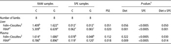
As shown in Table 1, the SPE purification procedure strongly reduced the values of the Folin–Ciocalteu assay in both the liver and plasma (P<0.0005). The dietary treatment had an effect in tendency on the values of the Folin–Ciocalteu assay performed in the liver samples, whereas no significant effect was found in plasma samples (P=0.056 and 0.322, respectively; Table 1). However, in both the liver and plasma samples, there was a Diet×SPE interaction (P=0.050 and 0.030, respectively; Table 1). This showed that, in both the liver and plasma, the effect of the dietary treatment on the Folin–Ciocalteu values was dependent on the SPE purification treatment of the samples. Indeed, the Folin–Ciocalteu values of samples that had not been SPE-treated (i.e. RAW samples) were higher in the liver of pasture-fed lambs compared with the tissues of concentrate-fed animals (+13.13%, P=0.029). Data of the Folin–Ciocalteu values of RAW-plasma samples had a higher variability (higher pooled standard error; PSE) compared with RAW-liver samples (Table 1). Nevertheless, a trend similar to that observed in the liver was evident for RAW plasma, with the Folin–Ciocalteu values being higher in tendency in samples from lambs in the G group compared with the C group (+4.27% P=0.088). Conversely, when the liver and the plasma samples were treated with SPE, the Folin–Ciocalteu assay showed no differences between the experimental treatments (Table 1).
Effect of the dietary treatment (C=concentrate; G=grass) and of the SPE treatment on the Folin–Ciocalteu and FRAP values of the liver and plasma samples (least square mean values and PSE)

SPE=solid-phase extraction; FRAP=ferric-reducing antioxidant power; PSE=pooled standard error; GAE=gallic-acid equivalents.
1 P-values of the test of the fixed effects performed with the MIXED procedure statistical analysis. The fixed effects tested were dietary treatment (Diet; C or G), SPE treatment (SPE; RAW samples or SPE samples) and their interaction (Diet×SPE).
2 Expressed as mg of GAE/g of the liver, or as mg of GAE/ml of the plasma.
3 Expressed as mmol of Fe2+ equivalents/g of the liver, or as mmol of Fe2+ equivalents/ml of the plasma.
a,b,cWithin a row, different superscript letters indicate significant differences between least squares means (P<0.05) tested using the Tukey’s adjustment for multiple comparisons.
FRAP assay of the lamb tissues
Table 1 also shows that the SPE purification procedure strongly reduced the FRAP assay values in both the liver and plasma (P<0.0005). Dietary treatment had a significant effect on the values of the FRAP assay performed in the liver and plasma samples (P=0.001 and 0.009, respectively; Table 1). Furthermore, in both the tissues, the Diet×SPE interaction was significant (P=0.001 and 0.014; Table 1). This showed that, in both the liver and plasma, the effect of the dietary treatment on the FRAP values was affected by SPE treatment of samples. Indeed, as shown in Table 1, the values of the FRAP assay performed on the RAW samples were much higher in both the liver and plasma from pasture-fed lambs compared with the tissues from concentrate-fed animals (+20.03%, P<0.0005 for the liver samples and +12.27%, P=0.002 for the plasma samples). However, when the liver and plasma samples had been treated with SPE, the FRAP assay showed no difference between the experimental treatments (Table 1).
Analysis of feed samples and animal tissues by LC–MS
Representative chromatograms of grass and concentrate diets are given in Supplementary Figure S1. Under the LC–MS conditions used in the present study, phenolic compounds should elute between 5 and 13 min (Wright et al., Reference Wright, Gibson, Spencer, Lovegrove and Gibbins2010). In grass samples, only a few peaks and with relatively low intensity were observed in the range where phenolic compounds tend to elute (see Supplementary Figure S1). These compounds were flavonols and most were present as glycosides (Table 2). The mass m/z (M-H−) of 755.211 (C33H45O20) could be assigned to kaempferol-3-O-glucosyl-rhamnosyl-galactoside, 563.1438 (C26H28O14) to kaempferol-xylosyl-rhamnoside, 593.1549 (C27H30O15) to kaempferol-3-O-rutinoside and 489.1054 (C23H22O12) to kaempferol-3-O-acetyl-glucoside. The m/z value (M-H−) at 285.043 was assigned to kaempferol and m/z (M-H−) 609.1506 to rutin, which is a glycoside of quercetin. None of these compounds could be detected in LC–MS chromatograms from the concentrate diet.
LC–MS analysis of grass diet (see Supplementary Figure S1)

LC–MS=liquid chromatography–mass spectrometry.
1 Although not confirmed by other means, it is likely that the flavonols are glycosylated at carbon 3.
The liver and plasma extracts, which had been treated with SPE, were analysed for the same phenolic compounds as found in the grass diet or for possible metabolites arising from their degradation. No masses corresponding to phenolic compounds could be detected in the liver or plasma samples from the grass-fed lambs (see Supplementary Figures S2 and S3). Similarly, liver and plasma chromatograms from the concentrate-fed lambs also contained no detectable phenolic compounds.
Discussion
Several previous studies have demonstrated that pasture feeding can positively affect the antioxidant status of the animal tissues. For instance, Descalzo et al. (Reference Descalzo, Rossetti, Grigioni, Irurueta, Sancho, Carrete and Pensel2007), found that the muscle from the cattle raised on pasture had a higher overall antioxidant status, in terms of FRAP values, than meat from the grain-fed animals. Furthermore, using the same lambs in the present study, Luciano et al. (Reference Luciano, Biondi, Scerra, Vasta, López-Andrés, Valenti, Lanza, Priolo and Avondo2012) showed an improved oxidative stability of the muscle from lambs raised on pasture. In agreement with these observations, in the present study, we found that the total antioxidant status (FRAP and Folin–Ciocalteu values) of the liver and plasma samples, which had not been subjected to SPE purification (RAW samples), were increased by pasture feeding. It is known that diets based on the consumption of herbage at pasture result in much higher intakes of natural antioxidants and can also positively affect the endogenous antioxidant defences of the animal tissues compared with conventional concentrate-based diets (Descalzo and Sancho, Reference Descalzo and Sancho2008). Both FRAP and Folin–Ciocalteu assays measure the overall antioxidant status and are not specific for particular antioxidants, but react with a wide spectrum of antioxidant compounds (Benzie and Strain, Reference Benzie and Strain1996; Georgé et al., Reference Georgé, Brat, Alter and Amiot2005). Therefore, the improved FRAP and Folin–Ciocalteu values found here in the RAW samples from grass-fed animals simply suggest a positive effect of pasture on the animal antioxidant status, which is likely to be due to pasture feeding improving several different antioxidant defences and thus confirming previous findings.
However, the specific objective of the present study was to assess whether phenolic compounds present in ryegrass could contribute to improving the antioxidant status of the liver and plasma of grazing lambs. Therefore, we have used the FRAP and Folin–Ciocalteu assays also for the liver and plasma samples, which had been subjected to an SPE purification treatment to selectively isolate and purify phenolic compounds, while removing other interfering compounds (Lόpez-Andrés et al., Reference López-Andrés, Luciano, Vasta, Gibson, Biondi, Priolo and Mueller-Harvey2013). This explains the much lower FRAP and Folin–Ciocalteu values found in the SPE-treated liver and plasma extracts compared with the RAW samples. Interestingly, after the SPE purification treatment, differences in the overall tissue antioxidant status between pasture- and concentrate-fed lambs disappeared, as similar Folin–Ciocalteu and FRAP values of the liver and plasma were found between G and C lambs. On the basis of these results, it would appear that the increased antioxidant capacity of the liver and plasma consequent to pasture feeding was not because of the phenolic compounds that might have been transferred from grass to the animal tissues.
To further test our hypothesis, we first analysed the composition of phenolic compounds in the herbage grazed by the animals. We then analysed lamb liver and plasma samples for the same compounds and for possible metabolites. Ryegrass (L. perenne) is a member of the Poaceae family and is one of the most important pasture grass species for animal feeding (Cai et al., Reference Cai, Stewart, Inoue, Yuyama and Hirata2011). There is surprisingly little information available on the polyphenols that are present in this plant. Several studies analysed simple phenolic acids in ryegrass (Hartley and Jones, Reference Hartley and Jones1976, Reference Hartley and Jones1977) and two recent reports also described the presence of flavonoids (Tu et al., Reference Tu, Rochfort, Liu, Ran, Griffith, Badenhorst, Louie, Bowman, Smith, Noel, Mouradov and Spangenberg2010; Qawasmeh et al., Reference Qawasmeh, Obied, Raman and Wheatley2012). As plants belonging to the Poaceae family appear to have a narrower range of polyphenolic compounds than plants belonging to the Fabaceae family (Harborne, Reference Harborne1967; Marais et al., Reference Marais, Mueller-Harvey, Brandt and Ferreira2000; Regos et al., Reference Regos, Urbanella and Treutter2009; Reynaud et al., 2010), therefore it would be interesting to evaluate whether feeding of forage legumes would lead to larger antioxidant effects in the lamb tissues.
The chromatogram of the grass sample (Supplementary Figure S1a) showed only few peaks and with relatively low intensities between 5 and 13 min. These compounds were identified mainly as kaempferol glycosides (Table 2), which occur in many plants (Chopin and Dellamonica, Reference Chopin and Dellamonica1988). Tu et al. (Reference Tu, Rochfort, Liu, Ran, Griffith, Badenhorst, Louie, Bowman, Smith, Noel, Mouradov and Spangenberg2010), recently identified several flavonols in L. perenne, including some of the compounds identified in the present study, that is, kaempferol-3-O-rutinoside, rutin and kaempferol-3-O-glucosyl-rhamnosyl-galactoside. However, these authors did not report the presence of kaempferol, kaempferol-O-acetyl-glucoside and kaempferol-xylosyl-rhamnoside found in the present study (Table 2).
It is likely that when L. perenne is ingested phenolic compounds are metabolised first in the rumen, then in the liver and finally they may be excreted as conjugated compounds in urine (Martin, Reference Martin1982). Indeed, it is known that some phenolic acids of forage cell walls can be degraded to hippurate, which is excreted in the urine, and this compound has been used as an indicator of detoxification load (Foley et al., Reference Foley, Iason and McArthur1999). It is also known that absorbed, low molecular weight phenolics, such as orcinol and quinol, can contribute to the digestible energy content of the diet without necessarily contributing to its nutritional value (Iason and Murray, Reference Iason and Murray1996). Although it has been reported that dietary flavonoids (quercetin, genistein, catechin) are absorbed by humans (Paganga and Rice-Evans, Reference Paganga and Rice-Evans1997; Day et al., Reference Day, Mellon, Barron, Sarrazin, Morgan and Williamson2001; Manach et al., Reference Manach, Williamson, Morand, Scalbert and Rémésy2005), no phenolic compounds or any of the above metabolites could be detected, in our study, in the liver or plasma from lambs fed the grass diet. Three possible explanations may be offered to explain these findings. First, the phenolic compounds from ryegrass were not absorbed in the digestive tract of lambs or they were quickly excreted in the urine. Indeed, the bioavailability of these polyphenols in ruminants is as yet unknown and, in monogastric animals, it has been reported that most dietary polyphenols are quickly eliminated in both the urine and bile after ingestion (Scalbert et al., Reference Scalbert, Morand, Manach and Rémésy2002). Second, flavonoids from grass could have been absorbed in the gastrointestinal tract, but had already been excreted by the time the sampling was carried out. In the present study, the blood and liver samples were taken at least 12 h after the last grass feeding and no information is available on the turnover of polyphenols in ruminants. Third, the concentration of phenolic compounds in L. perenne was too low for appreciable deposition into the animal tissues.
However, it is noteworthy that that the results of the present study are not directly comparable to those of other authors who studied the bioavailability of dietary phenolic compounds in ruminants and reported that some phenolic compounds were transferred from the diet to the animal tissues. For example, Gladine et al. (Reference Gladine, Rock, Morand, Bauchart and Durand2007), administered a single acute dose of plant extracts (rosemary, grape, citrus or marigold) directly into the rumen of sheep. The authors found that, after administration, epicatechin was detected in the plasma of animals dosed with grape extract, whereas naringenin was detected in the plasma of lambs dosed with citrus extract. However, some phenolic compounds commonly found in rosemary (rosmarinic acid) or in marigold (vanillic, caffeic, chlorogenic, p-coumaric and ferulic acids) were not detected in the plasma of sheep. A study by Moñino et al. (Reference Moñino, Martínez, Sotomayor, Lafuente and Jordán2008) found that the muscle of lambs receiving the milk of ewes fed with a rosemary-rich concentrate contained some of the phenols that were present in the diet of the ewe. However, of the numerous phenolic compounds present in rosemary leaves, only three were actually found in lamb meat (rosmarinic acid, carnosol and carnosic acid). Clearly, these plant extracts and aromatic plants contained relatively higher concentrations and different phenolic compounds (Zheng and Wang, Reference Zheng and Wang2001) than ryegrass.
Conclusions
In conclusion, the results of this study confirm the positive effect of a pasture-based diet on the overall antioxidant status of ruminant tissues compared with a concentrate-based diet. However, it was not possible to attribute this effect to the transfer of phenolic compounds from herbage to the animal tissues, as suggested by the fact that when the liver and plasma extracts were concentrated to isolate phenolics, there was no difference in the overall antioxidant capacity between the pasture- or concentrate-fed animal tissues. Furthermore, with the analytical procedures adopted in the present study, none of the phenolic compounds present in L. perenne or their metabolites could be detected in the liver or plasma samples from grass-fed lambs. Therefore, it appears that the improvement of the tissue antioxidant status in lambs consequent to a grass-based diet could not be linked to the intake of polyphenols from grass. Therefore, the phenolic compounds present in L. perenne appear to have a negligible impact on the animal antioxidant status relative to other antioxidant mechanisms induced by grass feeding. Further studies are needed to understand the bioavailability of flavonoids and to identify their role in the global antioxidant status of ruminants. Furthermore, it would be of interest to study the possible antioxidant effects of a wide range of dietary polyphenols, especially if present at high concentrations in other commonly used forages, such as forage legumes.
Acknowledgements
The authors gratefully acknowledge funding from the European Community financial participation under the Seventh Framework Programme for Research, Technological Development and Demonstration Activities, for the Integrated Project LOWINPUTBREEDS FP7-CP-IP 222623. The views expressed in this publication are the sole responsibility of the authors and do not necessarily reflect the views of the European Commission. Neither the European Commission nor any person acting on behalf of the Commission is responsible for the use that might be made of the information contained herein.