Introduction
Vestibular schwannoma has an incidence which approaches 20 per million per year,Reference Stangerup, Tos, Thomsen and Caye-Thomasen1 and accounts for approximately 8 per cent of intracranial tumours.Reference Lalwani2 In Denmark, sporadic unilateral vestibular schwannomas smaller than approximately 20 mm (extrameatally) are followed by yearly magnetic resonance imaging (MRI). If the tumour is cystic, larger than approximately 20 mm or expanding, surgery or radiotherapy is recommended. Patient age, psychology and co-morbidity may influence this management strategy. The aim of surgery is total tumour removal and minimal post-operative morbidity. However, complete tumour removal may increase the risk of facial nerve paresis.Reference Arriaga, Luxford, Atkins and Kwartler3–Reference Martin, Fox, Ho, Holder, Walsh and Irving6 Consequently, the surgeon may intentionally leave residual tumour tissue behind in order to preserve facial nerve function. Furthermore, fragments of tumour are sometimes left behind due to adherence to the brainstem, cerebellum, blood vessels or other cranial nerves. Based on the surgeon's assessment, the extent of tumour removal may be divided into three groups: total, near-total and subtotal excision.Reference Freeman, Ramsden, Saeed, Alzoubi, Simo and Rutherford7–Reference Sanna, Falcioni, Taibah, Donato, Russo and Piccirillo10 ‘Subtotal excision’ is defined as excision of less than 95 per cent of the original tumour. ‘Near-total excision’ is defined as excision of more than 95 per cent of the original tumour, while the term ‘total excision’ is used when the surgeon estimates that the entire tumour has been excised.
The aim of this study was to assess the occurrence of residual vestibular schwannoma intra-operatively and at MRI one year after surgery, as well as the need for revision surgery and the facial nerve function in relation to the extent of tumour removal. In addition, patient age, gender and primary treatment strategy were related to the occurrence of residual tumour.
Materials and methods
Since 1975, all patients with a unilateral cerebello-pontine angle tumour resembling a vestibular schwannoma had been registered prospectively in a database at one centre in Denmark (a country of 5.5 million inhabitants). Of the 2008 patients registered between January 1976 and January 2008, 1143 were operated upon via the translabyrinthine approach. Parameters in the vestibular schwannoma database included: age; sex; tumour size; facial nerve function (according to House–Brackmann staging); MRI before and one year after surgery; treatment strategy; and extent of tumour removal, assigned by the surgeon as either total excision, near-total excision (i.e. tiny fragments of tumour capsule, less than a few millimetres, left behind) or subtotal excision (i.e. remnant larger than a few millimetres).
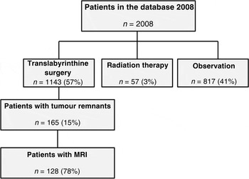
Residual tumour was retained in 165 patients operated upon using the translabyrinthine approach (14 per cent). Of these 165 patients, 25 (15 per cent) died of causes unrelated to their vestibular schwannoma, before the first follow-up MRI could be performed. Patients treated in the 1970s and 1980s did not receive MRI scanning, even if residual tumour was present, as this imaging modality was not available at that time; these patients underwent MRI scanning when it became available. In 12 patients (7 per cent), MRI was not performed because of emmigration, claustrophobia, or patients declining a follow-up MRI scan performed to exclude tumour growth. Thus, MRI scans taken at least one year after surgery were available for 128 patients (78 per cent of patients with residual tumours) (Figure 1).
Flow diagram showing the study population. MRI = magnetic resonance imaging

The occurrence and amount of residual tumour one year post-operatively were evaluated on 1.5-tesla MRI scans using T1-weighted sequences with a slice thickness of 1 mm, with gadolinium enhancement and fat suppression. Subsequently, yearly MRIs were performed in all tumour remnant patients to check for tumour remnant growth.
The indication for revision surgery or radiotherapy depended on tumour remnant growth, subsequent size and patient symptoms. Choice of management strategy was further influenced by age, comorbidity and psychological factors. Until 2002, patients with total tumour excision only received post-operative MRI scanning if they had unexpected symptoms (n = 891 patients). After 2002, a routine MRI scan was performed on all patients one year after surgery (n = 252 patients).
Fisher's exact test was used to compare subgroups regarding the presence of residual tumour on the follow-up MRI. The chi-square test was used to compare the occurrence and extent of incomplete tumour resection between groups. One-way analysis of variance, with Bonferroni correction for multiple comparisons, was used to compare pre-operative tumour size and age. A p value of less than 0.05 was chosen to indicate statistical significance. All calculations were made using the SPSS version 17.0 software program. The reported tumour sizes represent the largest extrameatal diameter, and do not include the intrameatal part of the tumour.
Results
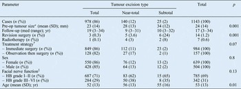
Details of the surgical outcome of the 1143 translabyrinthine cases are provided in Table I.
Translabyrinthine surgery for vestibular schwannoma: patient data and outcomes

*Largest extrameatal diameter. †Two patients had primary radiosurgery and subsequent surgery, and are not included in the analysis of treatment stategy. ‡Data were missing for 16 patients. Pre-op = pre-operative; SD = standard deviation; med = median; yr = years; Revn = revision; HB = House–Brackmann
Total excision was achieved in 978 patients (86 per cent), following a median follow-up period of 19 years for revision surgery (range, 3–34 years). Three of these 978 patients (0.3 per cent) required revision surgery, following the unexpected finding of a tumour remnant at routine MRI scanning one year post-operatively and subsequently growth of that tumour remnant.
Near-total excision was achieved in 140 patients (12 per cent), following a median follow-up period of 9 years for revision surgery (range, 3–31 years), of whom 5 (3.6 per cent) required revision surgery.
Twenty-five patients (2 per cent) had subtotal excision, of whom 6 (24 per cent) required revision surgery (median follow-up time, 10 years; range, 3–32 years). In four cases of subtotal excision, debulking was the primary surgical goal.
There was a significant correlation between the need for revision surgery and the tumour remnant size (p = 0.001). The pre-operative tumour size was significantly smaller in patients undergoing total excisions than in those receiving near-total or subtotal excisions (23 mm versus 28 and 34 mm, respectively; p < 0.001).
Seven patients (0.6 per cent) received stereotactic radiotherapy because of a growing tumour remnant (Table I). One patient underwent radiotherapy and subsequent revision surgery. One of the 978 total excision patients (0.1 per cent) received radiotherapy for an unexpected, growing remnant. Radiotherapy was received by four patients (3 per cent) with near-total excisions and two patients (8 per cent) with sub-total excisions.
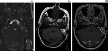
The results of MRI scans taken one year after surgery are given in Table II. Tumours which had undergone near-total excision were not visible in 65 per cent of cases. More surprisingly, 11 per cent of subtotally excised tumours were not visible one year after surgery (for an example, see Figure 2). The proportion of tumour remnants which were between 1 and 10 mm was 20 per cent in the near-total excision group and 17 per cent in the subtotal excision group. One patient (1 per cent) had a tumour remnant larger than 30 mm.
Magnetic resonance imaging (MRI) scans of a patient with a subtotally excised tumour not visible either one or two years post-operatively. (a) Pre-operative axial MRI scan; (b) axial, T1-weighted, contrast-enhanced MRI scan taken one year post-operatively; (c) axial, T1-weighted, contrast-enhanced MRI taken two years post-operatively.

Patients with non-total tumour excision: residual tumour size one year post-operatively*

* On magnetic resonance imaging scan. Pts = patients
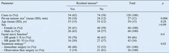
Patients undergoing near-total tumour excision were significantly older than those receiving total tumour excision (mean ages of 56 and 52 years, respectively; p = 0.01). The mean age of patients receiving subtotal tumour excision was 55 years. Age had no influence on the occurrence of residual tumour at MRI one year after surgery. Patients requiring revision surgery had a mean age of 43 years, and were significant younger than patients not needing revision surgery (p = 0.04).
Gender had no impact on the initial operation result or the occurrence of residual tumour (Table I and III). Six women and eight men required revision surgery.
Patients with non-total tumour excision: data by one-year post-operative residual tumour status*

* From magnetic resonance imaging scan. †Largest extrameatal diameter. ‡Facial nerve function data were missing for one patient. **One patient had primary radiotherapy and subsequent surgery and was thus excluded. Pre-op = pre-operative; SD = standard deviation; yr = years; HB = House–Brackmann
Of the 1143 patients who underwent surgery, 157 had initially been observed for tumour growth. These ‘wait and scan’ patients had significantly smaller tumours than the 984 patients who had undergone immediate surgery (mean largest extrameatal diameters of 10 and 27 mm, respectively). Two patients received radiotherapy prior to surgery, and were for that reason excluded from the analysis of treatment strategy. In the ‘wait and scan’ group, subtotal excision was seldom performed, but near-total excision was more frequent than in the immediate surgery group. Compared with the immediate surgery group, a significantly smaller proportion of the ‘wait and scan’ group had tumour remnants identified on follow-up MRI scans (48 versus 19 per cent, respectively; p = 0.03).
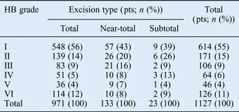
Facial nerve function one year after surgery, according to the House–Brackmann grading system, is given in Table IV. For statistical purposes, facial nerve function outcome was divided into good (i.e. House–Brackmann grade I–II) or poor (House–Brackmann grade III–VI). There were no statistically significant differences in facial nerve function between patients undergoing total, near-total or subtotal tumour excision (good facial nerve outcomes were seen in 71, 62 and 65 per cent, respectively) (p = 0.13). There was also no statistically significant difference in facial nerve function between patients with and without residual tumour on follow-up MRI (p = 0.17).
Facial nerve function one year post-operatively, by tumour excision type*

* Data were missing for 16 patients. HB = House–Brackmann; pts = patients
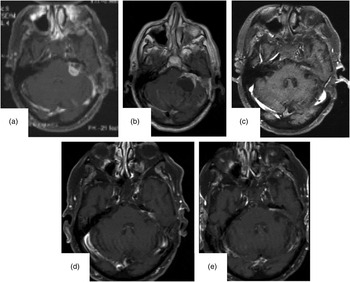
As described above, revision surgery was performed when growth of a tumour remnant was observed at the yearly follow-up MRIs performed in the patients with incomplete tumour removal. As a case example, magnetic resonance images from a patient requiring revision surgery are shown in Figure 3.
Magnetic resonance imaging (MRI) scans for a 74-year-old patient with a 38 mm vestibular schwannoma operated upon without complications, achieving near-total excision (according to the surgeon). (a) Axial MRI taken one year post-operatively showing an 18-mm remnant. (b) Axial MRI taken two years post-operatively showing cystic tumour growth to 36 mm; the patient had gait disturbance and left hemi-paresis, and underwent repeated surgery. (c) Axial MRI taken one year after re-operation; subsequently, the patient was observed for growth of the tumour remnant. (d) Axial MRI taken two years after re-operation, showing no change. (e) Axial MRI taken three years after re-operation, showing no change.

Discussion
To date, the present study is the largest case series investigating residual tumour after translabyrinthine resection of vestibular schwannoma.
The study found that 58 per cent of all tumour remnants were not visible on follow-up MRI one year after surgery. Surprisingly, 11 per cent of subtotally excised tumours were likewise not visible on the follow-up MRI. Other studies have found similar regression in near-totally excised tumoursReference Godefroy, van der Mey, de Bruine, Hoekstra and Malessy8, Reference Lye, Pace-Balzan, Ramsden, Gillespie and Dutton11 but not in sub-totally excised tumours. A possible explanation for the observed tumour disappearance may be post-operative tumour necrosis due to devascularisation, as a result of disruption of tumour feeding vessels during surgery. Studies of staged tumour resections have found that residual tumour remnants are relatively avascular.Reference Donzelli, Motta, Cavallo, Maiuri and De Divitiis12 Vascular endothelial growth factor is a potent mediator of angiogenesis; since this factor is related to vestibular schwannoma growth,Reference Cayé-Thomasen, Werther, Nalla, Bøg-Hansen, Nielsen and Stangerup13 it may help explain why some tumour remnants grow and others do not. Another possible explanation is detection limitations on MRI scans with contrast enhancement, even when employing 1-mm slices. Persistent, non-specific radiological enhancement within the post-operative field is common and this makes the diagnosis of tumour recurrence challenging. Nodular enhancement on the initial post-operative MRI increases the risk of future recurrence, compared with linear enhancement.Reference Carlson, Van Abel, Driscoll, Neff, Beatty and Lane14
The outcome of tumour remnants is controversial. The reported proportion of cases with regrowth requiring revision surgery varies between 15 and 56 per cent,Reference Lownie and Drake4–Reference Martin, Fox, Ho, Holder, Walsh and Irving6, Reference Sanna, Falcioni, Taibah, Donato, Russo and Piccirillo10, Reference Beatty, Ebersold and Harner15–Reference Silverstein, Rosenberg, Flanzer, Wannamaker and Seidman19 considerably larger percentages than in the present series, in which the need for revision surgery was very small (1.2 per cent). A study by Carlson et al. Reference Carlson, Van Abel, Driscoll, Neff, Beatty and Lane14 of MRI surveillance following vestibular schwannoma resection found that 12 of 203 patients experienced recurrence at a mean of 3.0 years following surgery. Patients receiving subtotal excision recurred earlier than those receiving total or near-total excision. The present study had a median follow up of 19 years (considering need for revision surgery).
In the current study, the size of the tumour remnant was the most important predictor of the need for revision surgery. We found that 0.3 per cent of patients with total excision, 3.6 per cent with near-total excision and 24 per cent with subtotal excision required revision surgery. Freeman et al. Reference Freeman, Ramsden, Saeed, Alzoubi, Simo and Rutherford7 reported revision surgery frequencies of the same order, with revision surgery required in 0.5 per cent of total excision, 1.6 per cent of near-total excision and 26 per cent of subtotal excision patients. El-Kashlan et al. Reference El-Kashlan, Zeitoun, Arts, Hoff and Telian17 reported a need for revision surgery in 39 per cent of subtotally excised and 6 per cent of near-totally excised vestibular schwannomas.
It has been suggested that female patients have a more aggressive course.Reference Freeman, Ramsden, Saeed, Alzoubi, Simo and Rutherford7 Most studies of revision surgery find a higher female to male ratio (1.5 women to 1 man).Reference Freeman, Ramsden, Saeed, Alzoubi, Simo and Rutherford7, Reference Sanna, Falcioni, Taibah, Donato, Russo and Piccirillo10 In our study, however, we found a higher proportion of male revision surgery patients. There was no difference between men and women regarding intra-operative and MRI assessment of tumour removal. A previous study found no correlation with progesterone and oestrogen receptors.Reference Curley, Ramsden, Howell, Healy and Lye20
Patients receiving initial observation and subsequent surgery had smaller tumours pre-operatively than patients undergoing immediate surgery without observation. Surprisingly, there were more near-total excisions in the observation plus surgery group, compared with the immediate surgery group, although the pre-operative tumour size was smaller in the former group. One possible explanation is that most patients in the observation plus surgery group were operated upon after approximately 1990, when the strategy of initial observation for smaller tumours was introduced, and when there was increasing awareness of the feasibility of retaining residual tumour tissue.
There was a non-statistically significant trend towards better facial nerve function in patients undergoing total tumour excision compared with those undergoing near-total or subtotal excision. This was probably because, in patients for whom near-total or subtotal excisions were chosen, tumours were more adherent to a relatively thinner or more distended facial nerve. This hypothesis is supported by the fact that the pre-operative tumour size was larger in near-total and subtotal excision patients, compared with total excision patients (27 and 37 mm versus 23 mm, respectively; p < 0.001).
• At surgery, residual vestibular schwannoma may intentionally be left to preserve facial nerve function or prevent complications
• Most residual tumour remnants disappear spontaneously, probably due to devascularisation
• Few patients with a small tumour remnant will require revision surgery
• Tumour remnant size is the most important predictor of revision surgery
On the whole, our study's facial nerve function results were comparable to those of other authors. Falcioni et al. Reference Falcioni, Fois, Taibah and Sanna21 found a lower prevalence of good facial nerve function (i.e. House–Brackmann grade of I–II): 65 per cent of total resection patients and 50 per cent of partial resection patients.Reference Falcioni, Fois, Taibah and Sanna21 On the other hand, Arriaga and ChenReference Arriaga and Chen22 and Gjuric et al. Reference Gjuric, Wigand and Wolf23 found a somewhat higher prevalence of good facial nerve function; however, the tumour sizes in their study population were smaller.
Conclusion
Based on follow-up MRI scans one year after translabyrinthine surgery for vestibular schwannoma, we conclude that a large number of residual tumours disappear spontaneously, probably due to devascularisation. In this study, very few patients with residual tumour required revision surgery. The size of the tumour remnant is the most important predictor of the need for revision surgery.