Introduction
Alongside cognitive symptoms, the majority of patients with dementia in Alzheimer's disease (AD) or mild cognitive impairment (MCI) show neuropsychiatric symptoms (NPS) (Lyketsos et al., Reference Lyketsos2011). Common NPS include among others depression, apathy, agitation, delusions, and hallucinations. Individual NPS tend to form discernible subgroups (Van Der Linde et al., Reference Van Der Linde, Dening, Matthews and Brayne2014). There is evidence of a high frequency of NPS in MCI (Apostolova and Cummings, Reference Apostolova and Cummings2008) and AD (Lyketsos et al., Reference Lyketsos2011), yet it is reported to vary considerably. A review (Cerejeira et al., Reference Cerejeira, Lagarto and Mukaetova-Ladinska2012) reported frequencies of NPS across studies in MCI (up to 85%) and dementia (up to 98%). Higher rates of NPS in clinical than in community-based settings were found in both dementia (91–96% vs. 56–98%) and MCI (75 vs. 43%) (Apostolova and Cummings, Reference Apostolova and Cummings2008). A meta-analysis of NPS in AD (Zhao et al., Reference Zhao2016) found a wide range for the frequency of individual NPS in AD across studies, for instance for apathy (19–88%), delusions (9–59%), hallucinations (6–41%), or aberrant motor behavior (0.8–70%). In a meta-regression, however, they found an overall heterogeneous correlation of different factors to the frequency of NPS in AD, such as study setting or evaluation method.
An increasing frequency of NPS along increasing severity of AD is described (Lopez et al., Reference Lopez2003), but conflicting results were found. Some authors (Starr and Lonie, Reference Starr and Lonie2007) reported a weak relationship between NPS and cognitive performance in AD, whereas others failed to detect an association between NPS and cognitive performance, regarding cognitive symptoms and NPS as independent dimensions of dementia (Lam et al., Reference Lam, Leung, Lui, Leung and Chiu2006). Some individual NPS seem to be associated with the level of cognitive decline: depression is reported to predominate in MCI (Ismail et al., Reference Ismail2017), apathy is described as most common and stable throughout different stages of AD, and psychotic symptoms are found to be highly prevalent in more severe stages of AD (Craig et al., Reference Craig, Mirakhur, Hart, Mcilroy and Passmore2005).
The most consistently reported subgroups in studies with factor analyses are depression, psychosis, and hyperactivity (Van Der Linde et al., Reference Van Der Linde, Dening, Matthews and Brayne2014), though number and composition of subgroups vary across studies. Most studies found between three and five subgroups (Cerejeira et al., Reference Cerejeira, Lagarto and Mukaetova-Ladinska2012). Some individual NPS in these subgroups, such as depression, hallucinations, and delusions, appear as stable across studies. Other NPS, however, such as motor disturbance, night-time behavior or anxiety, seem to be less specific and were included in different subgroups or overlapped across them. Motor disturbance for instance co-occurred with depression, anxiety, delusions, hallucinations, and night-time behavior in a subgroup labelled “mood/psychosis/psychomotor agitation” in one study (Zuidema et al., Reference Zuidema, De Jonghe, Verhey and Koopmans2007), in another study (Aalten et al., Reference Aalten2008), however, with agitation, euphoria, disinhibition, and irritability in a subgroup labelled “hyperactivity.”
Most studies into the frequency of NPS in MCI and dementia analyzed samples with fewer than 500 subjects. They vary, however, in setting, rating tools, and included patients with dementia of varying etiologies and severity (Cerejeira et al., Reference Cerejeira, Lagarto and Mukaetova-Ladinska2012). Only few studies included MCI and AD in one setting. Unlike ours, they were community based (Lyketsos et al., Reference Lyketsos, Lopez, Jones, Fitzpatrick, Breitner and DeKosky2002; Okura et al., Reference Okura, Plassman, Steffens, Llewellyn, Potter and Langa2010) or based on a small sample for MCI (n = 28) and AD (124) (Hwang et al., Reference Hwang, Masterman, Ortiz, Fairbanks and Cummings2004). We found only two larger studies (Zuidema et al., Reference Zuidema, De Jonghe, Verhey and Koopmans2007; Aalten et al., Reference Aalten2008) with sample sizes between 1,000 and 2,000 subjects that analyzed the subgroups of NPS in different stages of dementia. These two studies, however, diverged in several aspects. They included outpatients from multiple centers (Aalten et al., Reference Aalten2008) or nursing-home patients (Zuidema et al., Reference Zuidema, De Jonghe, Verhey and Koopmans2007) and described diverging frequencies for depression (37% vs. 20%), apathy (56% vs. 34%), and delusions (19% vs. 14%) as well as diverging solutions for their NPS subgroups. One study (Aalten et al., Reference Aalten2008) revealed the subgroups hyperactivity, affective, apathy, psychosis, and a subgroup with euphoria and motor disturbance, the other one (Zuidema et al., Reference Zuidema, De Jonghe, Verhey and Koopmans2007) revealed the subgroups agitation/aggression, psychosis, depression, psychomotor agitation, and apathy. In addition, their conclusions on NPS subgroups across different stages of dementia varied. One study concluded that the profile did not vary with severity (Zuidema et al., Reference Zuidema, De Jonghe, Verhey and Koopmans2007), whereas the other one (Aalten et al., Reference Aalten2008) found some differences, since a subgroup with euphoria and motor disturbance appeared only in severe dementia, while the psychosis subgroup disappeared. Neither study included patients with MCI.
The aim of this cross-sectional study was to describe the frequency of NPS and identify their subgroups in a large cohort of subjects with MCI and AD of varying severity. Our study differs from previous studies particularly by the size of its clinical sample with more than 2,000 enrolled subjects. We, thus could study a group of robustly diagnosed AD patients larger than in any comparable study we know of. To date, only few studies into the frequency and the subgroups of NPS included subjects with MCI and AD of varying severity in one setting. On the background of the mentioned inconsistencies, the varying results across studies and especially the mostly small sample sizes of previous studies, we expect to contribute valuable findings to the literature.
Material and methods
Material
This is a cross-sectional registry-based clinical study assessing frequency and subgroups of NPS in subjects with MCI and different stages of AD (mild, moderate, and severe). The study is based on the Norwegian Registry of persons with cognitive symptoms (NorCog), which to date includes 4,571 subjects. The registry was appointed the status of a combined national research and quality registry in 2013 and is run by the Norwegian National Advisory Unit on Ageing and Health. Inclusion and collection of data are based on patients' and relatives' informed consent in writing. Throughout Norway, 26 memory clinics participate in collecting data. The registry is approved by the Norwegian Data Protection Authority and the Regional Committee for Medical and Health Research Ethics (REC 2016/592). In this study, we included subjects with MCI or AD for whom Neuropsychiatric Inventory-Questionnaire (NPI-Q) score (Kaufer et al., Reference Kaufer2000) and Mini-Mental State Examination (MMSE) (Folstein et al., Reference Folstein, Folstein and Mchugh1975) were available.
Examinations and diagnoses
Three groups of subjects were included: MCI; mild, and moderate-severe AD. The subjects were examined following a common protocol. Diagnostic assessment consisted of medical history, standard assessment scales, physical examination, blood samples, and structural imaging (computerized tomography or magnetic resonance imaging (MRI)). In some subjects, 18-fludeoxyglucose positron emission tomography or single photon emission computed tomography, and cerebrospinal fluid (CSF) analysis of beta-amyloid total tau and phosphorylated tau protein were used to aid in the diagnosis. In these subjects, CSF was examined for cells, protein, and glucose. Protein electrophoresis and analysis of antibodies for Borrelia Burgdorferi were also performed.
Clinical assessment
The physical examination included measurement of height, weight, and blood pressure, a preliminary neurological examination with emphasis on signs of cortical damage such as aphasia or anopsia and other signs such as hemiparesis, sensory loss, or ataxia. The psychiatric evaluation was conducted with emphasis on cognitive symptoms such as memory impairment, especially short-term memory, loss of orientation, and concentration.
Neuropsychiatric and cognitive assessment
NPS were examined by the NPI-Q. Cognitive tests included tests from the Consortium to Establish a Registry for Alzheimer's Disease (CERAD) (Morris et al., Reference Morris1989), such as the MMSE, verbal fluency test (Isaacs and Kennie, Reference Isaacs and Kennie1973), and ten word list (Atkinson and Shiffrin, Reference Atkinson and Shiffrin1971). Further, cognitive tests were the clock drawing test (CDT) (Shulman, Reference Shulman2000), and the trail-making test, a and b (TMT A and B) (Reitan, Reference Reitan1955).
The NPI-Q is an informant-based questionnaire, providing a brief assessment of neuropsychiatric symptomatology in clinical practice settings. It is based on the NPI (Cummings et al., Reference Cummings1994). The NPI-Q is widely used and validated (Kaufer et al., Reference Kaufer2000). The questionnaire includes 12 neuropsychiatric items: delusions, hallucinations, agitation/aggression, depression, anxiety, elation/euphoria, apathy/indifference, disinhibition, irritability/lability, motor disturbance, night-time behaviors, and appetite/eating. For each of the 12 NPI-Q items, the interview provides an initial screening question with response options “Yes” (present) and “No” (absent). If the response is “Yes,” the informant rates the severity of the symptoms on a three-point-scale (mild, moderate, and severe). NPS were considered as present if at least one NPI-Q item had a score > 0. The Norwegian version of the NPI-Q has been validated (Rogne and Ulstein, Reference Rogne and Ulstein2012).
MMSE (Folstein et al., Reference Folstein, Folstein and Mchugh1975) assesses global cognitive functioning. It provides 20 items concerning orientation, registration and recall, attention, language, following commands, and figure copying. The maximum score is 30. A lower score indicates worse cognitive functioning and a score <24 have been considered to reflect cognitive impairment (O'connor et al., Reference O'connor1989). The Norwegian version (MMS-NR) has been validated (Engedal et al., Reference Engedal, Haugen, Gilje and Laake1988).
The verbal fluency test (Isaacs and Kennie, Reference Isaacs and Kennie1973) measures impairment in verbal production, semantic memory, and language. Subjects are asked to name as many animals as possible in 1 minute. The score is the total number of different animals named.
The ten word list from the CERAD battery (Atkinson and Shiffrin, Reference Atkinson and Shiffrin1971), evaluates the ability to remember newly learned information. A list of ten words is presented in three trials in random order and the subject tries to recall all ten. The scoring represents the total number of correct words learned. The maximum number of correct responses is 30 for the three trials.
The CDT (Shulman, Reference Shulman2000) assesses visuoconstructive abilities. The subjects were presented with a circle on a piece of paper and asked to draw the face of a clock, reading “10 after 11.” We used the scoring method as described by Shulman (0–5, where 5 is best).
The TMT (parts A and B) (Reitan, Reference Reitan1955) evaluates attention, executive function, and processing speed. In part A (TMT-A) the subject is asked to draw a line between dots in increasing order (1–25). In part B (TMT-B), the ability to shift between multiple tasks is tested. In addition to the task of part A, the subject is asked to alternate between numbers and letters. Performance is scored from 0 to 4 (0 = cannot complete, 1 = slower than –2 standard deviations (SD) of the norms, 2 = between –1 and –2 SD, and 3 = better than –1 SD).
The subjects’ functional status in activities of daily living (ADL) was assessed by Lawton and Brody's scales for ADL (Lawton and Brody, Reference Lawton and Brody1969): they measure personal ADL (P-ADL) and instrumental ADL (I-ADL). The P-ADL consists of six items, which can be scored from 1 to 5, giving a minimum score of 6 and a maximum score of 30. The I-ADL scale includes eight items, which can be scored from 1 to 5. The items of the I-ADL have different maximum scores, resulting in a minimum score of 8 and a maximum score of 31. On both scales, higher scores indicate greater dependence in the performance of the ADL.
In all participating clinics, the Norwegian versions of the MMSE, verbal fluency test, ten word list, CDT, TMT A and B, and Lawton and Brody's scales were used in standardized interviews according to a common protocol, which was used in previous studies (Tangen et al., Reference Tangen, Engedal, Bergland, Moger and Mengshoel2014; Persson et al., Reference Persson, Braekhus, Selbaek, Kirkevold and Engedal2015).
A diagnosis of MCI was made in accordance with the Winblad criteria (Winblad et al., Reference Winblad2004): (1) the person is neither cognitively normal nor suffering from dementia; (2) there is evidence of cognitive deterioration shown by either objectively measured decline over time and/or subjective report of decline by self and/or informant in conjunction with objective cognitive deficits; and (3) ADL are preserved and complex instrumental functions are either intact or minimally impaired (Winblad et al., Reference Winblad2004).
A diagnosis of AD was made in accordance with the ICD-10 criteria (World Health, 1992): (1) Presence of a dementia. (2) Insidious onset with slow deterioration. (3) Absence of clinical evidence, or findings from special investigations, to suggest that the mental state may be due to other systemic or brain disease. (4) Absence of a sudden, apoplectic onset, or of neurological signs of focal damage.
Meetings were held annually among collaborating clinicians to establish and retain harmonized procedures and diagnostic inter-rater reliability.
Dementia stages in AD
We used MMSE to define categories of dementia severity, as has been done previously (Folstein et al., Reference Folstein, Folstein and Mchugh1975; Lopez et al., Reference Lopez2003; Aalten et al., Reference Aalten2008), as mild, moderate, and severe (MMSE scores ≥20; 19–10; <10). The moderate and severe groups were combined into one moderate–severe group, since the severe group consisted of only 12 patients.
Statistical analysis
Demographic and clinical patient characteristics were described as means and SD or frequencies and percentages, as appropriate. ANOVA was applied to compare continuous variables across patient groups. χ 2-test was used for comparison of categorical variables across patient groups.
To identify subgroups of NPS, factor analysis of NPI-Q items was performed with principal component extraction method and varimax rotation. An oblique rotation was considered as well but due to weak correlations among the factors a varimax method was chosen. The number of components was identified by the Kaiser's criterion of eigenvalues equal to or higher than 1. Cronbach's α was calculated to assess the consistency of the factors. We adhered to the following cut-offs (Hinton, Reference Hinton2004): high (α≥0.75), moderate (0.5<α<0.75), and low consistency (α≤0.5).
All analyses were performed in SPSS v 22. Results with p values below 0.05 were considered statistically significant.
Results
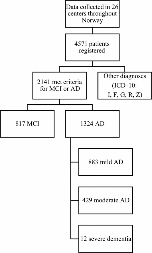
The total sample consisted of 4,571 subjects, with MCI or AD in 2,141 subjects (43.2% male). Of these 817 had MCI, and 1,324 AD (883 mild, 429 moderate, and 12 severe). A recruitment chart is given in Figure 1. The clinical and demographic characteristics of the groups are shown in Table 1.
Recruitment flowchart.

Sample characteristics

SD = standard deviation.
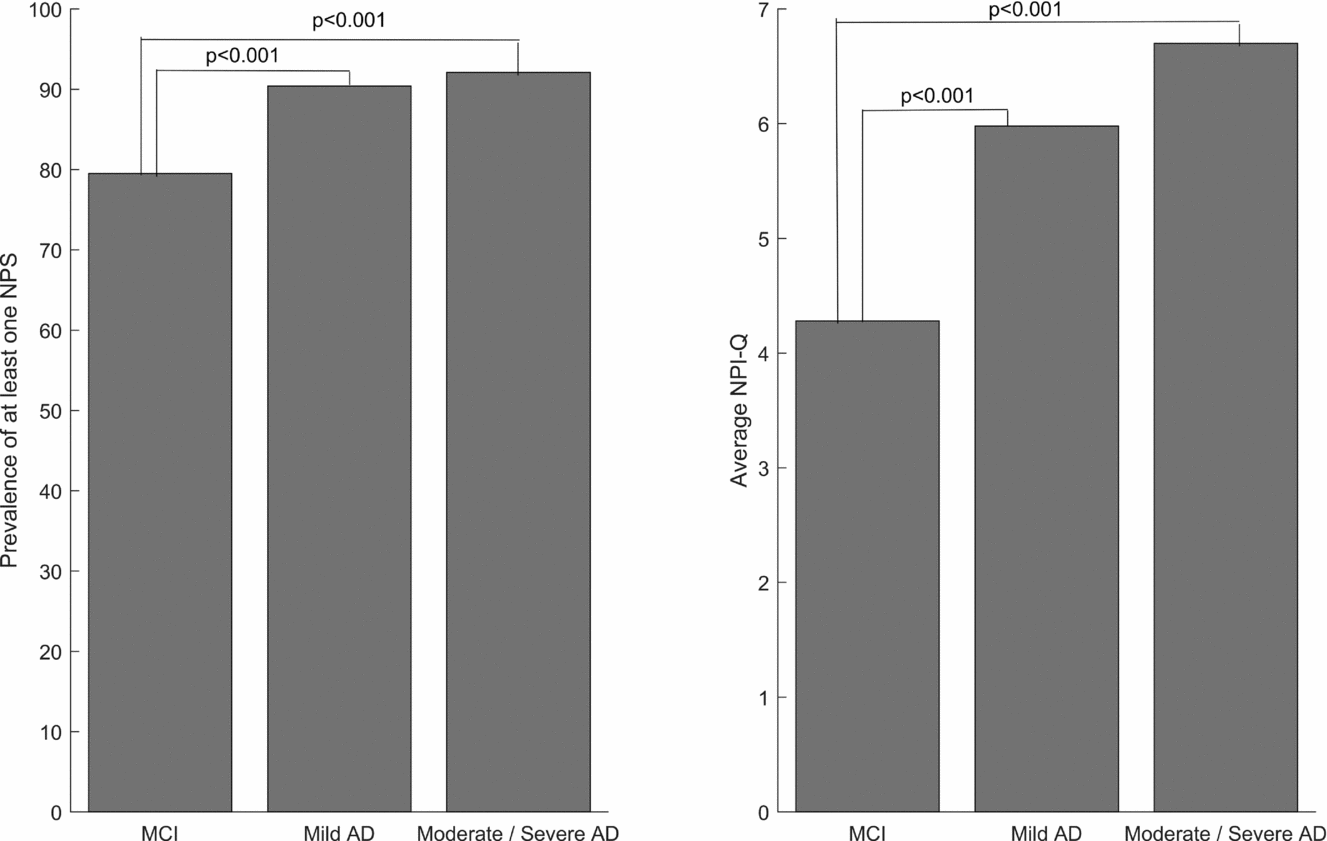
At least one NPS was found in 87.2% of the patients (AD 91.2%, MCI 79.5%). The NPS frequency and the NPI-Q total sum score increased significantly from MCI to mild AD (p < 0.001, χ 2-test) and increased slightly, though non-significantly further from mild to moderate–severe AD, Figure 2. The number of patients with two or more NPS was significantly higher in patients with AD than in those with MCI (AD 79.9%, MCI 65.3%; p < 0.001, χ 2-test).
Frequency of NPS and average NPI-Q sum score.

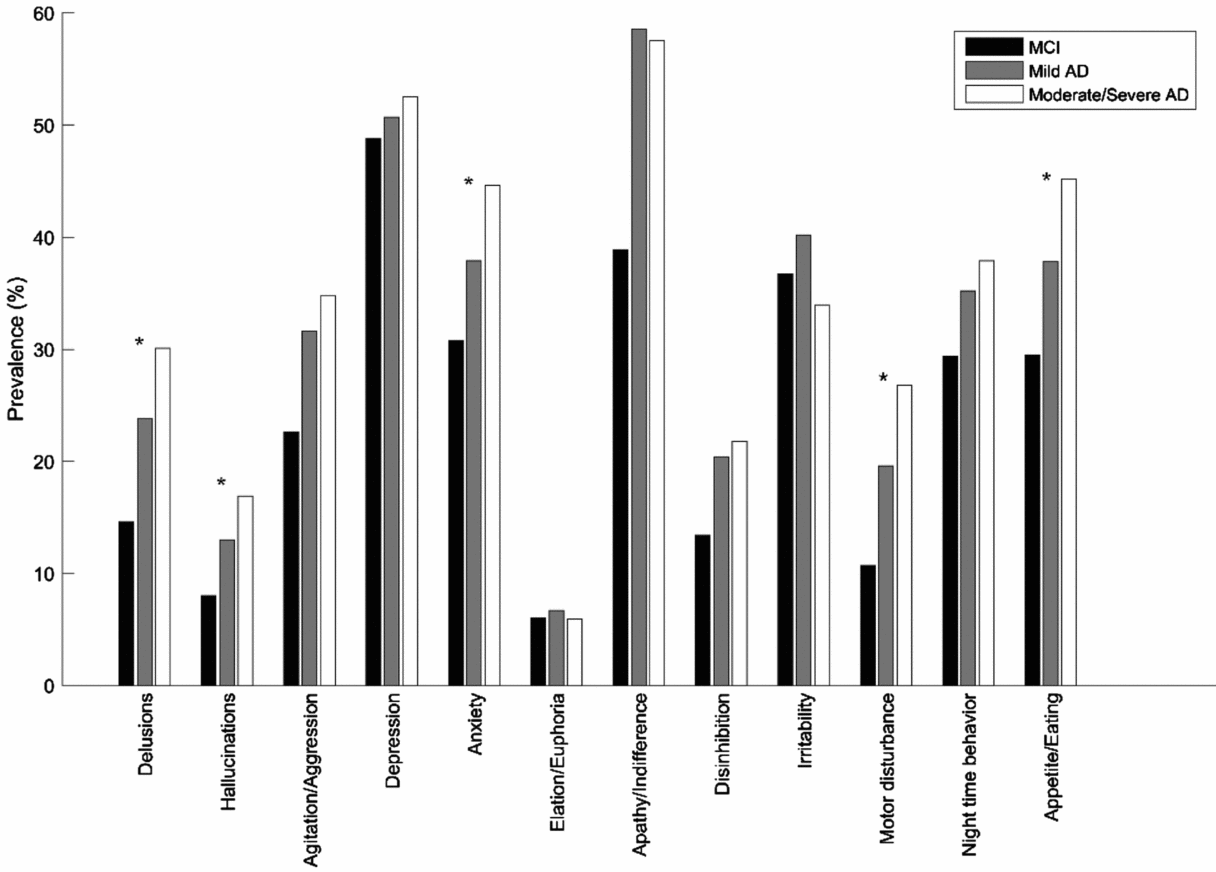
The most common NPS in MCI was depression, followed by apathy. In AD, apathy was the most common NPS and was significantly more common than in MCI (p < 0.001, χ 2-test). The frequencies of delusions and hallucinations increased with increasing severity of cognitive decline (p < 0.001, χ 2-test). Results for all individual NPS across different stages from MCI to moderate–severe AD are shown in Figure 3.
Distribution of individual NPS across different stages of cognitive decline from MCI to moderate-severe AD.

Factor analysis with principal component analysis
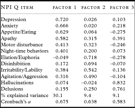
The results of the factor analysis of the entire data set are shown in Table 2. Three factors based on NPI-Q items were extracted, explaining 48.6% of the variance. Factor 1 contained the items depression, anxiety, disturbances in appetite, apathy, motor disturbance, night-time disturbance (30.1% of variance explained), factor 2 contained the items euphoria, disinhibition, irritability, and agitation (9.4% of variance explained), and factor 3 contained the items hallucinations and delusions (9.1% of variance explained). The consistency of the factors was considered moderate with Cronbach's α of 0.68 for factor 1, 0.64 for factor 2, and 0.58 for factor 3. We labelled factor 1 the depression subgroup, factor 2 agitation subgroup, and factor 3 psychosis subgroup.
Results of the factor analysis for the total sample

The factor analysis stratified for the groups MCI to moderate–severe AD (data not shown) showed an unchanged factor composition for MCI and mild AD. The consistency of the subgroups was moderate for depression (Cronbach's α in MCI 0.70, in mild AD 0.64), agitation (Cronbach's α in MCI 0.67, in mild AD 0.65) and psychosis (Cronbach's α in MCI 0.53, in mild AD 0.52). While in moderate–severe AD the factor composition of the subgroups depression and agitation was slightly different, the consistency of both subgroups remained nearly unchanged with a moderate consistency for depression (Cronbach's α 0.65) and agitation (Cronbach's α 0.69). The depression subgroup contained depression and anxiety; the agitation subgroup still contained agitation, disinhibition, irritability, and in addition disturbances in night-time behavior, appetite and eating, and apathy, while it no longer contained euphoria. The psychosis subgroup showed an unchanged composition and a now moderate consistency (Cronbach's α 0.67). An additional factor appeared, containing euphoria and motor disturbance and was labelled elation subgroup. Its consistency, however, was low (Cronbach's α 0.24).
Discussion
In this cohort of more than 2,000 subjects with MCI or AD of varying severity, the frequency of NPS was high in all groups. It increased significantly from MCI to mild AD with a slight further, yet non-significant increase in moderate–severe AD. The following neuropsychiatric subgroups were identified: depression, agitation, psychosis, and elation. Across different levels of cognitive decline from MCI to moderate–severe AD, the subgroups depression, psychosis, and agitation remained relatively stable, whereas the elation subgroup appeared only in moderate–severe AD. Taken together, the available evidence suggests that the pathology of AD contributes to NPS in AD, although it should be noted that not all MCI patients will develop AD or other types of dementia.
Our study supports findings that indicate a high frequency of NPS in both MCI (Apostolova and Cummings, Reference Apostolova and Cummings2008) and AD (Lyketsos et al., Reference Lyketsos2011). We could show that the frequency of NPS was higher in AD than in MCI. This is generally in line with results of community-based studies (Lyketsos et al., Reference Lyketsos, Lopez, Jones, Fitzpatrick, Breitner and DeKosky2002; Okura et al., Reference Okura, Plassman, Steffens, Llewellyn, Potter and Langa2010). These two studies, however, analyzed smaller samples than we did (n = 856, n = 682) and described frequencies of NPS for MCI (44%, 43%) and AD (57%, 75%) that were considerably lower than ours. Moreover, they included dementia of varying etiologies (Lyketsos et al., Reference Lyketsos, Lopez, Jones, Fitzpatrick, Breitner and DeKosky2002; Okura et al., Reference Okura, Plassman, Steffens, Llewellyn, Potter and Langa2010). Where different stages of dementia were analyzed (Okura et al., Reference Okura, Plassman, Steffens, Llewellyn, Potter and Langa2010), the results were partly different from ours, as the frequency of NPS slightly decreased in severe dementia, whereas it slightly increased in this stage in our study. Another clinical study (Hwang et al., Reference Hwang, Masterman, Ortiz, Fairbanks and Cummings2004) found frequencies of NPS in MCI (75%) and mild AD (89%) that were comparable to ours, however, in a sample (n = 202) much smaller than ours. They found a considerably lower frequency of NPS (12%) in a cognitively unimpaired control group (n = 50). Higher frequencies of NPS in clinic versus community-based settings have been described (Apostolova and Cummings, Reference Apostolova and Cummings2008; Cerejeira et al., Reference Cerejeira, Lagarto and Mukaetova-Ladinska2012) and may originate from a referral bias, since patients in clinical treatment may be more impaired than those in a community setting.
The number of subjects in different levels of dementia severity in our study differed from that in previous studies. The two larger studies (Zuidema et al., Reference Zuidema, De Jonghe, Verhey and Koopmans2007; Aalten et al., Reference Aalten2008), that performed a factor analysis in different stages of dementia, analyzed a considerably higher number of moderate to severely impaired patients than we did (n = 1,378, n = 1,793) and a comparable (n = 1,015) (Aalten et al., Reference Aalten2008) or much smaller (n = 59) (Zuidema et al., Reference Zuidema, De Jonghe, Verhey and Koopmans2007) number of subjects with mild dementia. They did so, however, regardless of dementia subtype and the proportion of subjects with AD is unclear. A clinical study (Hwang et al., Reference Hwang, Masterman, Ortiz, Fairbanks and Cummings2004), that assessed the frequency of NPS in MCI and AD, analyzed considerably fewer subjects in both MCI (n = 28) and mild AD (n = 124) and did not include moderate or severe stages of AD.
The most frequent NPS in our MCI group was depression (49%, Figure 3). A high frequency of depression in MCI (27%) has been reported previously in a population-based study (Geda et al., Reference Geda2008). A meta-analysis (Ismail et al., Reference Ismail2017) reported the pooled prevalence of depression in patients with MCI (32%), which varies across clinic (40%) and community-based settings (25%). The interaction of depression and MCI is widely discussed and depression seems to be associated with a higher risk of conversion to dementia (Mourao et al., Reference Mourao, Mansur, Malloy-Diniz, Castro Costa and Diniz2016). It is, however, unclear if depression in MCI is an early manifestation of neurodegeneration or a reaction to the experience of cognitive impairment. Depression and MCI can have overlapping symptoms (Ismail et al., Reference Ismail2017). Depression was the second most frequent NPS in mild (51%) and moderate–severe AD (53%) (Figure 3). The interaction of depression and dementia is complex: depression can appear as “pseudodementia” (i.e. reversible cognitive impairment induced by depression). On the other hand, depression may also be a symptom of the underlying AD pathology. A clinicopathological study describes depression as independent of neuritic plaques and fibrils in MCI and mild AD (Mccutcheon et al., Reference Mccutcheon2016). The most frequent NPS in our AD groups was apathy (58%), which was significantly more frequent than in MCI (39%). Its frequency in moderate–severe AD (58%) was almost as it was in mild AD (59%) (Figure 3). Overall, our frequencies for depression and apathy in AD concur with the findings of a recent meta-analysis (Zhao et al., Reference Zhao2016), reporting frequencies of apathy (49%) and depression (42%). We found delusions and hallucinations were significantly more frequent in moderate–severe AD than in mild AD and MCI (Figure 3). Some authors (Lyketsos et al., Reference Lyketsos2001), though, describe the increase of hallucinations as weak, whereas in our study their frequency was almost twice as high in moderate severe AD than it was in MCI.
The subgroups of NPS we found are both clinically plausible and overall in line with previous findings (Van Der Linde et al., Reference Van Der Linde, Dening, Matthews and Brayne2014). Across studies, however, rating methods for NPS differed. Most studies used the NPI (Cerejeira et al., Reference Cerejeira, Lagarto and Mukaetova-Ladinska2012), but those using other instruments (e.g. Behavioral Pathology in AD Scale, Neurobehavioral Rating Scale, Behavior Rating Scale for Dementia) found comparable results. We found two studies, comparable to ours, that performed a factor-analysis of NPI-Q items. One of these (Johnson et al., Reference Johnson, Watts, Chapin, Anderson and Burns2011) reported a factor solution, that was partly comparable to ours, albeit some divergent NPS were included into their subgroups, labelled “mood” (anxiety, apathy, and depression), “psychosis” (irritability, delusions, hallucinations, and agitation) and “frontal symptoms” (euphoria and disinhibition). However, they analyzed and compared dementia with varying etiologies such as AD, Vascular dementia (VaD), and dementia with Lewy Bodies (DLB). Their AD group was considerably larger than ours (n = 2,474). Another study (Travis Seidl and Massman, Reference Travis Seidl and Massman2016) suggested a factor solution with only two subgroups, labelled Negative/Oppositional (agitation, irritability, apathy, depression, disinhibition, and delusions) and Anxiety/Restlessness (night-time behavior, anxiety, hallucinations, and appetite). Here (Travis Seidl and Massman, Reference Travis Seidl and Massman2016), however, the items motor disturbance and euphoria were excluded from the factor analysis, and the AD sample (n = 256) was considerably smaller than ours. Neither of these analyzed the subgroups in MCI or different stages of dementia.
Minor differences in the subgroups of NPS exist between our study and other studies that used the NPI. One study (Lyketsos et al., Reference Lyketsos2001) described a psychosis subgroup similar to ours, and an affective subgroup (depression, irritability, anxiety, and euphoria), that partly resembled our depressive subgroup, but included irritability and euphoria, which we included into the agitation subgroup. That study, though, used latent class analysis, was community based and examined a much smaller sample of AD patients (n = 198). In another study (Aalten et al., Reference Aalten2003), motor disturbances have been included in the agitation subgroup, which in our analysis have been included in the depression subgroup. That study, however, analyzed a smaller sample and their patients met criteria for varying types of mostly mild dementia, such as AD, VaD, mixed AD and VaD, frontal dementia (FTLD), dementia in Parkinson's disease (PDD) and DLB.
The stratified factor analysis for different stages form MCI to moderate–severe AD showed some changes in both the composition and the number of NPS subgroups. In our MCI and mild AD groups, apathy was included in the depressive subgroup. Apathy and depression can overlap clinically, and there is an ongoing discussion about the status of apathy as a syndrome distinct from depression (Mortby et al., Reference Mortby, Maercker and Forstmeier2012). The definitions of depression and apathy often overlap (Cummings et al., Reference Cummings2015). Apathy and depression may or may not occur together. In moderate–severe AD apathy was included in the agitation subgroup, which has been reported previously (Archer et al., Reference Archer2007). Patients with severe AD often show shifting symptoms, especially between agitation and apathy. Some authors (Archer et al., Reference Archer2007) speculate that co-occurrence of apathy and agitation may be a sign of shared disturbances in the frontal lobe. In our moderate–severe AD group, an additional subgroup was identified, which we labelled elation. Its consistency, however, was low (Cronbach's α 0.24) and this finding should be confirmed or rejected in further studies.
In the few studies that applied factor analyses of NPS in different stages of cognitive decline, we found no agreed upon suggestions for the stability of a factor across different levels of cognitive decline. In different stages of cognitive decline all of our subgroups maintained two or more NPS, which may be looked upon as core symptoms and which we labelled our subgroups after. These NPS were described as relatively specific before (Van Der Linde et al., Reference Van Der Linde, Dening, Matthews and Brayne2014). At any stage of cognitive decline our subgroups were in line with generally suggested compositions of NPS subgroups (Van Der Linde et al., Reference Van Der Linde, Dening, Matthews and Brayne2014). Therefore and because of their almost invariant consistency, we conclude that they are stable across different levels of cognitive decline. Since the elation subgroup appeared only in moderate–severe AD, we regard it as stage dependent, which is in line with the findings of a previous study (Aalten et al., Reference Aalten2008).
There is evidence of a generally high prevalence of NPS across other neurodegenerative diseases, although it varies across diagnoses with reported rates for Huntington's disease (HD) (98%) (Paulsen et al., Reference Paulsen, Ready, Hamilton, Mega and Cummings2001), DLB (95%) (Bjoerke-Bertheussen et al., Reference Bjoerke-Bertheussen, Ehrt, Rongve, Ballard and Aarsland2012), VaD (70%) (D'onofrio et al., Reference D'onofrio2012), and Parkinson's Disease (PD) (60%) (Aarsland et al., Reference Aarsland1999b). A recent review (Cerejeira et al., Reference Cerejeira, Lagarto and Mukaetova-Ladinska2012) indicates varying predominant NPS across dementias of different etiologies, such as depression and anxiety in VaD, psychotic symptoms in DLB, and disinhibition and apathy in FTLD. A study (Aarsland et al., Reference Aarsland2007) into NPS in PDD found depression as the most common NPS in this diagnosis. In HD, depression, and agitation were reported as the most frequent NPS (Paulsen et al., Reference Paulsen, Ready, Hamilton, Mega and Cummings2001). Although these findings may suggest a heterogeneous profile of individual NPS across varying types of dementia, there is some evidence for an overall comparable constellation of subgroups of NPS in AD, PDD, VaD, and DLB (Johnson et al., Reference Johnson, Watts, Chapin, Anderson and Burns2011).
We see some limitations to our study design. We selected subjects with MCI and AD in a clinical setting, and the results may be subject to referral bias and thus not generalizable to the general population. A cross-sectional design fails to detect fluctuations and trajectory of NPS, so that the severity-related associations may not be entirely accurate. We did not apply histopathological examinations for the diagnosis of AD, misdiagnosis thus is possible. We did not adjust for medication, which can influence NPS. The NPI-Q, though validated (Kaufer et al., Reference Kaufer2000) and often used, has shortcomings, as it provides informant-based data, so we may be prone to over-reliance on proxy report. However, studies that used instruments other than the NPI-Q yielded overall comparable results. Analyses conducted at the participating centers may be under-represented in the NorCog registry. Diagnostic routine may vary slightly across the participating centers: all of them collected blood samples; in some cases computered tomography and MRI of the brain were conducted by the general practitioner, nevertheless in accordance to the research protocol; few analyzed CSF and these data were not available.
Some of our findings may be influenced or explained by the specific questions in the NPI-Q. A part of the question about hallucinations (“Does the patient have hallucinations such as false visions or voices”) could have been affected by a part of the question about delusions (“Does the patient have false beliefs?”) or vice versa. These items may be difficult to separate for an informant not trained in psychiatry. The slightly lower Cronbach's α in the psychosis subgroup may possibly be explained by this. The inclusion of motor disturbance in the depression subgroup may be explained by the wording in the NPI-Q (“Does the patient engage in repetitive activities such as pacing around the house, handling buttons, wrapping string, or doing other things repeatedly?”). Motor disturbance, however, is reported to be a key symptom in severe depression, that increases with age (Parker et al., Reference Parker, Roy, Hadzi-Pavlovic, Wilhelm and Mitchell2001). In the NPI-Q, there are no guidelines to distinguish the items apathy (“Does the patient seem less interested in his/her usual activities or in the activities and plans of others”) and depression (“Does the patient seem sad or say that he/she is depressed?”), which may contribute to the sometimes difficult disentanglement of these items. Since we did not include a healthy control group, we could not directly compare the frequencies of NPS in AD and MCI with the normal population. However, previous studies, including a Norwegian study (Aarsland et al., Reference Aarsland, Larsen, Karlsen, Lim and Tandberg1999a), have reported much lower frequencies of NPS in cognitively healthy controls, supporting the conclusion that NPS are associated with the underlying disease.
Strengths of this study include the inclusion of a large sample size assessed with a common standardized protocol and the inclusion of subjects across the full spectrum from MCI to severe AD, which allows us to study different stages of cognitive decline, although the number of patients with severe AD was low. Since frequency and severity of NPS tend to increase with disease progression this will inevitably influence the findings.
Taken together, in our study the frequency of NPS was high in both MCI and AD. It increased only slightly with the severity of AD. Although the subgroups depression and agitation showed changes in their composition, we regard them as stable across different levels of cognitive decline, since they maintained at least two NPS. Our psychosis subgroup was invariant across different levels of cognitive decline, while the elation subgroup was dependent on moderate–severe AD. The observed subgroups can thus be considered reflections of non-cognitive syndromes that occur alongside cognitive disturbances in MCI and AD, regardless of level of cognitive decline. The relevance of the subgroup elation can be debated, since its consistency was low.
Conflict of interest
Siafarikas N, Selbaek G, Fladby T, Šaltytė Benth J, Auning E: Declare no conflict of interest. Dr Aarsland has received research support and/or honoraria from Astra Zeneca, H. Lundbeck, Novartis Pharmaceuticals, Eisai, and GE Health, and serves as paid consultant for H. Lundbeck and Axovant.
Description of authors’ roles
Siafarikas, N: Idea of the study, data analysis, and manuscript writing. Selbæk, G and Fladby, T: Protocol/project development, data collection, and manuscript editing. Šaltytė Benth, J: Data analysis and manuscript editing. Auning, E: Protocol/project development and manuscript editing. Aarsland, Dag: Idea of the study, Protocol/project development, and manuscript writing/editing.
Acknowledgments
Dag Aarsland is a Royal Society Wolfson Research Merit Award Holder and would like to thank the Wolfson Foundation and the Royal Society for their support. This study represents independent research partly funded by the National Institute for Health Research (NIHR) Biomedical Research Unit for Dementia at South London and Maudsley NHS Foundation Trust and King's College London. The views expressed are those of the author and not necessarily those of the NHS, the NIHR or the Department of Health.
Supplementary material
To view supplementary material for this article, please visit https://doi.org/10.1017/S1041610217001879