Introduction
Acute supraglottitis is a life-threatening condition that can progress into sudden airway obstruction. This condition may present with non-specific signs; its high mortality rate (up to 20 per cent) results from misdiagnosis and inappropriate management.Reference Khilanani and Khatib1 Supraglottitis associated with acute submandibular sialadenitis is an extremely rare presentation. Prompt diagnosis of this co-morbidity and recognition of submandibular sialadenitis are essential for the optimal management of this potentially fatal condition.Reference Carey2
This report describes the case of a patient who presented with acute exacerbation of a chronic condition. The presence of additional worrying features required prompt diagnosis and urgent investigation and treatment.
Case report
A 70-year-old Caucasian woman with a background of insulin-dependent diabetes, hypertension, chronic renal failure, unstable angina and triple coronary artery bypass grafting, was admitted as an emergency under the care of ENT surgeons. She reported a four-week history of a tender, right-sided submandibular neck swelling that fluctuated in size. She had previously attended the department's acute referral clinic and had subsequently undergone ultrasound scanning of the neck. Unfortunately, the swelling had reduced at that time, and mild hypervascularity of the right submandibular gland was the only remarkable finding. There was no improvement, and the swelling had increased in size in the two days prior to admission. The patient also complained of a sore throat and difficulty in swallowing, along with symptoms of breathlessness and a change in voice quality. She was not stridulous, but was intermittently febrile. She denied any relieving factors and the pain worsened on palpation.
Clinical examination revealed a smooth, tender, firm swelling (15 × 8 cm in size) in the submandibular region, with no changes to the overlying skin. She had mildly restricted neck movement, but no palpable cervical lymphadenopathy. Generalised oedema of the floor of mouth was seen on inspection of the oral cavity. Flexible nasal endoscopy revealed gross swelling of the right vestibular fold and arytenoid mucosa. The vocal folds were unaffected and functional. The airway was not imminently compromised.
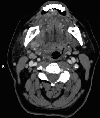
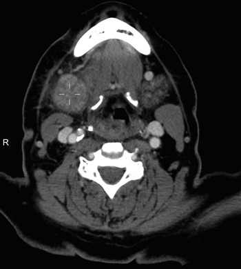
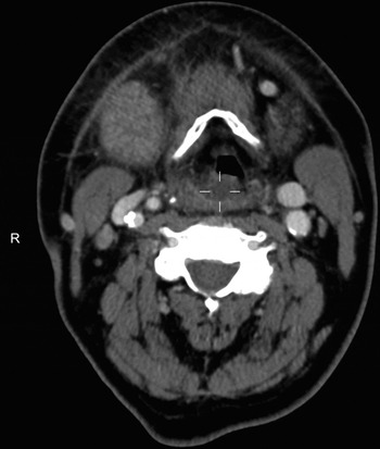
An urgent computed tomography (CT) scan of the neck and chest showed right submandibular sialadenitis with extensive adjacent inflammatory changes secondary to an impacted 3-mm right Wharton's duct calculus (Figures:1 and 2). An inflamed, oedematous right supraglottis and aryepiglottic fold (consistent with supraglottitis) was also seen (Figure 3). No infective collections were seen.
Axial computed tomography image showing a submandibular gland duct calculus measuring 3 mm in diameter. It is situated within the floor of the mouth directly posterior to the mandibular lateral incisor.

Axial computed tomography image showing heterogenous enhancement of the right distal submandibular gland without gross extraglandular ductal dilatation. There is marked asymmetry and enhancement compared with the left side.

Axial computed tomography image showing that the right aryepiglottic fold is oedematous, with narrowing of the supraglottic airway. The right submandibular gland is enlarged, with associated periglandular fat stranding consistent with inflammation.

A decision for medical treatment was taken and the patient was treated with regular intravenous dexamethasone and clindamycin (she had a known penicillin allergy). She was closely monitored, particularly for airway obstruction, and her condition improved over the subsequent few days: the swelling reduced, her voice improved and the pain decreased. The calculus became palpable and was removed under local anaesthesia. The patient was finally discharged with a course of oral antibiotics.
Discussion
Obstructive sialadenitis
Obstructive sialadenitis is the most common benign disorder of the salivary glands. Important aetiologies include duct calculi and stenosis, but there are several others, including mucous plugs, foreign bodies, neoplasia, ductal polyps and immunological conditions (e.g. Sjögren's syndrome).Reference Meulemans, Hens and Vander Poorten3
Sialolithiasis is the formation of a calculus in the salivary glands or ducts. It accounts for up to 60 per cent of cases of salivary gland obstruction.Reference Marchal, Dulguerov, Becker, Barki, Disant and Lehman4 The submandibular gland is particularly susceptible to sialolithiasis because of has more viscous mucus and more alkaline secretions compared with other glands.Reference Lustmann, Regev and Melamed5 The submandibular gland is affected in 80–90 per cent of cases, whilst the parotid and sublingual and/or minor salivary glands are implicated in 5–10 per cent and 0–5 per cent of cases, respectively.Reference Bodner6
Sialoliths are believed to form during periods of secretory inactivity. First, sialomicroliths accumulate and cause obstruction, forming atrophic foci. Bacterial proliferation at these atrophic foci stimulates inflammation, swelling and fibrosis that can affect larger ducts. Further calcium precipitation from solution is caused by salivary flow disturbance; this accumulates on degenerating cell membranes to produce an obstructive sialolith.Reference Harrison7
A Wharton's duct stone typically presents with recurrent periprandial pain and a tender submandibular swelling. Additional systematic symptoms such as fever and malaise indicate the development of a superadded infection. If infected, purulent discharge into the oral cavity occurs.Reference Capaccio, Torretta, Ottavian, Sambataro and Pignataro8 With progression, the area of infection and inflammation can develop into an abscess, requiring emergency care.
Treatment of obstructive submandibular sialadenitis involves rehydration, analgesia, warm compresses, sialogogues, gland massage and antibiotics to treat infections. These measures are used with the aim of spontaneous calculus expulsion. For impacted calculi and recurrent problems, the previous mainstay was intra-oral sialithotomy (with the associated risk of ductal stenosis development) or sialadenectomy (with the risk of damaging the surrounding nerves, poor cosmesis and potentially missed calculi).Reference Meulemans, Hens and Vander Poorten3 Marchal et al. refuted the claim that a chronically inflamed gland cannot regain its normal function by demonstrating a normal histopathological appearance in half of the glands excised for chronic sialolithiasis.Reference Marchal, Kurt, Dulguerov, Becker, Oedman and Lehmann9
Technological advances over the past 20 years have guided preference towards gland-sparing approaches such as sialendoscopy; shock-wave lithotripsy, interventional radiology techniques, endoscopically assisted calculi extraction and transoral surgical stone removal. The increasing use of these techniques, either separately or in combination, has resulted in sialadenectomy being avoided in up to 95 per cent of cases.Reference Meulemans, Hens and Vander Poorten3
Infection and inflammation within the head and neck
Infection and inflammation within the head and neck can present with specific signs and symptoms, as well as site-dependent complications. When dealing with inflammation in this area, the site and surrounding anatomy must be considered because the fascial layers of the neck are important barriers to infection and guide local spread.Reference Rana and Moonis10
Infections are broadly divided into superficial neck infections and deep neck space infections. Superficial neck infections are usually caused by cellulitis, lymphadenitis or abscess. Most commonly, superficial neck infections are seen as cervical lymphadenitis in the paediatric population. If bilateral, they are normally caused by viral upper respiratory tract infections, but unilateral lymphadenitis is usually bacterial. Superficial neck abscesses usually originate from lymphadenitis.
Deep neck space infections are associated with life-threatening complications such as airway compromise, venous thrombosis, sepsis and continued extension. They often present with fever, pain and swelling, but can also display odynophagia, dysphagia, trismus and a change in voice quality.
The deep cervical fascia is divided into three discrete layers: superficial, middle and deep. The superficial layer encloses the strap muscles and sternocleidomastoid muscle. The trachea, thyroid, carotid sheaths, oesophagus and other viscera are enclosed by the middle (or visceral) layer of deep cervical fascia. The deepest layer of the deep cervical fascia encloses the paraspinal muscles and perivertebral space. The retropharyngeal (anterior) and danger (posterior) spaces are located between the visceral and deep layers. They are separated by the alar fascia, which is derived from the deep fascia.
Infections of the floor of mouth can be caused by sialadenitis and/or sialolithiasis, but can also result from odontogenic infection. A severe manifestation of this is Ludwig's angina, which was described by von Ludwig in 1836: he recounted an induration of the connective tissues of the neck, involving the tissues covering the small muscles between the larynx and floor of mouth.Reference von Ludwig11 This results in diffuse cellulitis, submandibular fasciitis, and involvement of the laryngeal and muscular connective tissues. It is considered a maxillofacial emergency because the airway can rapidly become compromised, with up to 75 per cent of patients requiring tracheostomy.Reference Rana and Moonis10
Supraglottitis is cellulitis of the aryepiglottic folds, the epiglottis and adjacent tissues, usually caused by the direct epithelial invasion of pathogens. This is a similarly dangerous condition because progressive oedema and inflammation of the epiglottis and surrounding structures can quickly cause airway obstruction. Mortality rates for this condition range from 7 per cent to 20 per cent and are directly related to the development of airway compromise.Reference Carey2, Reference Bansal, Miskoff and Lis12
In the case described in this report, it is most likely that an intense acute infective process commencing at the submandibular gland caused gross local inflammation. In much the same way as in Ludwig's angina, it may have spread inferoposteriorly towards the base of tongue and supraglottis.Reference Ellis and Mahadevan13
• A thorough medical history is essential for diagnosing patients presenting with neck symptoms
• Increased airway vigilance is required when managing patients with non-typical histories
• Obstructive submandibular sialadenitis can cause intense localised inflammation, leading to supraglottitis
• Prompt recognition of this condition and airway management are crucial
The treatment of supraglottitis involves airway management, intravenous steroid administration and prompt intravenous antibiotic administration. Early liaison with the anaesthesia team is essential because some patients may need to have their airway secured by endotracheal intubation. If this fails, then a surgical approach (e.g. tracheostomy, cricothyroidotomy) is the second line treatment. If possible, it is preferable to perform imaging (especially CT scanning) to map out the affected area and find the source of infection (as in our case). It is also used for planning potential surgical intervention (in cases of abscess formation).
Conclusion
A complete and thorough medical history is essential to establish the nature and progression of disease in patients presenting with neck symptoms. To our knowledge, this is the first reported case of submandibular sialolithiasis resulting in ipsilateral supraglottitis.
It must be emphasised that increased vigilance is required when assessing patients with non-typical medical histories. This was especially true in this case, in which the patient was investigated for a chronic pathology, but displayed acute atypical symptoms.
This case report highlights that obstructive sialadenitis caused by an impacted calculus can cause superadded infection resulting in supraglottitis. Prompt airway management and monitoring of these patients are crucial.
Acknowledgements
We would like to thank Dr A Yong, consultant radiologist at Ninewells Hospital, Dundee, for providing copies of the CT images, along with verbal and written interpretations.