Introduction
The discovery of scanning tunneling microscope (STM) in the beginning of the 1980s was, to a large extent, an engineering feat,Reference Binnig, Rohrer, Gerber and Weibel1 but STM has revolutionized the field of surface science with enormous impact in the basic and applied science of semiconductors and catalysis. STM is conceptually very simple and relies on the measurement of a small tunneling current (on the order of nA) flowing between an atomically sharp metal tip, which is raster-scanned at a subnanometer distance to the surface to be imaged. Atomic-scale control of the tip position is achieved through the use of piezoelectric materials such as lead zirconate titanate, which change their physical dimensions when a voltage is applied across the material. Also, great care must be taken to decouple the scanning tip from acoustic and ground vibrations that would otherwise smear the image.Reference Wiesendanger2
In this article, we focus on examples of unique STM applications to surface chemistry studies. As we shall see, together with information obtained from other microscopy and spectroscopy tools, as well as theory, STM is closer and better at providing insight to fundamental phenomena. This review will focus on examples of unique STM capabilities, including imaging the structure of active sites (e.g., steps, kinks, vacancies, and special atomic geometries in compounds); imaging dynamic processes such as diffusion of adsorbates and reactions; and surfaces with a high density of adsorbates under high pressures of gases.
Such insight can be used to answer, and investigations are indeed very often motivated by, problems in catalysis, the industrial application of which generates enormous economic value. Surprising perhaps to the outsider, atomic-scale insight into phenomena such as those listed previously can often be applied directly to understand the performance of catalysts employed in real-life applications. Examples are plenty, but in the present energy context, one might mention the three-way car catalyst,Reference Bowker3 desulphurization of crude oil (see later),Reference Besenbacher, Brorson, Clausen, Helveg, Hinnemann, Kibsgaard, Lauritsen, Moses, Nørskov and Topsøe4 and fuel cells.Reference Wieckowski and Koper5
Imaging the structure of active sites
In the following examples, we will show how we are approaching a point where fundamental insights into surface structure and reactivity obtained by STM, in combination with other techniques, are enabling the design of new superior catalysts operating under technically relevant conditions, which can help meet today’s global energy challenges.
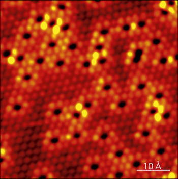
The first example deals with steam-reforming catalysts, which are typically composed of Ni nanoclusters supported on a porous oxide. Steam reforming is a widely used catalytic process to yield carbon monoxide and hydrogen from water and a carbon source (methane, higher alkanes, etc.). Hydrogen, when produced, forms the feedstock in ammonia synthesis. One of the challenges of existing Ni-based catalysts is their rapid passivation by graphite, formed at high pressures and temperatures. While gold and nickel are immiscible in the bulk, high resolution STM images revealedReference Besenbacher, Chorkendorff, Clausen, Hammer, Molenbroek, Nørskov and Stensgaard6 that they form a surface alloy where Au atoms substitute Ni atoms on the top surface layer of Ni(111). These Au atoms perturb the electronic structure of the nearby surface Ni atoms, as revealed by their higher tunneling probability, giving brighter spots in the STM image in Figure 1.Reference Besenbacher, Chorkendorff, Clausen, Hammer, Molenbroek, Nørskov and Stensgaard6 This has the surprising effect of lowering the tendency of the surface to bind carbon and form graphite. Motivated by these fundamental surface science findings, a high-surface-area, MgAl2O4-supported Ni catalyst (with 16.5 wt% Ni) modified with 0.3 wt% Au, was synthesized.Reference Besenbacher, Chorkendorff, Clausen, Hammer, Molenbroek, Nørskov and Stensgaard6 The activity of the steam reforming reaction of n-butane and water to yield H2 and CO on this high surface area Au-Ni catalyst was compared to that of the pure Ni catalyst. The conventional Ni catalyst deactivated rapidly, whereas the conversion factor for the Au-Ni catalyst remained almost constant, indicating that on the new Au-Ni catalyst, graphite formation is significantly reduced.Reference Besenbacher, Chorkendorff, Clausen, Hammer, Molenbroek, Nørskov and Stensgaard6
Scanning tunneling microscopy image of a Ni(111) surface with Au atoms (black areas) alloyed into the surface. Both Au and Ni atoms, in fact, form part of the topmost crystal layer, but due to electronic effects affecting the tunneling probability, Au atoms are imaged as black dots and Ni atoms as red peaks. Ni atoms lying next to Au are imaged brighter due to modifications to their electronic structure caused by the Au atoms.Reference Besenbacher, Chorkendorff, Clausen, Hammer, Molenbroek, Nørskov and Stensgaard6

Another example of rational design of new catalysts, based on information provided by STM studies, concerns ethylene (C2H4) dissociation on Ni(111).Reference Vang, Honkala, Dahl, Vestergaard, Schnadt, Lægsgaard, Clausen, Nørskov and Besenbacher7 The reactivity of catalytic surfaces is often dominated by very reactive low-coordinated atoms, such as those at step edges, but little knowledge exists about the influence of such atoms on the selectivity in reactions of larger molecules involving multiple reaction pathways. Using STM experiments and density functional theory (DFT) calculations, it was shown that the activation of C2H4 on Ni(111) follows the trend of higher reactivity for decomposition at step edges as compared with the higher coordinated terrace sites.Reference Vang, Honkala, Dahl, Vestergaard, Schnadt, Lægsgaard, Clausen, Nørskov and Besenbacher7 The step-edge effect is considerably more pronounced for C–C bond breaking than for C–H bond breaking, and thus steps play an important role in bond-breaking selectivity.
A third example illustrates the unique capabilities of STM to resolve reaction intermediates and active sites in the hydrodesulphurization (HDS) process, which is employed in the production of practically all combustion products derived from crude oil to abate sulphur emissions. The HDS catalyst is known to consist of 2–3 nm wide, single-layer MoS2 nanoparticles, mainly “promoted” with Co or Ni and supported on porous alumina. “Promotion” is a term used in catalysis to describe the enhancement effect seen when very small amounts of a “promoter” compound are added to the main catalyst formulation. In this particular case, Co and/or Ni are known to enhance the performance of the oxide-supported MoS2 nanocluster catalyst, where Co and/or Ni are a component in the real industrial catalyst. The exact atomistic mechanism behind such promotion effects, however, is rarely known, but its use is a result of “black-box” type combinational screening.
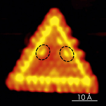
Since only the edges of the S-Mo-S layers in MoS2 are known to be catalytically active, their structure can only be studied on dispersed MoS2 nanoclusters, in this case grown on the chemically inert Au(111) gold surface. Atomically resolved STM images revealed a hitherto unknown electronic “brim” state with metallic character at the edges of the active MoS2 nanoclusters, which imparts unusual chemical characteristics. This state produces the bright band near the edges in the STM images, as shown in Figure 2.Reference Vang, Honkala, Dahl, Vestergaard, Schnadt, Lægsgaard, Clausen, Nørskov and Besenbacher7Figure 2 shows the reaction intermediate, cis-butene-thiolate (C4H7S-, imaged as “bananas”), formed by partial hydrogenation of thiophene (C4H4S) coordinated through the terminal sulfur atom to the metallic brim. Based on this and otherReference Lauritsen, Nyberg, Nørskov, Clausen, Topsøe, Lægsgaard and Besenbacher8, Reference Tuxen, Kibsgaard, Gøbel, Lægsgaard, Topsøe, Besenbacher and Lauritsen9 insights gained from STM studies combined with theoretical DFT calculations, chemical engineers at Haldor Topsøe A/S synthesized a new type of superior HDS catalyst, the BRIM catalyst. The new catalyst formulation enables a higher degree of sulfur removal than any existing catalyst on the market.
Scanning tunneling microscopy image of a molybdenum disulphide cluster. The bright yellow “brim” is a metallic edge state that influences the catalytic reactivity. Yellow “bananas” (marked by dashed ovals) inside the brim are adsorbed partially reacted thiophene.Reference Vang, Honkala, Dahl, Vestergaard, Schnadt, Lægsgaard, Clausen, Nørskov and Besenbacher7

Diffusion of atoms and molecules
Diffusion is an important step in any surface reaction, as the adsorbates move on the surface to encounter other reactants and surface active sites. Insight into adsorbate diffusion is thus important for an atomic-scale understanding of catalyst action in general. While simple site-to-site diffusion is a relatively straightforward and well understood thermally activated process, recent studies have revealed more complicated situations where diffusion can be mediated by a minority species or otherwise influenced by the presence of co-adsorbates.
Diffusion has been studied extensively in the past using a large variety of techniques that analyze areas or volumes of material containing a large number of diffusing species, host atoms, and geometries. However, fundamental insights into diffusion phenomena are difficult to obtain by such space-averaging techniques. STM on the other hand makes it possible to image single atoms and to follow their spatial evolution as a function of time, thus providing truly atomistic information.
The study of water diffusion on Pd(111) by Mitsui et al.Reference Mitsui, Rose, Fomin, Frank Ogletree and Salmeron10 provides an example of the unique type of information provided by STM. Sequential images (i.e., a series of time-lapse STM images of the same surface area) visualized the random walk displacement of water molecules hopping from site to site, with rates that increased with temperature. When diffusion was too fast to capture molecular single hops between neighboring sites, the tip motion was programmed to follow individual water molecules. In this manner, the diffusion activation barrier was measured. As the water coverage increased, the molecules were also seen interacting with each other, forming strongly bound pairs that diffused as a unit. Surprisingly, the diffusion was found to be faster, by several orders of magnitude than that of water monomers, a result explained by the peculiar geometry of the molecular pair, with a donor molecule bound to a Pd substrate atom and an acceptor molecule bound to the first by a H-bond.Reference Ranea, Michaelides, Ramírez, de Andres, Vergés and King11 When, due to thermal fluctuations, the acceptor molecule binds to a neighboring Pd atom, a quantum-tunneling-assisted rotation exchanges H-bonding from one molecule to the other. This process reverses the role of donor and acceptor in the pair such that the unit advances by one lattice spacing once the new acceptor molecule detaches from the Pd atom.
Another example is the study of S diffusion on Re(0001) that was shown to occur in a correlated manner between pairs of S atoms.Reference Hwang, Zeglinski, López Vázquez-de-Parga, Ogletree, Salmeron and Denley12, Reference Dunphy, Sautet, Ogletree, Dabboussi and Salmeron13 Other molecules are also known to diffuse in pairs or even larger clusters, such as CO on Pd(111), as determined using STM.Reference Mitsui, Rose, Fomin, Ogletree and Salmeron14 STM is uniquely suited to study diffusion phenomena.
Reactions
Ertl et al. demonstrated that it is possible to obtain atomic-scale insight into surface reactions in real space and time.Reference Wintterlin, Völkening, Janssens, Zambelli and Ertl15 They studied the oxidation of CO on Pt(111) by performing titration experiments in which an O-covered surface was exposed to CO while monitored by STM. As the CO coverage increased, at 247 K, the O coverage remained constant, and no reaction took place. However, O formed large (2 × 2) islands due to CO-O repulsion. After 140 sec, the (2 × 2) O islands started to shrink, while islands with a c(4 × 2) structure simultaneously nucleated and grew. This was interpreted as CO reacting with the pre-adsorbed O to form CO2, which desorbs and leaves behind vacancies for CO molecules to adsorb from the gas phase. The STM studies revealed that the reactants are not randomly distributed, but rather two different phases exist on the surface, and the reaction occurs only at the boundary between these two phases with a rate proportional to the edge length along the phase boundary. Based on STM experiments carried out at different temperatures, the authors were also able to determine the activation energy for the reaction to be 0.49 eV, in excellent agreement with the 0.51 eV value found in a previous molecular beam study, where a beam of CO molecules was directed toward an oxygen covered surface.Reference Campbell, Ertl, Kuipers and Segner16 The important finding here is that the STM study confirmed a macroscopic measurement and, in addition, provided a detailed atomic-scale view of the reaction.
High coverage surfaces: The temperature-pressure problem
We now examine the important topic of surfaces under high coverage, which are the conditions of most “real” catalyst surfaces during operation. High coverage can be obtained in high vacuum (i.e., below 10–9 bar [10-4 Pa]) by cooling down to a temperature where eventually even weakly bound species will remain adsorbed for long times, even after the gas phase has been pumped away. Here thermodynamics and kinetics combine to create the so-called pressure gap. This refers to the fact that the surface phases generated in vacuum may not represent thermodynamic equilibrium structures at higher pressures and temperatures. It is useful to consider the entropic contribution to the free energy under pressure p, G = kT ln(p/p 0) (p 0 = 1 bar = 105 Pa, k is the Boltzmann constant, and T is temperature), which at room temperature and one bar (105 Pa) is about 0.35 eV, a non-negligible energy. For this reason, efforts to build STM instruments that operate under controlled gas environments at relevant pressures have been under way since the early 1990s.Reference McIntyre, Salmeron and Somorjai17–Reference Rasmussen, Hendriksen, Zeijlemaker, Ficke and Frenken19 The results obtained so far largely justify the effort with the discovery of new phases and phenomena, some of which are briefly reviewed here.
An example is that of NO on the Rh surface, a metal catalyst used in automobile converters to reduce NO and oxidize CO. At pressures of up to 4 Pa at room temperature, it was found that both CO and NO form 2 × 2 periodic structures on Rh(111), with a coverage of 0.75 monolayer (ML).Reference Rider, Hwang, Salmeron and Somorjai20 As the pressure increased beyond 4 Pa, however, NO formed a new phase with 3 × 3 periodicity, not known to exist before. This phase is slightly denser, with 0.78 ML, than the 2 × 2 structure. Images obtained during the addition of CO to this surface revealed that CO displaces NO from the hollow sites first, and later, when all the sites have been filled by CO, the NO molecules on the top sites are also displaced.Reference Rider, Hwang, Salmeron and Somorjai21 These results were later verified spectroscopically by x-ray photoelectron spectroscopy experiments under similar gas pressures.Reference Requejo, Hebenstreit, Ogletree and Salmeron22
Stability of surfaces at high gas pressures
Finally, we examine the question of the stability of surfaces under ambient pressures and near room temperature and above. It is known from surface science studies in vacuum that adsorbates can alter the free energy of a surface and drive it to reconstruct. Since catalytic reactions occur at the surface, it is obviously important to know the structure of the surface on which reactants diffuse, meet, and react. Again, such insight is crucial for a detailed understanding of any catalyst, including the ones employed in energy applications.
The Pt(100) and Pt(110) surfaces are notorious for reconstructing when adsorbates are adsorbed.Reference Thostrup, Christoffersen, Lorensen, Jacobsen, Besenbacher and Nørskov23, Reference Thostrup, Kruse Vestergaard, An, Lægsgaard and Besenbacher24 The well-known 5 × 1 structure of Pt(100), consisting of a hexagonal overlayer of Pt atoms on top of the (100) surface, breaks down into islands of square symmetry upon CO adsorption.Reference Behm, Thiel, Norton and Ertl25, Reference Ritter, Behm, Potschke and Wintterlin26 At atmospheric pressure in the case of H2, O2, and CO, Pt(110) undergoes extensive restructuring, with the surface changing from a 1 × 2 missing row to multiple nested missing rows, vicinal (111) facets, and large terraces in atmospheres of each of these gases, respectively.Reference McIntyre, Salmeron and Somorjai17
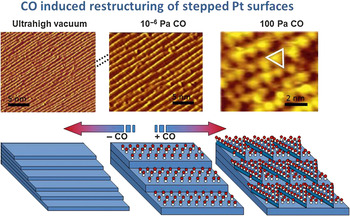
More recently, stepped (111) vicinal surfacesReference Tao, Dag, Wang, Liu, Butcher, Bluhm, Salmeron and Somorjai27 were shown to reconstruct dramatically under CO when the pressure reached around 100 Pa. As the CO pressure increased from ultrahigh vacuum, first the terraces and steps doubled in width and height. Then at 100 Pa, the coverage of CO approached 1 ML, an extremely dense concentration where the separation between CO molecules becomes that of the substrate, 2.78 Å, much smaller than the 3.7 Å separation at a coverage of 0.68 ML obtained in high vacuum . The high density is accommodated by a large restructuring of the substrate metal atoms that form clusters, providing a high density of low coordinated Pt atoms. This geometry alleviates the CO-CO repulsion by allowing tilting of the edge-bound CO molecules, which is not possible on the flat terrace.Reference Tao, Dag, Wang, Liu, Butcher, Bluhm, Salmeron and Somorjai27 STM images corresponding to these observations are shown in Figure 3. This is the surface that is active during reaction conditions, not the ones observed in vacuum or even moderately low CO pressures. The implications of these findings for understanding catalytic reactions are obvious.
Left: Scanning tunneling microscopy images of Pt(557) in ultrahigh vacuum (1 × 10⁻⁹ Pa), showing the monoatomic steps and terraces running diagonally from top right to bottom left. Center: Under ∼10−6 Pa of CO, the steps double in height and the terraces in width due to the 0.5 monolayer of CO. Right: Under 100 Pa of CO, the surface breaks up into roughly triangular clusters (one is marked by a triangle) that provide a high density of low coordinated Pt atoms. The CO coverage is now close to 1 monolayer. This is illustrated by the schematic figures at the bottom. The C and O atoms of the CO molecules are represented by white and red balls, respectively.Reference Tao, Dag, Wang, Liu, Butcher, Bluhm, Salmeron and Somorjai27

Conclusion
The last 25 years of application of scanning tunneling microscopy (STM) to surface chemistry studies have revolutionized the field. Very important and otherwise difficult or impossible to observe phenomena have been discovered. Reactive sites and new phases have been determined for the first time. STM has also made it possible to follow dynamic processes of diffusion and reaction. Important new developments, such as the design of STM setups that can operate at high gas pressures, are opening the new field of surface science under realistic environmental conditions,Reference Lægsgaard, Österlund, Thostrup, Rasmussen, Stensgaard and Besenbacher28 helping to bridge the pressure-temperature and materials gap in our understanding of surface phenomena. Such setups, however, are still quite rare, but the future will likely see their more widespread use in academia and industry, since local atomistic insight into catalysis can, in practicality, only be obtained with probe techniques. As we have seen in this review, surprising new details emerge from such studies, which shift the current understanding of catalysis. Significant challenges, however, lie ahead in approaching “real-life” conditions in terms of ever higher pressures and temperatures as well as in the synthesis of more realistic catalyst model systems, which incorporate, for example, promoter and substrate effects. Catalysts are known to exhibit transient phases during reaction, so we also see a move toward in operando analysis where the catalyst is monitored under working conditions. Here again, STM and other scanning probe techniques will play a significant role.
Acknowledgments
F.B. and P.T. were supported by the Villum Foundation, the Carlsberg Foundation, the Danish National Research Foundation, and the European Research Council. M.S. was supported by the Office of Science, Office of Basic Energy Sciences of the US Department of Energy under Contract No. DE-AC02–05CH11231.