Fistula between the right pulmonary artery and the left atrium is a rare type of pulmonary arteriovenous fistulas. The physical examination of newborns reveals continuous murmurs, central cyanosis, signs of heart failure, and tiredness on feeding. In older children, exertional dyspnoea and clubbing may also be present. The left atrium, left ventricle, and pulmonary artery may appear larger than normal on two-dimensional echocardiography. Colour Doppler ultrasonography shows continuous flow patterns. By following the continuous flow on colour Doppler ultrasonography, the origin, course, and opening of the fistula can be identified. Although echocardiography is sufficient to make the diagnosis in most cases, catheter angiography is a good method for finalising the diagnosis and selecting the most appropriate treatment method. We present a case diagnosed by fetal echocardiography as an arterial venous malformation who developed signs of heart failure and cyanosis in the newborn period.
Case report
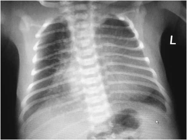
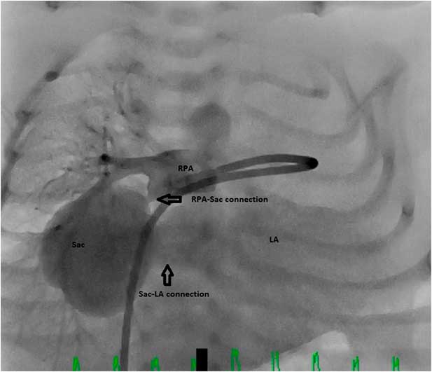
A 26-year-old pregnant woman was referred for fetal echocardiography after detection of cardiomegaly on obstetric ultrasonography performed in the 20th week of gestation. On fetal echocardiography, we suspected the presence of a fistula or arteriovenous malformation that carried blood from the aorta to a 10×12 mm sac behind the left atrium, which formed a wide union with the left atrium (Supplementary videos 1 and 2). Continuous-wave Doppler demonstrated continuous flow pattern. The case was followed with fetal echocardiography for fetal heart failure at regular intervals until birth. Although dilatation occurred in the left heart chambers during the intrauterine period, no finding suggestive of growth retardation or congestive heart failure was observed. He was born in the 38th week of gestation and weighed 3200 g. Physical examination revealed central cyanosis and a continuous murmur in the right hemithorax. The oxygen saturation was measured as 85%. The anterior–posterior chest radiogram showed that pulmonary vascularity and the cardio-thoracic ratio (0.76) was increased (Fig 1). The echocardiographic study revealed a sac behind the left atrium sized 22×26 mm, a fistula that carried blood from the right pulmonary artery to this sac, and a wide communication between the sac and the left atrium (Supplementary videos 3 and 4). The echocardiogram demonstrated no reverse flow in the distal right pulmonary artery. A patent ductus arteriosus with right to left shunting was present. As the patient manifested signs of congestive heart failure, it was necessary to treat the fistula in the newborn period. On postnatal day 15, the patient was taken to the catheter laboratory. Angiogram showed dilated pulmonary artery and contrast agent was carried from the right pulmonary artery to a sac localised via a narrow fistula, and the sac was widely communicated with the left atrium (Fig 2) (Supplementary video 5). Transcatheter closure was discussed for the patient; however, surgery was preferred because after the occlusion of the fistula, the sac that formed a wide communication with the left atrium would have been left behind. There would be a risk for systemic embolism until the sac undergoes involution. The surgery was performed on the 25th day of life. The mediastinum was entered through a midline sternotomy and the thymus was resected. The aorta and the caval veins were cannulated. A 2-mm arterial duct was closed before the institution of cardiopulmonary bypass. A 3-mm fistula was identified proximal to the lower lobe artery of the right pulmonary artery branch and was ligated. The right atrium was opened and a left atrial sump was placed via the patent oval foramen after the onset of ventricular fibrillation. Diastolic arrest was done with cold crystalloid cardioplegia given through the aortic root cannula. The patient was cooled down to 26°C. Interatrial septum was resected to enter the left atrium. The left atrium was inspected under total circulatory arrest. The mitral valve, the left atrial appendage, and the four pulmonary veins were identified to be normal. A 3.5-cm aneurysmal pouch with a wide opening was seen posterior to the right pulmonary veins. The pouch was closed with a running 7-0 Prolene suture. After a 14-minute period of circulatory arrest, perfusion was reinstituted. Interatrial septum was constructed with a bovine pericardial patch. The patient was rewarmed and the heart gained sinus rhythm after the removal of the aortic clamp. The weaning from bypass was uneventful with a low dose of dopamine and milrinone. Two chest drains were left in place and the sternum was closed with sternal wires in usual fashion. The postoperative course was uneventful. He was extubated the next day and left the ICU on the 2nd postoperative day. The patient was discharged on the 5th day after surgery.
The anterior-posterior chest radiogram showed that pulmonary vascularity and the cardio-thoracic ratio (0.76) was increased.

Angiogram showed contrast agent was carried from the right pulmonary artery to a sac localized via a narrow fistula, and the sac was widely communicated with the left atrium.

Discussion
Fistula between the right pulmonary artery and the left atrium was first described by Friedlich and colleagues in 1950 and is a rare cyanotic CHD.Reference Friedlich, Bing and Blount 1 These fistulas are three times more likely to be in males compared with females. The severity of symptoms depends on the amount of the right to left shunt through the fistula. In fetal life, it runs its course with enlargement of the left heart chambers, congestive heart failure, and intrauterine growth retardation. In the postnatal period, symptoms such as central cyanosis, polycythaemia, continuous murmur, and the signs of congestive heart failure are observed. In older children, exertional dyspnoea and clubbing may also be present. A fistula between the right pulmonary artery and left atrium may lead to serious complications such as systemic embolism, infective endocarditis, and brain abscesses.Reference Kawano, Hirano, Ikeno, Fuwa and Uchino 2 , Reference Zeebregts, Nijveld, Lam, van Oort and Lacquet 3 Transthoracic echocardiography, CT angiography, and in selected patients catheter angiography are used for diagnosis. In our case, cardiomegaly was identified in the 20th week of fetal life. The fetal echocardiography revealed continuous flow to a sac posterior to the heart, but origin could not be depicted at that time. The fetus was followed for cardiac failure with fetal echocardiography, and the diagnosis was confirmed with transthoracic echocardiography after birth. After birth, our case developed hepatomegaly, central cyanosis, tachypnea, and dyspnoea during feeding.
Although surgical treatment was mostly preferred until recently, a number of recent case reports describe transcatheter device closure.Reference Zeebregts, Nijveld, Lam, van Oort and Lacquet 3 , Reference Krishnamoorthy and Rao 4 – Reference Ece, Uner, Cuce and Balli 9 In our case, we elected for surgical repair of the fistula, as we thought that the patient would be at risk for systemic thromboembolism following transcatheter device closure of the fistula, which would leave the large aneurysmal sac with stagnant blood flow in direct communication with the left atrium. In patients who undergo transcatheter closure, the duration of hospital stay after the procedure is much shorter compared with surgery and complications such as phrenic nerve paralysis are avoided. However, at least 6 months of anticoagulant use should be considered to minimise the risk of systemic embolism after transcatheter closure of right pulmonary artery–left atrium fistulae.Reference Ece, Uner, Cuce and Balli 9 , Reference Ding, Xu, Liu, Yan and Qin 10 Patients do not require anticoagulant therapy after surgical closure. Besides this, surgery substantially reduces the risk of residual shunts. As is the case in our patient, potential embolic complications can be minimised by removing the sac in cases with a large sac. In our case, the infant was rather small, the fistula was associated with a wide sac, and there was a risk of thrombus formation in the sac. Therefore, the fistula and wide sac were closed with surgery. The patient was discharged from hospital on day 5 after surgery.
In conclusion, pulmonary arteriovenous malformations are among rare congenital cardiac diseases that cause cyanosis and heart failure in the newborn period. Heart failure may present in fetal and postnatal periods and surgical closure is a safe treatment method. We want to emphasise that arteriovenous malformations and fistulae should be considered in the differential diagnosis in cases with unexplained cardiomegaly or congestive heart failure in the fetal period and cyanosis or heart failure in any period of life after birth.
Acknowledgements
The authors thank Baskent University Medical School for its support.
Financial Support
This research received no specific grant from any funding agency and commercial or not-for-profit sectors.
Conflicts of Interest
None.
Ethical Standards
The authors assert that all work reported complies with the ethical standards of the Helsinki convention.
Supplementary material
To view supplementary material for this article, please visit https://doi.org/10.1017/S1047951117002669