Introduction
Quality control (QC) is an important component in the quality assurance programme of the radiotherapy chain. In radiation dose delivery using a linear accelerator, the quality of the photon beam is commissioned and checked by comparing the flatness, symmetry and penumbra, which are the main beam parameters of a linear accelerator.Reference Saminathan, Manickan, Chandraraj and Supe 1 – Reference Peiffert, Simon and Eschwege 6
The optimal level of deviation of the flatness and symmetry of a beam should be ±3% according to the World Health Organisation (WHO) report of quality assurance by national societies. 7 The field flatness and symmetry of a photon beam changes with the depth, field size and energy of a photon beam. Flatness and symmetry can be checked weekly, monthly or yearly. The computer-controlled radiation field analyser system is used as a dose profile verification tool. Owing to this time-consuming procedure, it is not feasible for medical physicists to use this as a daily dose quality assurance testing method in radiotherapy departments.Reference Kouloulias, Poortmans, Antypas, Kappas and Sandilos 8 , Reference Khan, Doppke and Hogstrom 9
Since the 1960s, radiochromic film has been an accurate dosimetric tool used to verify the photon beam properties for different field sizes and energies. Radiochromic film consists of a thin layer of colourless radiosensitive leuco-dye bonded onto a 100 μm thick Myler base. Radiochromic film is used for dosimetry measurements according to the procedure reported in the American Association of Physicists in Medicine (AAPM) Task group TG-55.Reference Niroomand‐Rad, Blackwell and Coursey 10 It has the ability to perform measurements over a wide range of doses, based on gafchromic MD-55 low-energy dependence.Reference Koji and Toshiyuki 11 – Reference Butson, Cheung and Yu 16
In this work, we commissioned and verified the dosimetric properties (symmetry, flatness and penumbra) of radiochromic EBT3 film (Film QA Pro software 2016, Ashland Advanced Materials, Bridgewater, NJ, USA) using different beam energies (6 and 15 MV), film scanning geometries (upper and lower side) and ionisation chamber at different dose levels (10 and 40 Gy). The purpose of this work is to commission a radiochromic film dosimetry system using the timely EBT3 film and carry out dosimetric evaluation on different characteristics of photon beams (e.g., flatness, symmetry and penumbra) in radiation dose delivery. Ionisation chamber measurement was used in the experimental verification of the commissioning process.
Materials and Methods
The QC data were collected at the time of the machine commissioning using the ionisation chamber at the Department of Radiation Oncology, Shaukat Khanum Memorial Cancer Hospital and Research Center (SKMH&RC), Lahore, Pakistan. A Varian Clinac DHX linear accelerator (Varian Medical System, Palo Alto, CA, USA) operating in photon mode at two different beam energies (6 and 15 MV) was used in this study. Three field sizes, namely 5×5, 10×10 and 15×15 cm2, were used to commission the photon beam data. The measurements were carried out at a depth of 10 cm in a commissioning water tank using a 3-D array ionisation chamber PTW M30001 (PTW, Germany, Freiburg).Reference Huq, Andreo and Song 17
Quality control procedure and dose profile measurement
For QC purpose, the gafchromic EBT3 film was used to verify the dose profile of photon beam. A Varian 2100 C/D linear accelerator (Varian Medical System) was used to produce the 6 and 15 MV photon beams, with the millennium 120 multi-leaf collimator. The maximum isocentric dose was 200 cGy (300 MU/minute) per fraction. Gafchromic EBT3 (Batch number A10171102) with sheet dimension of 20.3×25.4 cm2 was used, consisting of a single active layer with thickness of 30 µm. Yellow marker dye and decreased light sensitivity enable all benefits of multi-channel dosimetry. The active layer is between two 125 µm translucent polyester substrates. The film was handled according to the procedure described in the AAPM TG-55 report.Reference Niroomand‐Rad, Blackwell and Coursey 10
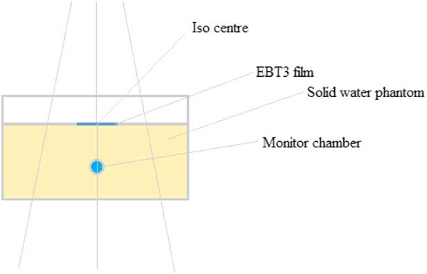
The Epson expression 10000 XL flatbed scanner (Seiko Epson Corporation, Nagano, Japan) was used to scan the film, as shown in Figure 1. The flatbed scanner worked in both transmission and reflective mode with a colour depth of 48-bits at 72 dpi resolution on the red–green–blue colour modes, by splitting the incident light into three primary colours, namely red, green and blue of specific wavelengths. The orientation of the film in each image was recorded at different channels; the film sheet is placed perpendicular to the scan direction, as the EBT3 film is sensitive to the film position on the scanner and the dose exposed on the film.
Phantom setup and region of interest on the piece of film.

The Film QA Pro software 2016 was used in this work. The software is commercially available and is a powerful quantitative analysis tool specially designed to check the intensity-modulated radiotherapy quality assurance. It is used to calculate the dose map produced on the film and is based on a triple channel dosimetry process and uses red, green and blue channels for gafchromic EBT3 image and to detect any errors.
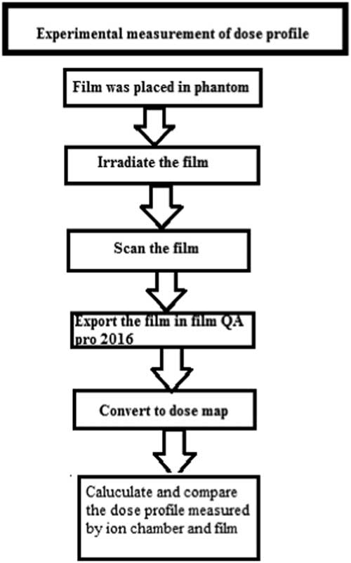
The radiochromic film was calibrated by choosing the film calibration part from case object menu. The scanned film was loaded by choosing the calibration tool section. The whole process to check beam profile can be seen in Figure 2. The region of interest was selected by using fiducial marks, and then checking the dose profile by choosing physics QA in software at the AAPM-47 protocol and analysed it using two channels, that is the red and green at resolution of 72 dpi and analysing colours for the red channel, red (beam darker) and for the green channel, green (beam darker).
The process to check the dose profile using EBT3 film.

The results of the ionisation chamber and gafchromic EBT3 film were compared by taking the difference between them using two different film scanning geometries and beam energies to check the linear accelerators performance. The misinterpretations because of measurement errors were minimised by comparing the results.
Results
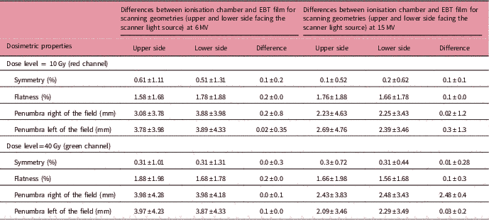
The symmetry, flatness, right and left penumbra were analysed on two different dose levels of 10 and 40 Gy. The difference between the symmetry of the photon beam measured using ionisation chamber and gafchromic EBT3 film was compared for different beam energies (6 and 15 MV), film scanning geometries at field sizes of 5×5, 10×10 and 15×15 cm2. From the results, the red channel represents the 10 Gy dose and the green channel represents the 40 Gy dose from photon beams.
Symmetry at a dose level of 10 Gy (red channel)
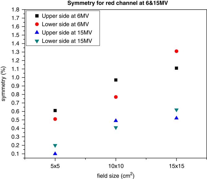
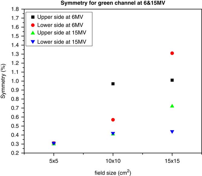
The difference between the reference symmetry of the photon beam measured using the ionisation chamber and the calculated symmetry of the photon beam using the gafchromic EBT3 film at 6 MV varied from 0.61±1.11% for the upper side scanning geometry. It ranged from 0.51±1.31% for the lower side. The difference between both scanning geometries was 0.1±0.2%. The symmetry at 15 MV in respective field sizes for the upper side ranged from 0.1±0.52%. For lower side, it varied from 0.2±0.62%, and the difference between both scanning geometries was 0.1±0.1%, as shown in Figure 3 and Table 1. Symmetry at 40 Gy (red channel) is shown in Figure 4 and Table 1 for both photon beam energies.
Relation of reference symmetry and calculated symmetry at the red channel for both scanning geometries of EBT3 film for energies (6 and 15 MV) and field sizes on the horizontal axis.

Relation of reference symmetry and calculated symmetry at the green channel for both scanning geometries of EBT3 film for energies (6 and 15 MV) and field sizes on the horizontal axis.

Dosimetric properties from 10 and 40 Gy dose values

Flatness at a dose of 10 Gy (red channel)
The difference measured between the reference flatness data from the photon beam measured using the ionisation chamber, and the EBT3 film at 6 MV varied from 1.58±1.68%. For the upper side scanning geometry, it ranged from 1.78±1.88% and the difference between both scanning geometries was 0.2%.
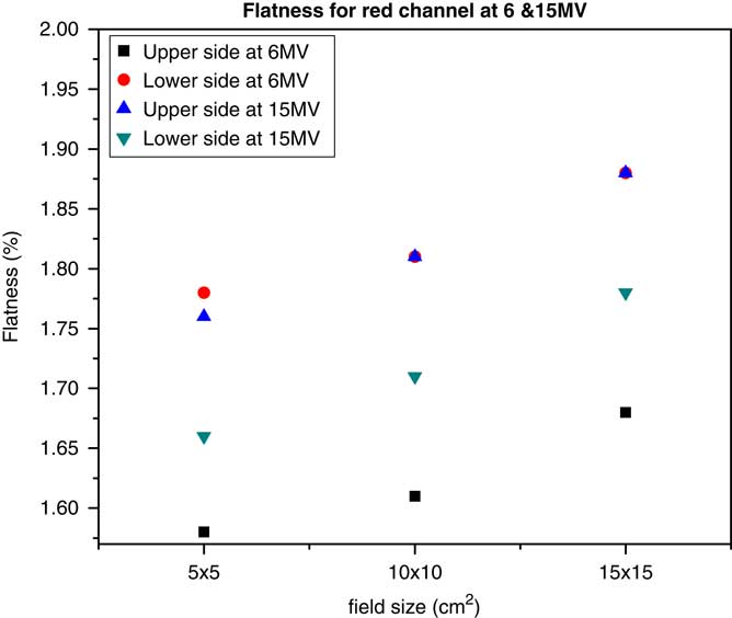
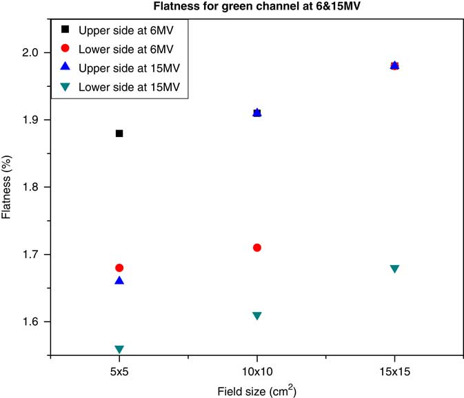
The flatness at 15 MV in various field sizes such as 5×5, 10×10 and 15×15 cm2 for the upper side scanning geometry was 1.76±1.88%. For the lower side geometry, it varied from 1.66±1.78%, and the difference between both geometries was 0.1%, as shown in Figure 5 and Table 1. The flatness at a dose level of 40 Gy (red channel) is shown in Figure 6 and Table 1 for both photon beam energies.
Relation of reference symmetry and calculated flatness at the red channel for both scanning geometries of EBT3 film for energies (6 and 15 MV) and field sizes on the horizontal axis.

Relation of reference symmetry and calculated flatness at the green channel for both scanning geometries of EBT3 film for energies (6 and 15 MV) and field sizes on the horizontal axis.

Penumbra of the field at a dose level of 10 Gy (red channel)
The difference measured between the reference right penumbra of the field measured from the ionisation chamber and the calculated right penumbra of the field from EBT3 film at 6 MV for various field sizes such as 5×5, 10×10 and 15×15 cm2 was compared. For the upper side scanning geometry of EBT3 film, the right penumbra of the field varied from 3.08±3.78 mm; for the lower side geometry, it ranged from 3.88±3.98 mm, and the difference between both geometries was 0.2±0.8 mm.
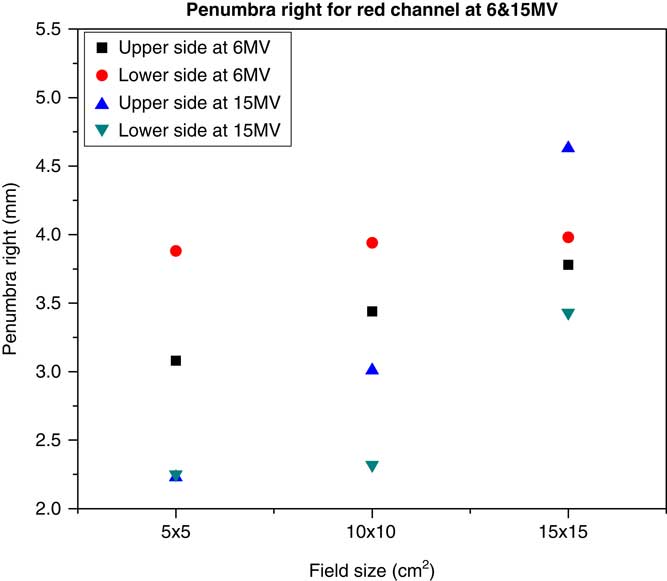
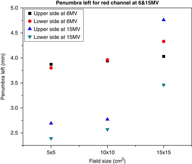
The right penumbra of the field at 15 MV in various field sizes such as 5×5, 10×10 and 15×15 cm2 for the upper side scanning geometry varied from 2.23±4.63 mm. For the lower side geometry, it ranged from 2.25±3.43 mm, and the difference between them was 0.02±1.2 mm, as shown in Figure 7 and Table 1. The left penumbra of the field at a dose level of 10 Gy (red channel) is shown in Figure 8 and Table 1 for both photon beam energies.
Relation of reference symmetry and calculated penumbra right of the field at the red channel for both scanning geometries of EBT3 film for energies (6 and 15 MV) and field sizes on the horizontal axis.

Relation of reference symmetry and calculated left penumbra of the field at the red channel for both scanning geometries of EBT3 film for energies (6 and 15 MV) and field sizes on the horizontal axis.

Penumbra of the field at a dose level of 40 Gy (green channel)
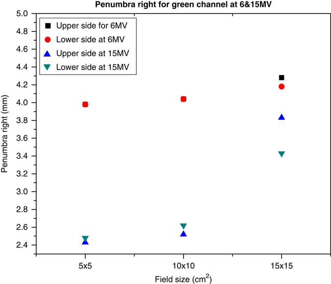
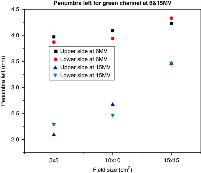
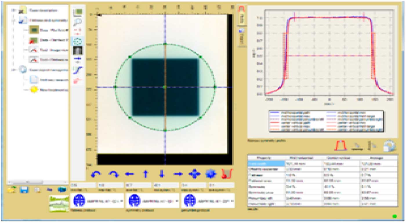
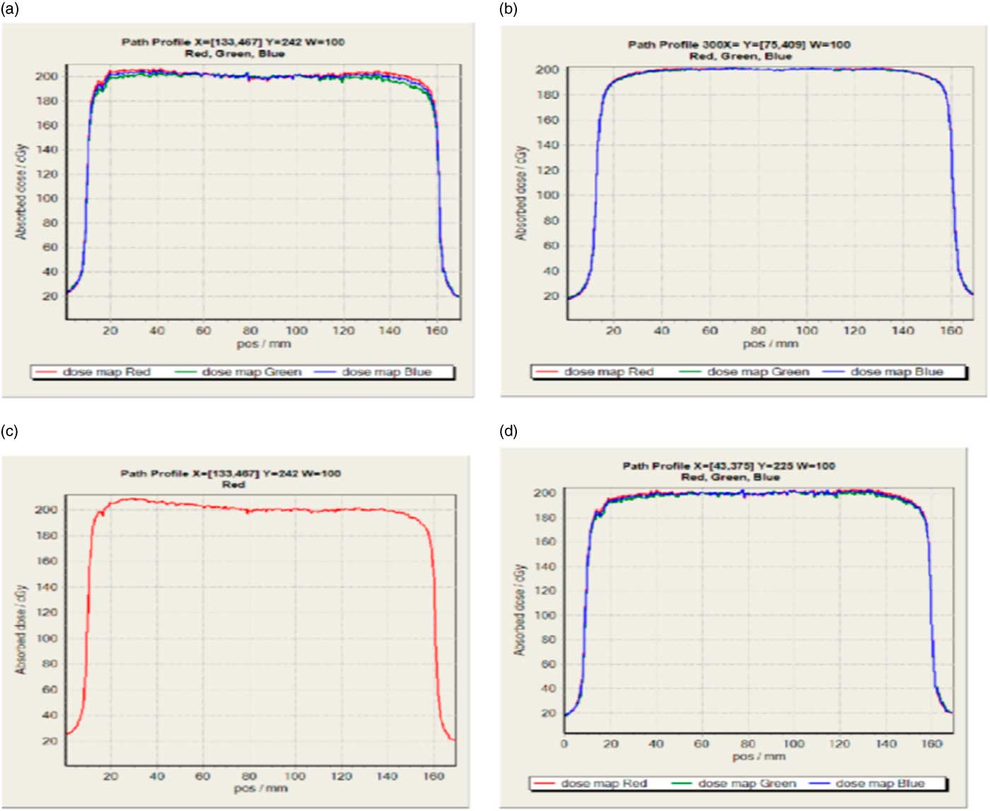
The difference between the reference penumbra on the left side of the field measured from the ionisation chamber and the EBT3 film at 6 MV was compared with various field sizes such as 5×5, 10×10 and 15×15 cm2. For the upper side scanning geometry of EBT3 film, the right penumbra of the field varied from 3.98±4.28 mm. For the lower side geometry, it ranged from 3.98±4.18 mm, and the difference between both geometries was 0.0±0.1 mm. The right and left penumbras of the field at a dose level of 40 Gy (green channel) are shown in Figures 9 and 10, for both photon beam energies, respectively. The dose profile of the film using QA Pro software and the results of the EBT3 film measured using this software are shown in Figures 11 and 12, respectively.
Relation of reference symmetry and calculated right penumbra of the field at the green channel for both scanning geometries of EBT3 film for energies (6 and 15 MV) and field sizes on the horizontal axis.

Relation of reference symmetry and calculated left penumbra of the field at the green channel for both scanning geometries of EBT3 film for energies (6 and 15 MV) and field sizes on the horizontal axis.

Dose profile of film using Film QA Pro software.

Results of EBT3 film measured after using Film QA Pro software 2016.

Discussion
The choice of the field sizes and energies was made to check the dosimetric properties such as symmetry, flatness and the penumbra on the right and left side of the field for photon beams. The difference in symmetry was measured using an ionisation chamber and calculated using EBT3 film for both scanning geometries. It was observed that the symmetry for the red channel at 10 Gy was 0.61 and 0.51% for 6 MV energy with a field size of 5×5 cm2. The symmetry of the photon beam decreased as the field size increased. For 15×15 cm2 the photon beam was less symmetric than the small field size. The comparison of the symmetry difference at both energies, 6 and 15 MV, revealed that 15 MV photon beam was more symmetric than 6 MV for both film scanning geometries of EBT3 film for the red channel, as shown in Figure 3. A similar trend was observed for the green channel, as shown in Figure 4 and Table 1.
The difference in flatness measured and calculated for both scanning geometries of EBT3 film for the red and green channels was 1.58 and 1.78%, respectively, for the 6 MV at a field size of 5×5 cm2. The flatness of the photon beam decreased as the field size increased. It was found that photon beam was less flattened for higher field size than the lower. The comparison of the flatness at the 6 and 15 MV energies showed that photon beam is more flattened at 6 MV for the upper side scanning geometry of EBT3 film than 6 MV for the lower side geometry of EBT3 film for the red channel, as shown in Figure 5. The same trend was observed for the green channel, as the beam was more flattened at 6 MV for both geometries of EBT3 film, as shown in Figure 6 and Table 1.
The acceptance tolerance of the beam symmetry and flatness should not exceed 3%. The beam is more symmetric and flattened at higher energies,Reference Nalbant, Kesen and Hatice 18 – Reference Zhang, Liengsawangwong and Lindsay 32 which is well in agreement with our results, as our symmetry and flatness value did not exceed 3% for red and green channels, and also symmetry and flatness were better at high energies except in some cases. The results are in agreement with those of Sahani et al.Reference Sahani, Sharma and Dash Sharma 33 and define the acceptance criteria of photon beam characteristics such as for symmetry, flatness and penumbra for pre-machine commissioning and for QC purposes at different energies and field sizes using International Electro Technical Commission (IEC 60976, 2008), which ensures that there is only a small negligible difference between calculated and measured data using two detectors.Reference Sahani, Sharma and Dash Sharma 33 , Reference Shende, Gupta, Patel and Kumar 34
The penumbra difference from the right side of the field measured using an ionisation chamber and calculated using EBT3 film for the upper side and lower side scanning geometry was 3.08 and 3.88 mm, respectively, for 6 MV at a field size of 5×5 cm2. The value of right penumbra of the field increased as the field size increased till 10×10 cm2 but for the field size of 15×15 cm2 the photon beam was less penumbral as compared with small field sizes. The same result was found for the left penumbra of the field at the green channel, but the penumbra was less than that at the red channel. These results can be seen in Figures 7–10 and in Table 1.
Henry et al. suggested that the penumbra is 3.9±5.6 mm for small field sizes. For large field sizes the penumbra varies from 4.7±8.0 mm measured by an ionisation chamber.Reference Godson, Ravikumar and Sathiyan 21 Penumbra increased at lower energies and decreased for higher energies as suggested by Arunkumar et al.,Reference Arunkumar, Supe, Ravikumar, Sathiyan and Ganesh 22 which agreed with our results.Reference Godson, Ravikumar and Sathiyan 21 , Reference Scott, Nahum and Fenwick 23 Our results for penumbra using gafchromic EBT3 are in agreement with Butson et al.Reference Butson, Cheung and Yu 16 and are within 3.6 mm for the photon beams, which also suggest that gafchromic EBT3 film has close matching results with ionisation chamber owing to low energy dependence of EBT3 film, which is a necessary property to find the penumbral dose measurement. High-energy photon beam penumbra can be accurately measured using gafchromic EBT film.Reference Butson, Cheung and Yu 16 , Reference Butson, Cheung and Peter 35
Conclusion
With experimental verification using ionisation chamber in the commissioning of film dosimetry, it is concluded that our EBT3 film dosimetry system is reliable with high spatial resolution that works well for measurement of absorbed dose. It is highly useable, low cost and have a long-time stability. The intrinsic variations in radiation response in this study are within the acceptable range given by WHO.
Acknowledgements
The authors would like to thank Stefano Martelli at Ryerson University for the English edition.