Introduction
Cavernous sinus tumours represent 1% of all intracranial neoplasms, and 41% of them are cavernous sinus meningiomas (CSM).Reference dos Santos, de Salcedo, Gutiérrez Diaz, Calvo, Samblás and Marsiglia 1 These tumours are located close to critical neurovascular structures, such as cranial nerves (CN) and the carotid artery. Total resection of a CSM is not always feasible and when possible is associated with high morbidity and mortality rates.Reference O’Sullivan, van Loveren and Tew 2 – Reference Sughrue, Rutkowski, Aranda, Barani, McDermott and Parsa 4 A studies of microsurgically treated CSM displayed recurrence rates ranging from 6% to 25% and progression rates ranging from 4.5% to 65%, with a mean 5-year progression-free survival (PFS) rate of 50%.Reference Nakamura, Roser, Jacobs, Vorkapic and Samii 5 The disappointing results of the microsurgical approach have led to the evaluation of a more conservative strategy, such as fractionated conformal radiotherapy (FCR) and stereotactic radiosurgery (SRS).
Here we report our experience with conformal radiotherapy (CR) in the treatment of CSM, aiming to evaluate local control, clinical response and radiation-induced toxicity.
Patients and methods
Patient population
A total of 18 patients with CSM, treated from 2011 to 2017, were retrospectively reviewed.
The term ‘cavernous sinus meningioma’ was used only for tumours arising primarily from the cavernous sinus. Histological confirmation was not required when imaging undertaken found the characteristic appearance of a CSM.
Procedures
A planning CT scan of the whole head was obtained with 3-mm slice thickness supplemented by image fusion with MRI sequences. Patients were immobilised with a thermoplastic mask and treated on supine position.
All patients were treated with three-dimensional conformal radiation, the unique available technique in our institution. Treatment was delivered on a Clinac 2100 (Varian) Linear Accelerator. Photon beam energy was 6 and 18 MV.
Follow-up criteria
A clinical recurrence was defined as the onset or worsening of neurologic symptoms.
A clinical response was defined as symptomatic and clinical improvement or resolution of a pre-existing CN deficit or symptoms.
An increase in tumour size (>25%) or the reappearance of enhancement visualised on MRI or computed tomography was considered as evidence of disease progression or recurrent disease.
A radiologic response was defined as a disappearance of the tumour or decrease of at least 25% of the tumour volume.
Results
Patient characteristics
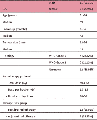
Our study population exhibited a male predominance: 11 male (61.11%) and 7 female (38.88%). Patients ranged in age from 31 to 74 years (median 59).
Clinical symptoms and signs
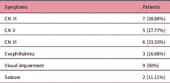
The patients’ clinical presentation included impaired vision, CN deficit, headache, ocular signs. The most common initial symptom was CN deficit, observed in 14 patients (77.77%). Trigeminal symptoms were present in five patients (27.77%). Exophthalmia was observed in three cases (16.66%). Visual acuity was impaired in nine patients (50%) and seizure was observed in two cases (11.11%). (Table 1)
Initial symptoms

Abbreviation: CN, cranial nerve.
Tumour size and extent:
Tumour size ranged from 13 to 56 mm with a median tumour size of 35 mm.
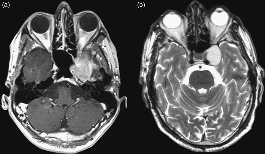
Only four tumours were confined to the cavernous sinus. Meningioma presented a petroclival extension in three cases. It extended to the sphenoid sinus in three cases, the orbit in three cases, the temporal fossa in five cases and the sphenoid greater wing in one case. Figure 1 shows axial T1-weighted image after Gadolinium administration (a) and T2-weighted image (b) showing a well-defined tumour involving the left cavernous sinus.
Axial T1-weighted image after gadolinium administration (a) and T2-weighted image (b) of two different patients showing a well-defined tumour involving the left cavernous sinus.

Treatment
Six patients (33.33%) were operated on prior to radiation. The resection was incomplete in four cases. In 12 patients (66.66%) radiotherapy (RT) was performed as first-line treatment.
Margins were generally 5 to 10 mm from gross tumour volume (GTV) for creation of a clinical target volume (CTV) and an additional 5 to 10 mm for planning target volume (PTV).
Radiation dose to the tumour volume varied from 50.4 Gy in 28 fractions to 54 Gy in 30 fractions, one fraction per day, given five days per week over 5 weeks. Dose was depending on tumour’s histology grade, the surgery prior to radiotherapy and the type of resection (complete or incomplete). Photon beam energy was 6 and 18 MV.
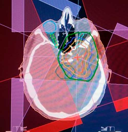
Figure 2 shows a left CSM treated by 3D-CR
Left cavernous sinus meningiomas (CSM) treated by 3D conformal radiotherapy.

Follow-up
Follow-up time ranged from 6 to 84 months, with a median of 43 months. (Table 2).
Patient characteristics

Response
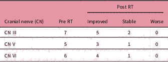
Among the 18 patients, patient’s signs and symptoms of disease remained unchanged in three cases (16.66%), 12 (66.66%) were improved and 1 patient (5.55%) exhibited tumour progression (exophthalmia). Two patients (11.11%) were lost to follow-up after the end of the treatment with radiotherapy. Table 3 represents CN function before and after radiotherapy. One patient lost to follow-up had V and VI CN deficit.
Cranial nerves affected prior to treatment and outcome after radiotherapy

The patient who presented a tumour progression had a Grade 2 extensive tumour. He underwent adjuvant radiotherapy (54 Gy) after incomplete resection. There was an initial decrease in size of the tumour but he presented with tumour growth and new-onset ptosis at 3-year follow-up.
Regarding the radiologic response to treatment, only seven patients underwent MRI which showed stability in six patients and tumour progression in one patient. Two patients were lost to follow-up. Nine patients underwent computed tomography showing a decreasing of the tumour volume.
Toxicity
No severe complications due to CR occurred in 17 patients.
A 73-year-old woman sustained a stroke during treatment. The patient was able to complete her treatment a few weeks later.
Discussion
Management of CSM poses a surgical challenge despite advances in microsurgical techniques. Fractionated CR given after incomplete resection of primary or recurrent CSM or as first-line treatment has already been shown to improve tumour control compared with outcomes in patients treated by surgery alone.Reference Maguire, Clough, Friedman and Halperin 6 – Reference Goldsmith, Wara, Wilson and Larson 8
Radiologic response
The radiologic response of CSM to CR has poorly been explored in the literature. In the few studies in which radiologic response after fractionated CR was delivered, the overall rates of reduction in size of CSM ranged from 18% to 31.6%.Reference Maguire, Clough, Friedman and Halperin 6 , Reference Nutting, Brada, Brazil, Sibtain, Saran and Westbury 7 , Reference Metellus, Regis, Muracciole, Fuentes, Dufour and Nanni 9 – Reference Milker-Zabel, Zabel-du Bois, Huber, Schlegel and Debus 11
In the study by Metellus et al.,Reference Metellus, Batra, Karkar, Kapoor, Weiss and Kleinberg 12 which evaluated long-term outcome of 53 patients with CSMs treated with fractionated CR, an overall decrease or disappearance of the lesion was seen in 30.2% of cases. In our study, MRI showed tumour stability in six patients and tumour progression in one patient.
The imaging response rate after SRS tends to demonstrate better radiologic control, ranging from 29% to 63%.Reference Metellus, Regis, Muracciole, Fuentes, Dufour and Nanni 9 , Reference Iwai, Yamanaka and Ishiguro 13 – Reference Liu, Wang, Sun, Wang and Liu 16 The selection of smaller tumour volumes may explain the greater radiologic response rate of meningiomas after SRS compared with fractionated CR.Reference Metellus, Batra, Karkar, Kapoor, Weiss and Kleinberg 12
No predictors of imaging response could be identified in the literature, due to the sparsity of relapse, infrequent imaging response and the small number of patients treated.Reference Metellus, Batra, Karkar, Kapoor, Weiss and Kleinberg 12
Clinical response, CN function
In the literature, clinical response rates after CR in patients treated for CSM varied from 20% to 63.2%.Reference Metellus, Regis, Muracciole, Fuentes, Dufour and Nanni 9 , Reference Brell, Villa, Teixidor, Lucas, Ferrán and Marín 10 In the study by Metellus et al.,Reference Metellus, Batra, Karkar, Kapoor, Weiss and Kleinberg 12 a clinical response was observed in 58.5% of patients.
An important clinical endpoint of treatment is the improvement or preservation of neurological function. Neurological deficits can be present for up to 70–80% of patients with cavernous sinus tumours as a consequence of the tumour or previous surgery.
Leber et al. showed that pre-existing nerve compression did not correlated with an increased risk of injury after radiation.Reference Leber, Bergloff and Pendl 17 Morita et al. also found that a major factor predicting improvement in CN function was in the non-previously operated patients.Reference Morita, Coffey, Foote, Schiff and Gorman 18
Neurological response rates are reported to be in the order of 20–60% after CR or stereotactic radiotherapy (SRT) and 20–80% for SRS.Reference Metellus, Batra, Karkar, Kapoor, Weiss and Kleinberg 12 , Reference Spiegelmann, Cohen, Nissim, Alezra and Pfeffer 19 , Reference Skeie, Enger, Skeie, Thorsen and Pedersen 20
In others studies, after SRS, neurological improvement has been reported in 20–69% of CSM patients, which is comparable to the results obtained with fractionated CR.Reference Spiegelmann, Nissim, Menhel, Alezra and Pfeffer 15 , Reference Liu, Wang, Sun, Wang and Liu 21 , Reference Pendl, Schrottner, Eustacchio, Ganz and Feichtinger 22
In our study, clinical response was observed in 61.53% of patients, which is comparable to results reported in the literature.
Tumour control
Conventional radiotherapy after subtotal microsurgery or as definitive treatment is a well-recognised approach with reported control rates of 80–95% at five years and PFS in the order of 80–90%.Reference Maguire, Clough, Friedman and Halperin 6 , Reference Pourel, Auque, Bracard, Hoffstetter, Luporsi and Vignaud 23 , Reference Mendenhall, Morris, Amdur, Foote and Friedman 24
In a study by Uy et al.,Reference Uy, Woo, Teh, Harnisch, Stevens and Lee 25 40 patients had benign meningiomas treated with intensity-modulated radiation therapy (IMRT). Follow-up ranged from 6 months to 71 months (median 30 months). Thirty-two tumours (80%) involved the skull base. Patients received 40–56 Gy (median: 50.4 Gy) at 1.7–2.0 Gy per fraction. The 5-year local control rate was 93%.Reference Uy, Woo, Teh, Harnisch, Stevens and Lee 25
SRS study report a radiographic response with a rate of 29–69% and PFS rates at 5 and 10 years of 80–100% and 73–98%.Reference dos Santos, de Salcedo, Gutiérrez Diaz, Calvo, Samblás and Marsiglia 1 , Reference Metellus, Regis, Muracciole, Fuentes, Dufour and Nanni 9 , Reference Maruyama, Shin, Kurita, Kawahara, Morita and Kirino 14 , Reference Spiegelmann, Nissim, Menhel, Alezra and Pfeffer 15 , Reference Pollock, Stafford, Link, Garces and Foote 26 , Reference Correa, Marta and Teixeira 27
Toxicity
In many studies, complications reported are rare, ranging from none to 2.6%.Reference Metellus, Regis, Muracciole, Fuentes, Dufour and Nanni 9 , Reference Brell, Villa, Teixidor, Lucas, Ferrán and Marín 10 Fractionated CR has proven to be relatively safe, and cranial neuropathies are rarely reported.Reference Metellus, Regis, Muracciole, Fuentes, Dufour and Nanni 9 , Reference Brell, Villa, Teixidor, Lucas, Ferrán and Marín 10 , Reference Milker-Zabel, Zabel-du Bois, Huber, Schlegel and Debus 11 Radiation-induced optic neuropathy is rarely reported in the literature.Reference Leber, Bergloff and Pendl 17 , Reference Parsons, Bova, Fitzgerald, Mendenhall and Million 28 On our study, no radiation-induced cranial or optic neuropathies were observed.
SRS targeting cavernous sinus usually causes delayed cranial neuropathies. Their incidence after SRS can be highly variable, ranging from 0% to 25% of cases.Reference Lee, Niranjan, McInerney, Kondziolka, Flickinger and Lunsford 29
Whether to treat CSM with 3D-CR, IMRT, SRS or SRT depends on the clinical features (tumour size, proximity to optic pathways…) and also on the equipment availability in the department. In the context of middle- or low-income countries with the transition to new RT techniques, 3D-CR is more available and is easier to implement than IMRT or SRS. Our results confirm its safe and effective use. So, in the era of SRT and SRS, and in a context of middle or low-income countries, CR can be a good and affordable therapeutic option in the treatment of CSM.
Conclusion
Based on our study findings, CR has proven to be a relatively safe and effective therapeutic option in the treatment of CSM, whether used as a first-line or as an adjuvant treatment. These results should be confirmed in studies with a larger number of patients and longer follow-up times.
CR may be particularly effective for tumours that are not amenable to SRS, owing to lesion size (>30–35 mm) and/or proximity to the optic apparatus (<3 mm).
Acknowledgements
None.
Financial Support
This research received no specific grant from any funding agency, commercial or not-for-profit sectors.
Conflicts of Interest
None.