Implications
Arginine is a common substrate for the synthesis of nitric oxide and polyamines, both of which affect angiogenesis and consequently foetal growth. In accordance, we report that arginine offered to pregnant gilts markedly enhanced foetal survival and total foetal weight. A novel finding was that the arginine supplementation positively affected the primary phase of myofiber formation. Based on the current knowledge of the important role of primary myofiber for total myofiber number, these results elucidate the potential of arginine to positively affect prenatal muscle development and ultimately postnatal growth, carcass composition and pork quality.
Introduction
Due to high prolificacy of sows, natural intrauterine growth retardation is common in pigs. Embryonic losses during early gestation accounts for 40% to 60% of the total number of fertilized ova (Foxcroft et al., Reference Foxcroft, Dixon, Novak, Putman, Town and Vinsky2006). When litter size increases, the uterine blood flow increases as well, but to a lower extent than the number of foetuses, resulting in a reduced uterine blood flow per foetus and thus lower nutrients and O2 supply (Père and Etienne, Reference Père and Etienne2000). This might explain why average birth weight (BtW) decreases with increasing litter size and its variability increases (Quiniou et al., Reference Quiniou, Dagorn and Gaudre2002; Berard et al., Reference Berard, Kreuzer and Bee2008). Maternal nutrition, especially the provision of an adequate amount of protein (Pond et al., Reference Pond, Maurer and Klindt1991; Schoknecht et al., Reference Schoknecht, Pond, Mersmann and Maurer1993) and of specific amino acids, plays a crucial role in placental growth and development (Schoknecht et al., Reference Schoknecht, Newton, Weise and Pond1994) as well as in the regulation of growth, development and survival of foetuses (Wu et al., Reference Wu, Bazer, Cudd, Meininger and Spencer2004).
One of the amino acids, that recently received special attention is l-arginine. This amino acid is not only required for protein synthesis but is also a common substrate for the synthesis of nitric oxide and polyamines via nitric oxide synthase and ornithine decarboxylase, respectively (Wu and Morrison, Reference Wu and Morrison1998). Evidence such as unusual abundance of l-arginine in porcine allantoic fluid during early gestation (Wu et al., Reference Wu, Bazer, Tuo and Flynn1996) suggests that both nitric oxide and polyamines are key regulators of angiogenesis and embryogenesis as well as placental and foetal growth (Reynolds and Redmer, Reference Reynolds and Redmer2001; Zeng et al., Reference Zeng, Wang, Fan, Yang, Zhou, Li, Yin, Wu and Wang2008). The potential of l-arginine for improving the reproductive performance of primiparous sows was recently demonstrated as dietary supplementation with 1% l-arginine HCl from day 30 of gestation to farrowing increased the number of piglets born alive by 22% and live litter BtW of piglets by 24% (Mateo et al., Reference Mateo, Wu, Bazer, Park, Shinzato and Kim2007).
Generally, average litter BtW decreases with increasing litter size (Quiniou et al., Reference Quiniou, Dagorn and Gaudre2002) and low BtW is associated with impaired myogenesis in utero resulting in lower total number of myofibers (TNF) (Gondret et al., Reference Gondret, Lefaucheur, Juin, Louveau and Lebret2006; Rehfeldt and Kuhn, Reference Rehfeldt and Kuhn2006). Myogenesis is a biphasic phenomenon in pigs. First, a primary generation of myotubes, called primary myofibers (P), is formed from days 35 to 55 of gestation, followed by a second generation, referred to as secondary myofibers (S), which develop between days 55 and 90 of gestation (Lefaucheur, Reference Lefaucheur2001). The S myofibers appear around each P myofiber, using them as a scaffold. In the pig, the TNF is considered to be fixed at day 90 of gestation (Lefaucheur et al., Reference Lefaucheur, Ecolan, Plantard and Gueguen2002). Results of a recent study did not reveal whether the increased litter size observed after l-arginine supplementation had an impact on myogenesis (Mateo et al., Reference Mateo, Wu, Bazer, Park, Shinzato and Kim2007). Thus, the aim of this study was to determine the effect of dietary l-arginine supplemented during early gestation to primiparous gilts on embryo survival and development. Furthermore, the impact of l-arginine on P and S myofiber formation as well as TNF of two muscles from male and female foetuses of low, medium and high foetal weight (FtW) at day 75 of gestation was assessed: a stage where all P myofibers are developed and the S myofibers are still in the process of being formed.
Material and methods
Animals and diets
All procedures involving animals were approved by the Swiss Federal Committee for Animal Care and Use. A total of 20 Swiss Large White gilts, originating from 10 litters, with initial body weight (BW) of 159 ± 9.3 kg at insemination were used in the study.
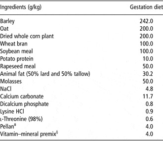
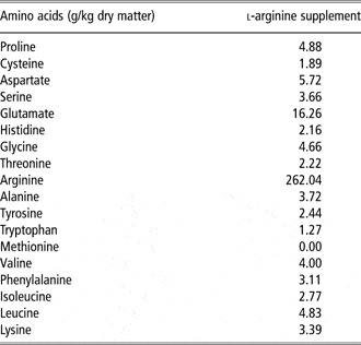
For oestrus stimulation, gilts were injected subcutaneously with 5 ml/day of Regumate® Porcine (Intervet International B.V., The Netherlands) for 18 days. At day 18, 5 ml Folligon® (Intervet International B.V.) and, 4 days later, 1.5 ml Chorulon® 1500 (Intervet International B.V.) were administrated via subcutaneous injection. During oestrus, all sows were artificially inseminated three times with the unfrozen semen of two Swiss Large White boars. Furthermore, at the start of oestrus, gilts were randomly allocated within the litter to either the control (C) or l-arginine (A) treatment. All gilts were offered daily 3 kg of a standard diet (Table 1) from mating to day 75 of gestation. From days 14 to 28, A-sows were offered daily in addition to the standard diet 100 g Progenos 28 (Trouw Nutrition, International, The Netherlands), which contains 26 g l-arginine HCL (Table 2). Sows were fed individually twice a day and had free access to water. At day 28, three C-gilts were excluded from the trial because they were non-pregnant. During the 14 days of arginine supplementation the diets were not isonitrogenous. However, over the whole experimental period, crude protein intake was only 1.2% higher in A- than C-sows. Pond et al. (Reference Pond, Maurer, Mersmann and Cummins1992) reported that even after severe dietary protein restriction (13% v. 0.5%) of the dams during gestation neither foetus survival at day 44 of gestation, nor litter size at birth differed. Consequently, it is quite unlikely in this experiment that the imbalance in dietary protein caused by the l-arginine supplementation for 14 days affected the foetal survival rate directly.
Ingredient and nutrient composition of the gestation, as fed basisFootnote †

† The energy, nutrient and amino acid contents/kg dry matter of the gestation diet were digestible energy, 12.1 MJ; total ash, 62.2 g; ether extract, 68.4 g; crude fibre, 94.0 g; proline, 10.3 g; cysteine, 3.1 g; aspartate, 13.5 g; serine, 7.0 g; glutamate, 30.2 g; histidine, 4.0 g; glycine, 7.4 g; threonine, 6.2 g; arginine, 9.6 g; alanine, 7.2 g; tyrosine, 5.1 g; tryptophan, 2.1 g; methionine, 2.5 g; valine, 8.0 g; isoleucine, 6.6 g; leucine, 11.8 g; lysine, 7.9 g.
‡ Binder that aids in pellet formation (Mikro-Technik, GmbH & Co. KG, Germany).
§ Supplied the following nutrients/kg of diet: all-trans retinol, 1.2 mg; cholecalciferol, 0.006 mg; vitamin E, 9.9 mg; riboflavin, 2.8 mg; vitamin B-6, 1.3 mg; vitamin B-12, 0.015 mg; vitamin K3, 0.2 mg; pantothenic acid, 102 mg; niacin, 10 mg; folic acid, 0.48 mg; Fe as Fe-sulfate, 84 mg; I as Ca(IO)3, 0.56 mg; Se as Na2Se, 0.2 mg; Cu as CuSO4, 9.2 mg; Zn as ZnO2, 81 mg; Mn as MnO2, 2.5 mg; choline, 196 g; biotin, 0.99 mg.
Amino acid composition of the l-arginine supplementFootnote †

† The nutrient content/kg dry matter of the arginine supplement (PROGENOS™ 28 (Trouw Nutrition International, The Netherlands)) were total ash, 248.2 g; ether extract, 9.1 g; and crude fibre, 4.4 g.
Blood sample collection and analyses
At day 24 of gestation, blood samples were collected 10 h after last feeding via jugular vein puncture into 9 ml heparinized tubes (Vacuette, Greiner Bio-one GmbH, Austria) and subsequently centrifuged at 1600 × g for 15 min at 20°C. Plasma was transferred to 2 ml microtubes (Treff AG, Degersheim, Switzerland) and stored at –20°C until analysis. The oestrone sulphate (E1S) concentration in the plasma samples was determined with a commercial radioimmunoassay kit (Estrone-sulfate DSL-5400®) modified for use with swine serum (Gaustad-Aas et al., Reference Gaustad-Aas, Ropstad, Karlberg, Hofmo and Dahl2002). Modification was carried out as follows: the standard curve was replaced by E1S diluted in pooled serum from castrated male pigs of approximately 30 kg BW. Dilutions of plasma samples with varying concentrations of E1S were performed parallel to the standard curve. In addition, plasma concentrations of amino acid were analysed by HPLC as described previously (Slocum and Cumming, Reference Slocum and Cumming1991).
Collection of foetuses and foetal tissues
At day 75 of pregnancy, all sows were sacrificed using approved necropsy procedures. Immediately after slaughter, the reproductive tract was recovered from each gilt and the number of corpora lutea (ovulation rate) and the number and weight of foetuses and mummies were recorded. From each litter the lightest (L), the heaviest (H) and the ones with a medium (M) FtW from both genders were selected. In three litters (one A- and two C-sows), three M-foetuses (one male and two female foetuses) were not available. In addition, the corresponding placentas were detached from the uterus matrix and weighed. Foetuses with an FtW lower than two times the standard deviation of the medium litter weight were excluded because they would be expected to be classified as runt piglets at birth (Gondret et al., Reference Gondret, Lefaucheur, Juin, Louveau and Lebret2006). The number of fertilized ova was estimated from the amount of foetuses and mummies.
From the selected foetuses, the weight of heart, liver, kidney and brain was determined. Brain to liver weight ratio was calculated to estimate the foetal growth retardation (Town et al., Reference Town, Patterson, Pereira, Gourley and Foxcroft2005). In addition, two morphologically and functionally distinct skeletal muscles were excised: the semitendinosus (ST), consisting of a light (STL) and dark portion (STD) and the rhomboideus (RH) muscles. In the adult pig, the STL is predominantly composed of fast-twitch glycolytic myofibers and the STD and RH of slow- and fast-twitch oxido-glycolytic myofibers. Both muscles were immediately frozen in isopentane cooled in liquid nitrogen for later histological analysis. These two muscles are, when compared to the longissimus muscle, of lower economic value, but since their myofibers are parallel to the longitudinal axis of the muscles, these muscles allow a better estimation of the TNF (Gondret et al., Reference Gondret, Lefaucheur, Juin, Louveau and Lebret2006).
Histological analyses
Histological analyses were performed on 10 μm-thick muscle transverse serial sections, which were prepared using a Cryotome (Shandon Inc., Pittsburgh, PA, USA). Immunocytochemical detection of type I MyHC (NCL-MHCs diluted: 1 : 20, Novocastra, Newcastle, UK) was realized according to the procedure described previously (Lefaucheur et al., Reference Lefaucheur, Ecolan, Plantard and Gueguen2002), which allowed the differentiation of the ST into the STD and STL portions. A section was stained using mATPase staining after preincubation at pH 4.35 to identify the muscle cross-sectional area (CSA), the TNF, the number of P and S myofibers and the secondary to primary myofiber ratio (S/P ratio) as described previously (Lefaucheur et al., Reference Lefaucheur, Ecolan, Plantard and Gueguen2002).
Statistics
Data were analysed using the MIXED procedure of SAS (version 9.1, SAS Institute Inc., Cary, NC, USA). The statistical model for the data on reproductive performance, E1S and amino acid concentrations in the plasma obtained in the sows included the diet as fixed and litter and sire as random effects. Data obtained from the selected foetuses were analysed using diet, FtW-class, gender and the two- and three-way interactions as fixed effects and litter and sire as random effects. The least square means (LSM) were separated using the PDIFF option. Differences with probability levels of P < 0.05 were considered significant. In tables, data are reported as LSM ± pooled s.e.
Results
Gestation performance
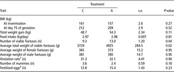
The BW at insemination and slaughter at day 75 of pregnancy, total weight gain and average daily feed intake during the experimental period did not (P > 0.11) differ between the two treatment groups of gilts (Table 3). The total number of foetuses and the total FtW were greater (P ⩽ 0.04) in A- than C-gilts, whereas the ovulation rate and the number of mummies did not (P ⩾ 0.18) differ. Likewise, the average FtW of females and males and the number of fertilized ova were similar (P ⩾ 0.23) in both groups.
Effect of l-arginine supplementation on the performance of sows assessed at day 75 of gestationFootnote *

* Values are LSM and pooled s.e. n = 7 for control treatment (C) and n = 10 for arginine treatment (A).
† Total number of corpora lutea counted in both ovaries.
‡ Estimated from the total number of foetuses and mummies.
Concentrations of E1S and amino acids in the plasma
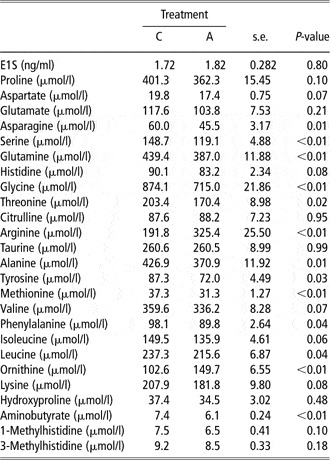
The l-arginine supplementation did not (P = 0.80) alter the E1S concentration in the plasma at day 24 (Table 4). After 10-day supplementation with l-arginine, the plasma concentrations of arginine and ornithine were 70% and 47% higher (P < 0.01 for each), respectively, in A- than C-gilts (Table 4). Conversely, plasma concentrations of asparagine, serine, glutamine, glycine, threonine, alanine, tyrosine, methionine, phenylalanine, leucine and aminobutyrate were up to 24% lower (P ⩽ 0.04) in l-arginine-supplemented gilts compared to control gilts.
Effect of l-arginine supplementation on the plasma concentrations of oestrone sulfate (E1S) and amino acids determined in gilts at day 24 of gestationFootnote *

* Values are LSM and pooled s.e. n = 7 for control treatment (C) and n = 10 for arginine treatment (A).
Foetal development
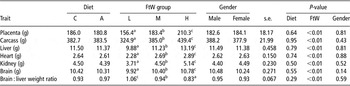
Although at day 75 of gestation the number of viable foetuses was greater in A- than C-gilts, the average FtW of the selected foetuses did not (P = 0.95) differ. Likewise, the weight of the placenta, liver, heart, kidney, brain and the brain : liver weight ratio were not (P ⩾ 0.29) affected by the maternal diet. Regardless of dietary treatment, the placenta, liver, heart, kidney and brain of L-foetuses were lighter (P < 0.01) compared to H-foetuses with intermediate values for M-foetuses (Table 5). The brain : liver weight ratio was higher (P < 0.01) in L- than H-foetuses with intermediate values for M-foetuses. These traits were not (P > 0.14) affected by gender and the two- and three-way interactions were never significant.
Effect of l-arginine supplementation, foetus weight group (FtW) and gender on weight of placenta, carcass and organs as well as brain : liver weight ratioFootnote *

a,b,c Within the FtW group LSM without a common superscript letter differ (P < 0.05).
* Values are LSM and pooled s.e.; n = 40 for control (C) and n = 59 for arginine treatment (A); n = 34 for low (L), n = 31 for medium (M) and n = 34 for high (H) FtW; n = 50 for male and n = 49 for female foetuses.
Muscle size and myofiber characteristics
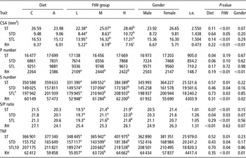
The TNF and consequently the muscle CSA of the ST and RH were not (P > 0.11) affected by the maternal diet (Table 6). However, the ST of foetuses from A-sows had more (P < 0.04) P myofibers and the S/P ratio tended (P < 0.07) to be lower than in the ST of foetuses from C-sows. The reason for these differences was the higher (P = 0.06) number of P myofibers, but not S myofibers, in the STD of foetuses from A-sows. In the RH, no effects (P > 0.19) of maternal diet were observed on the number of P and S myofibers. However, compared to foetuses from C-sows, the S/P ratio in the RH of foetuses from A-sows was lower (P < 0.01). Regardless of the dietary treatment, the CSA of the STD, STL and, consequently, the ST were smaller (P < 0.05) in L- than H-foetuses. In M-foetuses, the STD and ST CSA were smaller (P < 0.05), whereas the STL CSA was similar (P > 0.05) to H-foetuses. Because the P myofiber number in the STD and ST did not (P ⩾ 0.10) differ among FtW classes, the differences in the CSA found in L- and M- compared to H-foetuses were due to the lower S myofiber number, resulting in a lower S/P ratio and lower TNF (P < 0.05 for each). Similar to the ST, the CSA of the RH differed (P < 0.01) among the FtW classes, being smallest in L- and largest in H-foetuses with intermediate values in M-foetuses. These CSA differences can be explained by both the lower (P < 0.05) number of P and S myofibers and, consequently, the lower (P < 0.01) TNF in L- compared to M- and H-foetuses. Gender had only a small impact on myogenesis of the ST. Compared to male foetuses, females tended to have a greater (P = 0.07) CSA and S/P ratio in the ST and STD, respectively, whereas no (P > 0.10) differences were found in the S myofiber number as well as TNF of the ST, STD and STL. However, the STL of female M-foetuses had more P myofibers compared to female L- and H-foetuses, whereas male M-foetuses had fewer P myofibers compared to male L- and H-foetuses (FtW × gender interaction: P < 0.01). In the RH, female foetuses had fewer (P ⩽ 0.02) P and S myofibers and lower (P < 0.01) TNF compared to male foetuses. Consequently, the RH CSA of female foetuses was smaller (P < 0.01) compared to male foetuses.
Effect of l-arginine supplementation, FtW and gender on CSA, number P and S myofiber, S/P ratio and TNF of STD, STL, entire ST and RH muscleFootnote *

FtW = foetus weight group; CSA = gender on cross sectional area; P = number of primary; S = secondary; S/P = myofiber, primary-to-secondary myofiber ratio; TNF = total number of myofibers; STD = semitendinosus in the dark; STL = semitendinosus in the light; ST = semitendinosus; RH = rhomboideus.
a,b,c Within the FtW group LSM without a common superscript letter differ (P < 0.05).
* Values are LSM and pooled s.e.; n = 40 for control (C) and n = 59 for arginine treatment (A); n = 34 for low (L), n = 31 for medium (M) and n = 34 for high (H) FtW; n = 50 for male and n = 49 for female foetuses.
† The FtW × gender interaction is significant (P < 0.05).
Discussion
The number of offspring born per litter is of great economic importance to pig production. Thus, in the last decade, great efforts have been made to increase the prolificacy of sows (Bee, Reference Bee2007). Minimizing embryonic and foetal losses, which can amount to 40% to 60% of total prenatal loss (Foxcroft, Reference Foxcroft2007), is one of the major challenges in order to maximize the reproductive efficiency of the gestating sow. In this context, placental development and especially the formation of new blood vessels (angiogenesis) plays a key role (Reynolds et al., Reference Reynolds, Grazul-bilska, Killilea and Redmer1993). One highly effective way to enhance placental angiogenesis is by the modulation of the arginine-nitric oxide and polyamine pathways (Flynn et al., Reference Flynn, Meininger, Haynes and Wu2002; Wu et al., Reference Wu, Bazer, Cudd, Meininger and Spencer2004).
Arginine is a common substrate for nitric oxide and polyamine synthesis via nitric oxide synthase and ornithine decarboxylase, respectively (Wu et al., Reference Wu, Bazer, Tuo and Flynn1996). There is recent evidence that, under a standard nutritional regime, dietary l-arginine supplementation of 10 g/kg diet from days 30 to 110 of gestation affects the reproductive performance of gilts by increasing foetal survival and live litter BtW (Mateo et al., Reference Mateo, Wu, Bazer, Park, Shinzato and Kim2007). In accordance, we report that dietary l-arginine supplementation during early gestation increased the number of viable foetuses at day 75 of gestation without affecting the average FtW or foetal organ development. It is interesting to note that the effect on reproductive performance was similar to the one observed in the study of Mateo et al. (Reference Mateo, Wu, Bazer, Park, Shinzato and Kim2007), despite the slightly lower supplemental level of 0.8 g/kg diet and the fact that supplementation had been restricted to early gestation and solely to 14 days. Similarly, Ramaekers et al. (Reference Ramaekers, Kemp and van der Lende2006) found that 40 g/day of l-arginine, offered from days 14 to 28 of gestation, increased litter size by +0.8 piglet/litter and farrowing rate by 11.6%.
Already after 10 days of dietary l-arginine supplementation, in the A-sows the plasma concentration of arginine and its precursor ornithine was increased to a similar extent as reported by Wu et al. (Reference Wu, Pond, Flynn, Ott and Bazer1998) after a longer arginine supplementation period of 40 and 80 days. Wu et al. (Reference Wu, Pond, Flynn, Ott and Bazer1998) showed that decreased arginine and ornithine concentrations, combined with reduced activities of nitric oxide synthase and ornithine decarboxylase, resulted in reduced nitric oxide production and synthesis and concentrations of polyamines in the placenta and endometrium. Based on the current knowledge of the biological effects of nitric oxide and polyamines (Novaro et al., Reference Novaro, Gonzalez, Jawerbaum, Rettori, Canteros and Gimeno1997; Sooranna et al., Reference Sooranna, Morris and Steer1995; Wu et al., Reference Wu, Bazer, Cudd, Meininger and Spencer2004), one could hypothesize that elevated synthesis of nitric oxide and polyamines enhanced endometrial and placental angiogenesis and growth during the early stage of pregnancy. Accordingly, Hazeleger et al. (Reference Hazeleger, Ramaekers, Smits and Kemp2007) reported that vascularization of the placenta at day 35 of gestation was greater in gilts offered daily 40 g of l-arginine from days 16 to 28 of pregnancy. The greater placenta vascularization could promote an optimal intrauterine environment throughout gestation for a higher number of foetuses.
In pregnant sows each embryonic unit produces oestrone (Gaustad-Aas et al., Reference Gaustad-Aas, Ropstad, Karlberg, Hofmo and Dahl2002), which is conjugated with sulphate groups within the endometrium. By means of analysing the E1S levels in blood samples of gestating sows, Gaustad-Aas et al. (Reference Gaustad-Aas, Ropstad, Karlberg, Hofmo and Dahl2002) showed that the E1S concentration at day 24 of gestation positively correlated (r = 0.51) with litter size at term. Therefore, in this study the E1S plasma concentration at day 24 of gestation was used as an indicator for the number of viable embryos just after implantation, which occurs earlier between days 14 and 18 of pregnancy (Gadsby et al., Reference Gadsby, Heap and Burton1980). The plasma E1S concentrations did not differ between A- and C-sows, suggesting that l-arginine supplementation had no effect on the number of developing embryos. Because after day 40 of gestation the uterus is not able to reabsorb the dead embryos (Town et al., Reference Town, Patterson, Pereira, Gourley and Foxcroft2005) and no differences were found on the ovulation rate and number of fertilized eggs between the experimental groups, it may be concluded that l-arginine supplementation increased embryonic survival rate from days 24 to 40 of gestation resulting in a greater number of viable foetuses at day 75 of gestation. The data of this study do not allow us to evaluate the possible mechanisms on how dietary l-arginine would accomplish this, but the period of supplementation just precedes this time period.
It is a general belief that the most prominent factor determining muscle CSA is given by the number of the prenatally formed myofibers (Gondret et al., Reference Gondret, Lefaucheur, Louveau, Lebret, Pichodo and Le Cozler2005; Rehfeldt and Kuhn, Reference Rehfeldt and Kuhn2006). Results of recent studies demonstrated that TNF and myofiber size are correlated not only with muscle CSA but also with postnatal growth and adipose tissue deposition rate in pigs (Gondret et al., Reference Gondret, Lefaucheur, Louveau, Lebret, Pichodo and Le Cozler2005; Nissen et al., Reference Nissen, Jorgensen and Oksbjerg2004). Thus, a better understanding of exogenous factors affecting myogenesis of the developing foetus, such as nutrition of the dam during gestation, becomes an important prediction tool to foresee and improve the postnatal growth potential of the progeny. Dietary l-arginine supplementation had no effect on TNF and, consequently, on the muscle CSA, on both foetal muscles analysed. Nevertheless, it is interesting to note that ST of foetuses from sows supplemented with l-arginine expressed more P myofibers than did the ST of foetuses from C-sows. These findings disagree with results reported by Dwyer et al. (Reference Dwyer, Fletcher and Stickland1993), which suggested that P myofiber number is a fixed genetic component and its development is unaffected by conditions occurring in utero. The P myofibers are formed from days 35 to 55 followed by a second generation of S myofibers between days 55 and 90 of gestation. These S myofibers are grouped around individual P myofibers, using them as scaffold (Picard et al., Reference Picard, Lefaucheur, Berri and Duclos2002). In this study, dietary l-arginine supplementation (days 14 to 28) preceded the formation of P myofibers. Thus, it is plausible to assume that the observed effect on P myofiber numbers was not directly, but rather indirectly related to dietary l-arginine supply by improving the uterine environment, such as increased placental angiogenesis as reported by Hazeleger et al. (Reference Hazeleger, Ramaekers, Smits and Kemp2007), resulting in enhanced supply of nutrients of the developing foetuses. However, it is unclear why only the formation of P myofibers in the ST but not the RH was positively affected.
Although both muscles of foetuses from A-sows had more P myofibers, the number of S myofibers did not differ. A possible explanation could be that due to the greater number of pig conceptuses and the increasing nutrient demand in mid and late pregnancy of each foetus in A-sows, maternal energy supply per foetus was lower, which eventually may have delayed S myofiber formation. Considering that at day 75 of gestation the number of S myofibers is not fixed, yet the possibility still exists that foetuses from A-sows are able to compensate for the delayed formation of S myofibers during the remaining time of pregnancy. Moreover, as reported by Dwyer and Stickland (Reference Dwyer and Stickland1991), the contribution of a lower P myofiber number was found to be four times more important in causing a TNF reduction than a lower S/P ratio.
The BtW of piglets is closely related to their viability and survival rate after birth (Gardner et al., Reference Gardner, Hird and Franti1989; Quiniou et al., Reference Quiniou, Dagorn and Gaudre2002) and is determinant of the postnatal growth and carcass deposition rate of lean and adipose tissue (Gondret et al., Reference Gondret, Lefaucheur, Juin, Louveau and Lebret2006; Berard et al., Reference Berard, Kreuzer and Bee2008). Lower BtW has been associated with lower myofiber hyperplasia in the ST. Interestingly, in this study the FtW of L-foetuses from A- and C-sows did not differ as the diet × FtW interaction was not significant. Similarly, Mateo et al. (Reference Mateo, Wu, Bazer, Park, Shinzato and Kim2007) did not observe differences in BtW variations of piglets originating from larger (arginine supplemented) and smaller (control) litters. These findings are of great importance as the variability of the BtW as well as the percentage of low BtW piglets markedly increases with increasing litter size (Quiniou et al., Reference Quiniou, Dagorn and Gaudre2002). Thus, it can be assumed that the intrauterine environment was optimal also for the greater number of foetuses in the l-arginine group and the known negative effects of intrauterine crowding were not observed. In addition, it has to be mentioned that the number of viable foetuses in the A-sows was not excessively great, which might further explain the similar FtW of L-foetuses in both treatment groups.
Regardless of the maternal treatments, FtWs of the selected foetuses were closely related to the weights of the placentas. This relationship implies that blood flow and consequently nutrient and O2 supply from the dam to the foetuses differed among FtW classes, thereby impairing the development of organs such as the heart, liver, kidney and brain in lighter compared to heavier foetuses. Accordingly, Town et al. (Reference Town, Patterson, Pereira, Gourley and Foxcroft2005) reported a positive correlation between average FtW and average placenta, liver as well as brain weight at days 85 to 90 of gestation. Similar to the results obtained by Town et al. (Reference Town, Patterson, Pereira, Gourley and Foxcroft2005), brain : liver weight ratio was higher in L- compared to H-foetuses with intermediate values for M-foetuses. This finding, referred to as brain-sparing effect, confirmed that development of the brain depended also on FtW, but to a much lower extent than the other organs.
These observations raise the question how myogenesis is affected by overall foetal development. In agreement with the results reported by Handel and Stickland (Reference Handel and Stickland1987), the number of P myofibers in the ST was independent from the FtW, although the CSA decreased with decreasing FtW. Thus, it is plausible that the diameter of P myofibers was smaller in L- than H-foetuses providing less surface area for the attachment and fusion of myoblasts forming S myofibers and therefore resulting in a markedly lower S/P ratio (Wigmore and Stickland, Reference Wigmore and Stickland1983). By contrast, in the RH the lower S/P ratio observed in L-foetuses compared to M- and H-foetuses was related to the number rather than the size of P myofibers because L-foetuses formed fewer P as well as S myofibers in smaller muscle CSA. At day 75 of gestation, the TNF in both muscles were lower in L- than H-foetuses. Based on the results of Wigmore and Stickland (Reference Wigmore and Stickland1983), this difference is expected to persist until birth and might subsequently affect postnatal growth due to differences in the protein and fat deposition rate (Gondret et al., Reference Gondret, Lefaucheur, Juin, Louveau and Lebret2006; Berard et al., Reference Berard, Kreuzer and Bee2008).
In summary, the results of this study confirmed the potential of dietary l-arginine supply to enhance the reproductive performance of gilts. Of great interest is the fact that the greater number of viable foetuses had no negative impact on foetal development, especially on the foetuses with a low FtW. Exciting is the finding that l-arginine supply positively affected the primary phase of myofiber formation. Based on current knowledge, this could have important implications for postnatal muscle growth, carcass composition and, ultimately, pork quality.
Acknowledgements
The authors thank Prof. Michael Kreuzer for his essential support and assistance, Guy Maikoff and his team for taking care of the animals, Dr Paolo Silacci and his blood and muscle laboratory team for the help during sample collection and the analysis of the muscle samples, and Prof. Rupert Bruckmaier for analysing the E1S concentrations in the plasma. This study is supported by the Swiss State Secretariat for Education and Research grant COST C05,0126 and by Agroscope Liebefeld Posieux, Research Station ALP.