Introduction
Mitrofanovite, Pt3Te4, is a new platinum-group mineral (PGM) discovered in a low-sulfide disseminated ore in the East Chuarvy deposit, Fedorovo–Pana intrusion, Kola Peninsula, Russia (67°24′30″N, 36°03′00″E). The Fedorovo–Pana complex belongs to a group of Palaeoproterozoic layered intrusions distributed in the north-eastern part of the Fenno–Scandian Shield. Many of these intrusions contain Cu–Ni–PGE deposits (PGE = platinum-group elements), e.g. Penikat, Portimo, Monchegorsky (Monchepluton) and Koillismaa (Alapieti and Lahtinen, Reference Alapieti, Lahtinen and Cabri2002).
The Fedorovo-Pana intrusion (Fig. 1) is ~80 km by 0.5–8 km in size, the layering dips in a south-west direction. It consists of three separate blocks: Fedorova Tundra, West-Pana and East-Pana. Low-sulfide Cu–Ni–PGE deposits were discovered in the marginal taxitic rocks of Fedorova Tundra and in the lower zones of the layers of mafic rocks of West-Pana and East-Pana. The East Chuarvy deposit is located in the lower part of the East-Pana massif.
Schematic geological map of the Fedorovo–Pana intrusion.

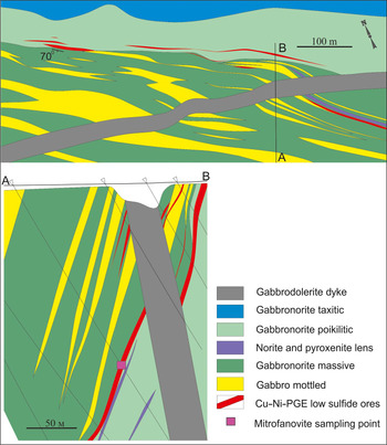
The PGE mineralisation on the East Chuarvy site was found by Pana JSC and the Geological Institute KSC RAS in the period 1995–2002. Further exploration in the East Chuarvy PGE deposit was performed in 2004–2006 by the Kola Mining Geological Company Ltd (Ward et al., Reference Ward, McLaughlin, Kalinin, Kazanov and Voitekhovich2008; Kazanov and Kalinin, Reference Kazanov and Kalinin2008). The location of the holotype material is shown on Fig. 2.
Geological map and cross-section (A–B) of the East Chuarvy deposit (adapted from Ward et al., Reference Ward, McLaughlin, Kalinin, Kazanov and Voitekhovich2008; Kazanov and Kalinin, Reference Kazanov and Kalinin2008).

The Main Ore body is associated with a contact zone of gabbronorite with a poikilitic or massive texture. PGE–Cu–Ni mineralisation was traced down to 350 m depth and to 900 m along the strike. The thickness of the mineralised body varies up to 16 m.
Low-sulfide ores are composed mainly of pyrrhotite, chalcopyrite and pentlandite (in order of their abundances) with an average total content of these sulfides in the ore of 0.6 wt.%. The PGE-rich phases are braggite, vysotskite, sperrylite, stillwaterite, moncheite, kotulskite, more than twenty species of PGM and PGE-bearing phases, and Pd-containing pentlandite (the average Pd content is 4900 ppm with variations from 1000 to 16,400 ppm – electron microprobe data).
Both the mineral and its name were approved by the Commission on New Minerals, Nomenclature and Classification of the International Mineralogical Association (IMA2017-112, Subbotin, et al., Reference Subbotin, Vymazalová, Laufek, Savchenko, Stanley, Gabov and Plášil2018). The name mitrofanovite (митрофановит) is in honour of Dr. Felix P. Mitrofanov (Феликс Петрович Митрофанов) (b. 1935, d. 2014), Academician of the Russian Academy of Sciences, a Russian geologist who was among the first to discover PGE mineralisation in the Fedorova–Pana complex, Russia.
Holotype material with mitrofanovite (polished section) is deposited in the Fersman Mineralogical Museum, Moscow, Russia, with registration number 5141/1.
Occurrence
Mitrofanovite was found in a specimen of mineralised olivine-bearing gabbronorite collected in the East Chuarvy deposit. It contains aggregates of disseminated pentlandite–pyrrhotite–chalcopyrite and grains or aggregates of various species of PGM (up to 0.2 mm across; Fig. 3a).
Back-scatter electron images of (a) individual grains and nest-like clusters of different PGMs (white and light grey) in Cu–Fe–Ni sulfides (Pn – pentlandite, Po – pyrrhotite and Ccp – chalcopyrite). The red outline marks the location of (b): mitrofanovite and associated minerals (mon – moncheite, kei – keithconnite, luk – lukkulaisvaaraite and rus – rustenburgite).

Mitrofanovite is associated with Au–Ag and Pt–Fe alloys, rustenburgite, moncheite, Pb-rich kotulskite, keithconnite, lukkulaisvaaraite, braggite, vysotskite; and rock-forming silicates: orthopyroxene, augite, olivine, amphiboles and plagioclase. The mineralised host-rock is fairly fresh. No signs of post-magmatic secondary alteration were found.
Genetic implications
Mitrofanovite belongs to low-sulfide Cu–Ni–PGE mineralisation in the lower layered East-Pana massif zone. This reef-type ore zone was probably formed as a result of segregation of sulfide droplets during the crystallisation of basic magma (e.g. Naldrett, Reference Naldrett2004).
Compared to the Kievey, Nort Kamennik and Fedorova deposits, the low sulfide PGE ores of the East Chuarvy deposit are 1.5–2.5 times richer in PGE content (mainly due to constant higher Pt content) at half the concentrations of Cu, Ni and S. Characteristic features of the mineralisation are the extremely variable content of sulfides and other minerals and a relative enrichment of Pt. In the ore of the East Chuarvy deposit, the average Pd/Pt ratio is 2.5, and in the ores of other deposits ranges from 4.4 to 6.7 (Kazanov and Kalinin, Reference Kazanov and Kalinin2008). Therefore, the origin of mitrofanovite could depend on the relative excess of Pt and possible deficiency of S during the formation of the PGE-mineralisation of the East Chuarvy deposit. On the basis of experimental studies the phase Pt3Te4 is stable up to 985°C (Bhan et al., Reference Bhan, Gödecke and Schubert1969).
Appearance, physical and optical properties
In polished section mitrofanovite (7 grains identified) forms anhedral grains (up to 20 μm × 50 μm) always in intergrowths with moncheite in aggregates with lukkulaisvaaraite, kotulskite, vysotskite, braggite, keithconnite, rustenburgite and Pt–Fe alloys hosted by a chalcopyrite–pentlandite–pyrrhotite matrix, Fig. 3b.
Due to the small grain size and intergrowths with other ore minerals, the physical properties of mitrofanovite could not be determined on natural or synthetic material.
Mitrofanovite is opaque with a metallic lustre. The powder of the synthetic analogue is grey in colour and has a grey streak. The mineral is brittle. Cleavage is good on {001}, parting was not observed. The density calculated on the basis of the empirical formula and cell dimensions of the synthetic analogue of mitrofanovite is 11.18 g/cm3.
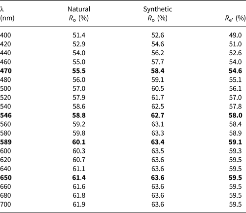
In plane-polarised light, mitrofanovite is bright white with medium to strong bireflectance, slight pleochroism, and strong anisotropy on non-basal sections with greyish brown rotation tints. It exhibits no internal reflections. Reflectance measurements were made in air relative to a WTiC standard on both mitrofanovite and its synthetic analogue using a J & M TIDAS diode array spectrometer attached to a Zeiss Axiotron microscope. The results are presented in Table 1. The only measurable grain of the natural material was a basal section (i.e. R o) but this does not match very well to the R o determined for the synthetic analogue in terms of its dispersion. It might be due to the small grain size and the difficulty of preparing a perfectly level surface in a multiphase area of the natural sample.
Reflectance data for mitrofanovite (grain 3 in Table 2) and its synthetic analogue.

Values required by the Commission on Ore Mineralogy are given in bold
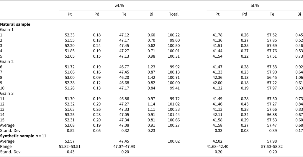
Electron-microprobe analyses for mitrofanovite and its synthetic analogue.

Chemical composition
Electron probe micro-analyses (EPMA) were performed with a CAMECA SX-100 electron probe microanalyser in wavelength-dispersive mode using an electron beam focused to 1–2 μm. Pure elements were used as standards. Concentrations were quantified on the PtMα, PdLα, TeLα and BiMα, with an accelerating voltage of 15 keV, and a beam current of 10 nA on the Faraday cup. Other elements were below the detection limit.
The EPMA results are given in Table 2, the EPMA data of natural material were collected on three different grains. The empirical formula, calculated for the mean composition on the basis of 7 atoms per formula unit is (Pt2.91Pd0.02)Σ2.93(Te4.02Bi0.05)Σ4.07 for mitrofanovite and Pt2.94Te4.06 for its synthetic analogue, with an ideal formula of Pt3Te4. The phase probably forms a narrow solid solution that may reflect slight non-stoichiometry in the formula.
Synthetic analogue
Synthetic Pt3Te4 was prepared in an evacuated and sealed silica-glass tube in a horizontal furnace in the Laboratory of Experimental Mineralogy of the Czech Geological Survey in Prague. To prevent loss of material to the vapour phase during the experiment, the free space in the tube was reduced by placing a closely fitting silica glass rod against the charge. The temperature was measured with Pt–PtRh thermocouples and is accurate to within ± 3°C. A charge of ~400 mg was carefully weighed out from the native elements. We used, as starting chemicals, platinum powder (Aldrich Chem. Co., 99.995% purity) and a tellurium ingot (Aldrich Chem. Co., 99.95% purity). The starting mixture was first melted at 1000°C for two days, quenched and then ground in an agate mortar under acetone and reheated to 400°C for five months. The sample was quenched by dropping the capsule in cold water.
X-ray crystallography
An attempt to extract the largest grain of mitrofanovite and to obtain single crystal X-Ray diffraction data was not successful. The small size of other grains of mitrofanovite intimately intergrown with moncheite and other PGM prevented its extraction and direct investigations by means of X-ray diffraction. Therefore, the relevant crystallographic and structural investigations were performed on the synthetic phase Pt3Te4. Powder X-ray diffraction data were also collected on synthetic Pt3Te4.
The powder X-ray diffraction pattern of synthetic Pt3Te4, used for structure refinement was collected in Bragg-Brentano geometry on a Bruker D8 Advance diffractometer, equipped with the LynxEye XE detector using CuKα radiation. The data were collected in the range between 5 and 100°2θ. The details of data collection and basic crystallographic data are in Table 3.
Powder diffraction data collection and Rietveld analysis of a synthetic analogue of mitrofanovite.

*This study; ** data from Cenzual et al. (Reference Cenzual, Gelato, Penzo and Parthé1990).
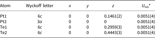
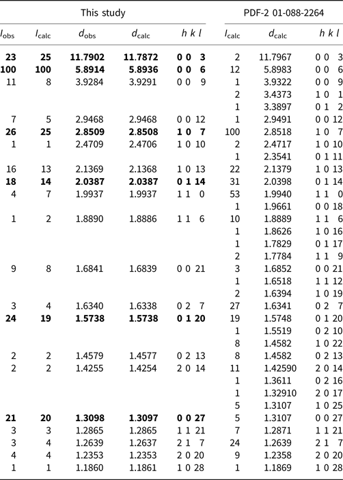
The crystal structure of Pt3Te4 was refined by the Rietveld method using the FullProf program (Rodríguez-Carvajal, Reference Rodríguez-Carvajal2006). The starting structural model for mitrofanovite (Pt3Te4) was taken from the work of Cenzual et al. (Reference Cenzual, Gelato, Penzo and Parthé1990). The pseudo-Voigt function was used to generate the shape of the diffraction peaks in the subsequent Rietveld refinement. The background was determined by the linear interpolation between consecutive breakpoints in the pattern. The refined parameters include those describing the peak shape and width, peak asymmetry, unit-cell parameters, three fractional coordinates, preferred orientation along [001] and isotropic displacement parameters. The final cycles of refinement converged to the residual factors: R Bragg = 0.110, R wp = 0.152 and R p = 0.104. The refinement indicated 9.5 wt.% PtTe2 impurity in the investigated sample of the synthetic analogue of mitrofanovite. The crystal structure data are given in Table 4, Fig. 4 shows the final Rietveld plot and Table 5 presents comparison of powder X-ray diffraction data of synthetic Pt3Te4 obtained in this work and those from the ICDD PDF-2 card no. 01-088-2264 (powder diffraction files from the International Centre for Diffraction Data, http://www.icdd.com/), which contains diffraction data calculated on the basis of the structure model of Pt3Te4 of Cenzual et al. (Reference Cenzual, Gelato, Penzo and Parthé1990). No measured powder diffraction data were published by Cenzual et al. (Reference Cenzual, Gelato, Penzo and Parthé1990). The differences in intensities (e.g. 006, 009 diffractions) can be explained by significant preferred orientation along [001] in the measured powder diffraction pattern of synthetic Pt3Te4.
Observed (red circles), calculated (solid line) and difference Rietveld profiles for the synthetic analogue of mitrofanovite. The upper reflection markers correspond to mitrofanovite and the lower markers to PtTe2 (9.5 wt.%) impurity.

Fractional coordinates and anisotropic displacement parameter (Å2) for synthetic mitrofanovite.

*Isotropic displacement parameters for all atoms were constrained to be equal.
Comparison of powder X-ray diffraction data for synthetic Pt3Te4 from this study and those from the ICDD PDF-2 card no. 01-088-2264, which were calculated on the structural model of Cenzual et al. (Reference Cenzual, Gelato, Penzo and Parthé1990). Reflections with I < 1 are not shown in the table.

ICDD – powder diffraction files from the International Centre for Diffraction Data, http://www.icdd.com/
The strongest lines are given in bold
Crystal structure
The trigonal structure model of Cenzual et al. (Reference Cenzual, Gelato, Penzo and Parthé1990) for Pt3Te4 was confirmed. The refined fractional coordinates (only the z coordinates for Pt1, Te1 and Te2 atoms) did not deviate by more than 0.01 from their starting values, although we reported a slightly lower c unit-cell parameter (32.363(1) versus 35.39 Å). This may be attributed to the different method of synthesis.
The crystal structure of mitrofanovite is depicted in Fig. 5. The Pt1 atom shows a trigonal pyramidal coordination by Te2 atoms at distances of 2.616(6) Å, whereas the Pt2 atom forms [Pt2Te6] octahedrons with Pt–Te1 bond distances of 2.656(5) Å. The mitrofanovite structure can be described as an intergrowth of the structural motifs found in PtTe (Cenzual et al., Reference Cenzual, Gelato, Penzo and Parthé1990) (i.e. layers of corner sharing trigonal pyramids [Pt1Te3]) and in the synthetic analogue of moncheite, PtTe2 (Gronvold et al., Reference Gronvold, Haraldsen and Kjekshus1960) (i.e. layers of edge-sharing octahedra [Pt2Te6]). In the mitrofanovite structure, these layers alternate along the c axis. The resulting layered structure of mitrofanovite consists of the stacking sequence ABBABBABB (A: [Pt2Te6] octahedra, B: [PtTe3] pyramids) within the unit-cell which yields the Pt3Te4 stoichiometry. As is indicated in Fig. 5a, the [Pt1Te3] pyramids form a double-layer with TePtPtTe sequence showing Pt–Pt contacts of 2.723(5) Å. This distance is a little shorter than that of 2.774 Å observed in the structure of elementary Pt (Owen and Yates, Reference Owen and Yates1933). The [Pt2Te6] octahedra and [Pt1Te3] pyramids are connected by a system of Te–Te bonds at 3.473(9) Å. This distance is significantly larger than that of 2.834 Å observed in native Te (Adenis et al., Reference Adenis, Langer and Lindqvist1989) and comparable to interlayer Te–Te distance of 3.543 Å found in vavřínite, Ni2SbTe2 (Laufek et al., Reference Laufek, Drábek, Skála, Haloda, Táborský and Císařová2007). The weak bonding of Te atoms parallel to the c axis is in perfect agreement with the observed cleavage along {001} and subsequently strong preferred orientation along [001] in the powder diffraction pattern of synthetic mitrofanovite.
(a) Crystal structure of mitrofanovite emphasising the [Pt2Te6] octahedra and [Pt1Te3] trigonal pyramids. (b) Detailed view along the c axis showing one layer consisting of [Pt2Te6] octahedra and [Pt1Te3] trigonal pyramids, respectively.

Mitrofanovite is most closely related to moncheite PtTe2 (Genkin et al., Reference Genkin, Zhuravlev and Smirnova1963; Gronvold et al., Reference Gronvold, Haraldsen and Kjekshus1960). Both minerals crystalise in layered structures containing slabs of edge-sharing [PtTe6] octahedra. The double layer with TePtPtTe sequence and short metal–metal contacts observed in the mitrofanovite structure is reminiscent of that in Bi tellurides, such as tsumoite, BiTe (Shimazaki and Ozawa, Reference Shimazaki and Ozawa1978). However, crystal structures of both minerals differ to a certain extent. The tsumoite structure contains an octahedral double layer Bi2Te3 with Bi in slightly eccentric octahedral coordination, whereas mitrofanovite shows a single octahedral layer with Pt in highly symmetrical octahedral coordination.
Proof of identity of natural and synthetic mitrofanovite
The structural identity between the synthetic Pt3Te4 and the natural material was confirmed by electron back-scattering diffraction (EBSD). We used a TESCAN Mira 3GMU scanning electron microscope combined with an EBSD system (NordlysNano detector, Oxford Instruments). The natural sample was prepared for investigation by re-polishing the surface with colloidal silica (OP-U) for 20 min to reduce the surface damage. The EBSD patterns were collected and processed using a proprietary computer program AZtec HKL (Oxford Instruments). The solid angles calculated from the patterns were compared with a synthetic Pt3Te4 match containing 46 reflectors to index the patterns. The Kikuchi patterns obtained from the natural material (four measurements on different spots on natural mitrofanovite) were found to match the patterns generated from the structure of synthetic Pt3Te4 provided by our crystal-structure determination (Fig. 6). The values of the mean angular deviation (MAD goodness-of-fit in the solution) between the calculated and measured Kikuchi bands range between 0.30° and 0.61°. These values reveal a very good match; mean angular deviations <1° are considered as indicators of an acceptable fit.
EBSD image of mitrofanovite; in the right pane, the Kikuchi bands are indexed.

The EBSD study, chemical identity and optical properties confirmed the correspondence between natural and synthetic materials and thereby legitimise the use of the synthetic phase for the complete characterisation of mitrofanovite.
Acknowledgements
The authors acknowledge Ulf Hålenius, Chairman of the CNMNC and its members for helpful comments on the submitted data. This work was supported by the Grant Agency of the Czech Republic (project No. 18-15390S to A.V.), through an internal project 331400 from the Czech Geological Survey and the Russian Foundation for Basic Research (project RFBR 18-05-70082, Arctic resources) and by the project No. LO1603 under the Ministry of Education, Youth and Sports, National sustainability program I of Czech Republic (to J.P.). The authors are grateful to Dr. Patricie Halodová (Czech Geological Survey) for EBSD measurements and Dr. Zuzana Korbelová (Institute of Geology AS CR, v.v.i.) for carrying out the electron microprobe analyses. C.J.S. acknowledges Natural Environment Research Council grant NE/M010848/1 Tellurium and Selenium Cycling and Supply. We are also grateful to Anatoly V. Kasatkin (Fersman Mineralogical Museum, Moscow) for his assistance in organising the work; Dr. Arkady A. Kalinin (Chief Geology Department Kola Mining Geological Company Ltd.) and Dmitry V. Zhirov (Geological Institute, KSC RAS) for their help in preparing the publication. The reviewer's comments of Andrei Barkov, Louis J. Cabri, Peter Leverett and the Editor Stuart Mills are greatly appreciated.