INTRODUCTION
Apple (Malus domestica) is cultivated over a large area of the world (Wang et al. Reference Wang, Xue and Lu2010a). The variety Nagafu No. 6 possesses excellent traits and is a favourite, well received by the consumer, but it is very susceptible to abiotic and biotic stresses. The genetic improvement of apple by conventional breeding is an extremely slow process due to the long juvenile period and high level of heterozygosity in this perennial fruit tree. However, genetic transformation is a promising alternative for improving plant properties and offers the possibility of introducing new characteristics, such as high quality and multiple disease resistance (Bolar et al. Reference Bolar, Norelli, Wong, Hayes, Harman and Aldwinckle2000; Faize et al. Reference Faize, Malony, Dupuis, Chevalier, Parisi and Chevreau2003; Belfanti et al. Reference Belfanti, Silfverberg-Dilworth, Tartarini, Patocchi, Barbieri, Zhu, Vinatzer, Gianfranceschi, Gessler and Sansavini2004). The TGA transcription factors are a recently discovered class of specific transcription factors in plants that bind to TGACG/AS-1 elements. The TGA transcription factor family is mainly related to stress tolerance and the physiological process of aging. Pathogenic bacteria, injury, plant hormones and other external factors can induce overexpression of the TGA gene (Shearer et al. Reference Shearer, Wang, DeLong, Despres and Fobert2009).
Zhang et al. (Reference Zhang, Qu, Du, Qiao, Cai, Guo and Zhang2012) identified and functionally characterized the crab apple (Malus hupehensis) basic leucine zipper (bZIP) transcription factor designated M. hupehensis TGA2 (MhTGA2) from M. hupehensis leaves treated with salicylic acid (SA). It was found that overexpression of the MhTGA2 gene in transgenic tobacco plants conferred enhanced resistance to sodium chloride (NaCl) at the germination stage and in seedlings, and to mannitol at the germination stage (Zhang et al. Reference Zhang, Qu, Du, Qiao, Cai, Guo and Zhang2012). In order to study the function of the MhTGA2 gene in woody plants and cultivate resistant apple plants, the MhTGA2 gene was transferred into Nagafu No. 6 using the Agrobacterium tumefaciens-mediated method to change the physiological and ecological characteristics of the plants. Another aim of the present study was to investigate whether the overexpression of the MhTGA2 gene enhanced the tolerance to environmental stress, particularly salt stress.
MATERIALS AND METHODS
Plant materials and bacterial strains
Nagafu No.6 apple plants were subcultured in vitro using the Murashige and Skoog (MS) medium supplemented with 6-benzylam-inopurine (6-BA, 0·5 mg/l, Sigma) and naphthalene acetic acid (NAA, 0·1 mg/l, Sigma). The plants were rooted in the half-strength MS medium supplemented with 3-indolebutyric acid (IBA, 0·3 mg/l, Sigma). All of the cultures were maintained under a 16 h light/8 h dark cycle and maintained at 25 °C throughout.
A. tumefaciens strain EHA105, provided by the Pomology Biotechnology Laboratory of Nanjing Agricultural University, China, was used for the transformation. The plant expression vector was pCAMBIA 1300, a plasmid with a broad-spectrum resistance MhTGA2 gene and the hygromycin (Hyg) B phosphotransferase gene conferring Hyg resistance.
A. tumefaciens-mediated transformation of Fuji apple
The fully extended top leaf of a rooted seedling that had been cultured for 35 days on the half-strength MS medium supplemented with 3-IBA (0·3 mg/l, Sigma) was used as the transformation material. The transformation experiments were performed as previously reported by Zhang et al. (Reference Zhang, Sun, Cong, Sheng, Yao and Cheng2006). The leaves were cut with three slices perpendicular to the main vein, but the leaf margin was not cut off. The leaves were exposed for 9 min to an Agrobacterium solution of an optical density (OD600) of 0·5. The excess bacteria were removed with a sterile absorbent paper, and the material was co-cultivated in MS regeneration media supplemented with 8 mg/l N-phenyl-No-1, 2, 3-thiazol-5-ylurea (TDZ) and 0·5 mg/l NAA in the dark for 3 days. The explants were then placed in the MS regeneration medium supplemented with 200 mg/l carbenicillin (Cb) to select against the Agrobacterium and 1 mg/l Hyg for selection of the transformants. The adventitious buds were transferred to the MS subculture selection medium supplied with 0·6 mg/l 6-BA, 0·1 mg/l NAA, 1 mg/l Hyg and 200 mg/l Cb. After 45 days, the subculture seedlings were inoculated in the half-strength medium supplied with 0·3 mg/l IBA and 200 mg/l Cb. The regeneration medium was replaced every 3 weeks. The culture conditions were a 16 h light/8 h dark cycle, a light intensity of 2000–3000 lx and a temperature of 23–25 °C.
Transplantation of transgenic plants
Five plants of the control and each transgenic plant were transplanted into pots (150 mm diameter) filled with an autoclaved substrate (peat: limestone: vermiculite at 3 : 1 : 1). Plants were grown under a shade in the field for 1 week and the shade was then removed. The phenotypic characteristics and growth conditions were monitored.
Polymerase chain reactions (PCRs) and reverse transcriptase– polymerase chain reaction (RT–PCR) detection of resistant plants
Polymerase chain reaction (PCR) and RT–PCR were performed using genomic DNA that was extracted from the leaves by the cetyltrimethylammonium bromide (CTAB) method. The total RNA was treated with DNase I (TaKaRa, Code No. D2215) according to the manufacturer's instructions and reverse transcribed using the M-MLV RTase cDNA Synthesis kit (TaKaRa, Code No. D6130), followed by PCR amplification. The concentration of total RNA was determined by Bio-photometer. The PCR and RT–PCRs included the following: 2·5 μl 10×PCR buffer; 1·5 μl magnesium chloride (MgCl2); 2 μl deoxyribonucleoside triphosphates (dNTPs); 1 μl (20–50 ng) template; 1 μl forward primer; 1 μl reverse primer; 0·15 μl rTaq enzyme and was then made up to 25 μl with sterile double-distilled water (ddH2O). The PCR and RT–PCR were performed under the following conditions: 94 °C for 4 min; 35 cycles at 94 °C for 30 s, 54 °C for 30 s and 72 °C for 1 min; and then 72 °C for 10 min. The forward primer sequence (5′–3′) was: ATCTCATGGCTGATGGCAG. The reverse primer sequence (5′–3′) was: CAGGAACATTTCACTCTCTTGG. The PCR products (10 μl) were separated by 1% agarose gel electrophoresis and viewed under a UV light after staining with ethidium bromide.
Determination of the leaf photosynthetic rate
Five plants of control and transgenic plant lines 1, 3 and 4 were transplanted and cultivated in pots for 4 months. Three uniform plants from each treatment were selected and the 4th, 5th and 6th mature leaves counting from the top of each plant were chosen for measuring the leaf net photosynthetic rate (Pn), the transpiration rate (Tr), stomatal conductance (Gs) and intercellular CO2 concentration (Ci) were determined for the control plants and transgenic lines 1, 3 and 4 at 11:00 h using a portable photosynthesis analyser (model LI-6400, LI-COR, USA).
Observation of leaf surface structure
The young, mature and fully expanded leaves, which came from 4-month-old control plants and transgenic line 3, were cultured in Petri dish with no saline conditions (water) and 200 mm salt stress for 7 days. Then the leaves were cut to a 7×7 mm size containing the main vein, fixed in glutaraldehyde, exposed to a vacuum, dried to the critical point and coated with metal. The surface of leaves, epidermal structure and stomatal changes were analysed using an X-650 scanning electron microscope (HITACHI, Japan), using the procedure described by Parsons et al. (Reference Parsons, Bole, Hall and Thomas1974).
Effects of different NaCl concentrations on transgenic apple leaves in vitro
The experimental materials were the leaves of the control and transgenic apple plants. The leaves were placed on Petri dishes that contained 1 ml saline (concentration of 0, 0·5, 1·0 and 1·5% of concentrated seawater) (Chen et al. Reference Chen, Zhang, Zhang, Du, Du and Qu2012). Each treatment with two blades of leaves in a Petri dish was repeated three times. All cultures were maintained under a 16 h light/8 h dark cycle at 25 °C) photoperiod for 7 days. The leaves were photographed daily to record the changes.
Statistical analysis
The data collected for Pn, Tr, Gs and Ci were statistically analysed by one way ANOVA using the SPSS17.0, to determine significant differences between control and transgenic plants.
RESULTS
Obtaining Hyg-resistant plants
A total of 158 Hyg-resistant buds were obtained and the resistant strains were transplanted after rooting. The roots of transgenic plants were more numerous and longer than those of control plants (Fig. 1).
(Colour online) Roots of wild and transgenic apple plants. WT, control; T, transgenic plants.

Confirmation of transgenic apple plants
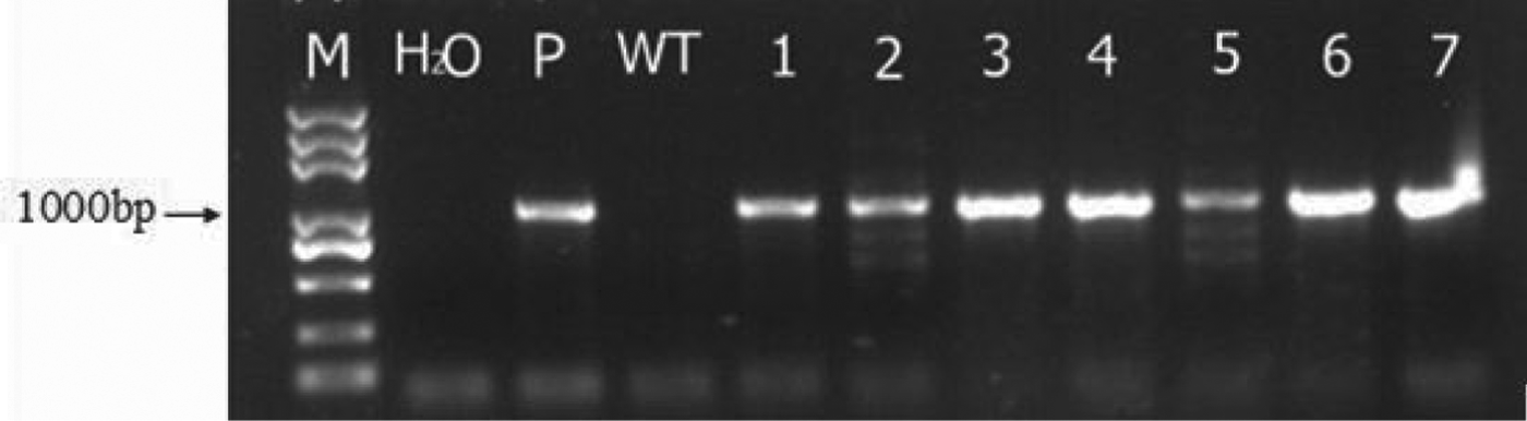
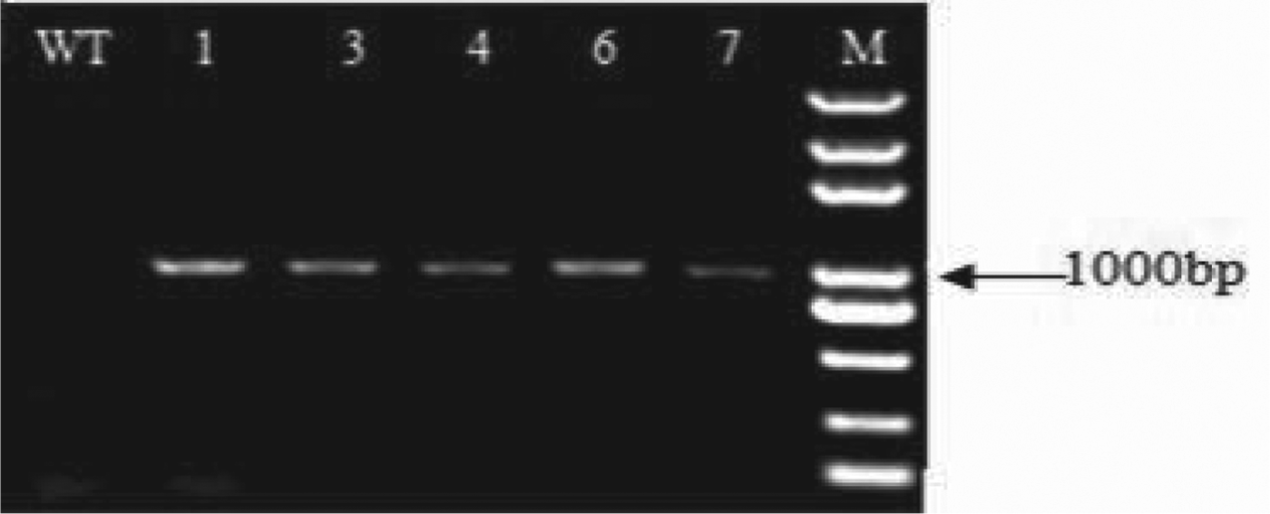
The surviving Hyg-resistant plants were analysed by PCR and RT–PCR. The DNA from the negative-control and H2O-treated plants produced no specific fragment (Fig. 2), whereas the positive control and seven transgenic lines amplified a 1002-bp target band: preliminary proof that the target gene had been successfully introduced into the genome of Nagafu No. 6. The RT–PCR assay also amplified the expected 1002-bp transcript of the transferred gene (Fig. 3). The results showed that these five apple lines were successfully transformed with MhTGA2 and that the gene was transcribed in the transgenic apple plants.
PCR analysis of transgenic apple plants. M, molecular size marker; H2O, blank control; P, positive control; WT, control; 1–7, transgenic plants.
RT–PCR analysis of transgenic apple plants.
Differences in photosynthetic parameters of control and transgenic apple plants
The net photosynthetic rate and instantaneous carboxylation efficiency (Pn/Ci: the CO2 assimilation capacity of the plants under the same conditions) were significantly higher (Table 1) in transgenic lines 1, 3 and 4 than in control plants (P<0·05). In addition, stomatal conductance was significantly greater in transgenic lines 1, 3 and 4 than in control plants (P<0·05). In transgenic plants, the intercellular CO2 concentration was significantly lower than in control plants (P<0·05) and the intercellular CO2 concentration changes were inversely proportional to the photosynthetic rate. The results showed that the net photosynthetic rate of the leaves of the transgenic plants were significantly improved, thus the intercellular CO2 concentration was significantly reduced.
Effects of gene transfer on the Pn, Pn/Ci, Gs and Ci values of transgenic and control lines

Means within a column followed by the same letter are not significantly different at P<0·05. Pn, leaf net photosynthetic rate; Tr, transpiration rate; Gs, stomatal conductance; Ci, intercellular CO2 concentration.
Changes in the leaf surface structure
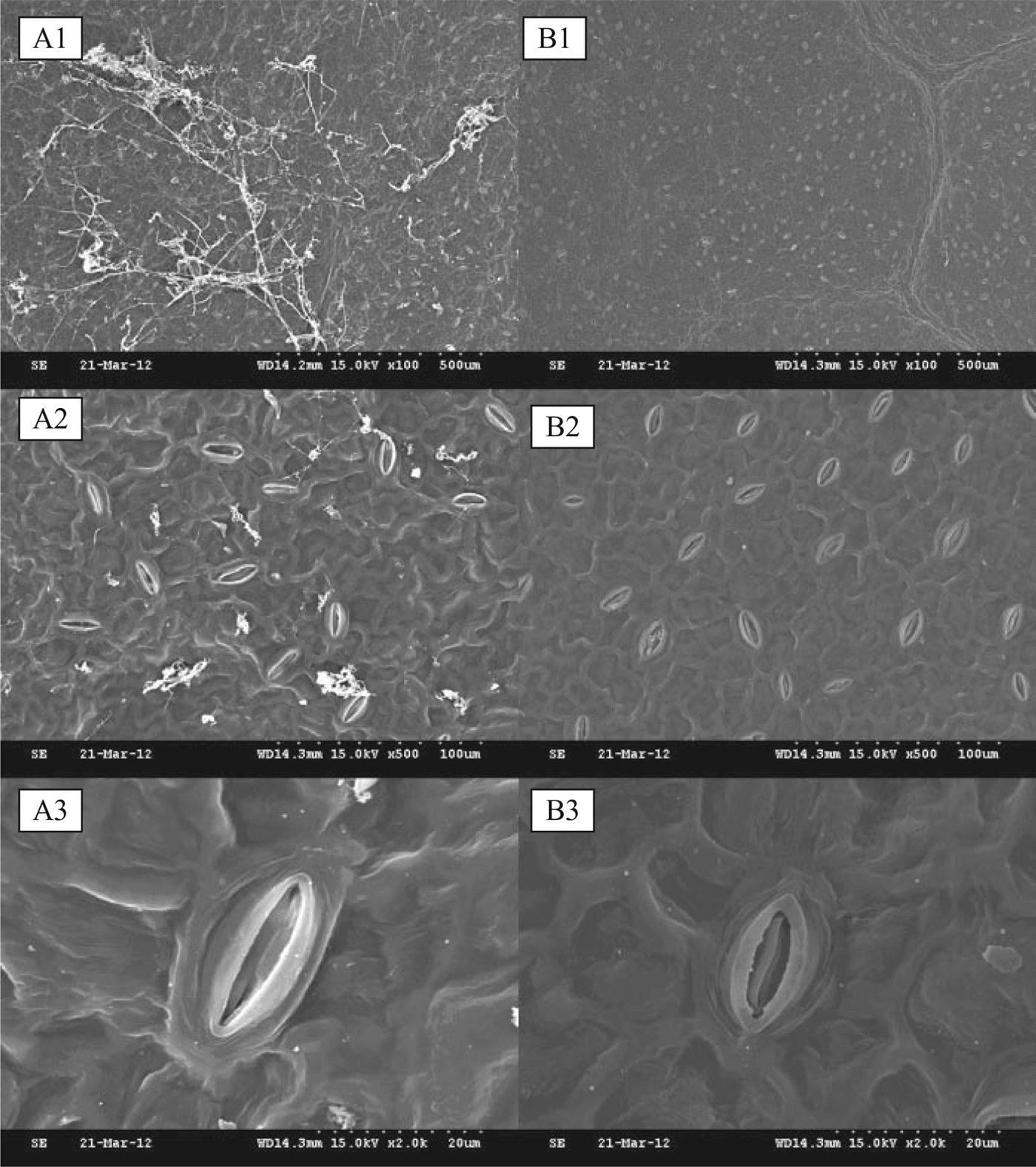
Under normal growth conditions, a large number of hyphae were attached to the surface of control leaves, and the stomatal distribution was uneven, whereas the mycelia attached to the surface of transgenic plants were relatively small, and the stomatal distribution in the leaves was relatively even (Fig. 4A1 and B1). The elongated stomata in the control plants were large and the density was lower, whereas the stomata in the transgenic plants were smaller and the density was greater (Fig. 4A2, A3 and B2, B3).
Leaf surface structure of transgenic and control plants under non-saline conditions. A1, the mycelia attached to the surface of control plants (×100); A2, stoma of control plants surface (×500); A3, stoma of control plants surface (×2·0 K). B1, the mycelia attached to the surface of transgenic plants (×100); B2, stoma of transgenic plants surface (×500); B3, stoma of transgenic plants surface (×2·0 K).
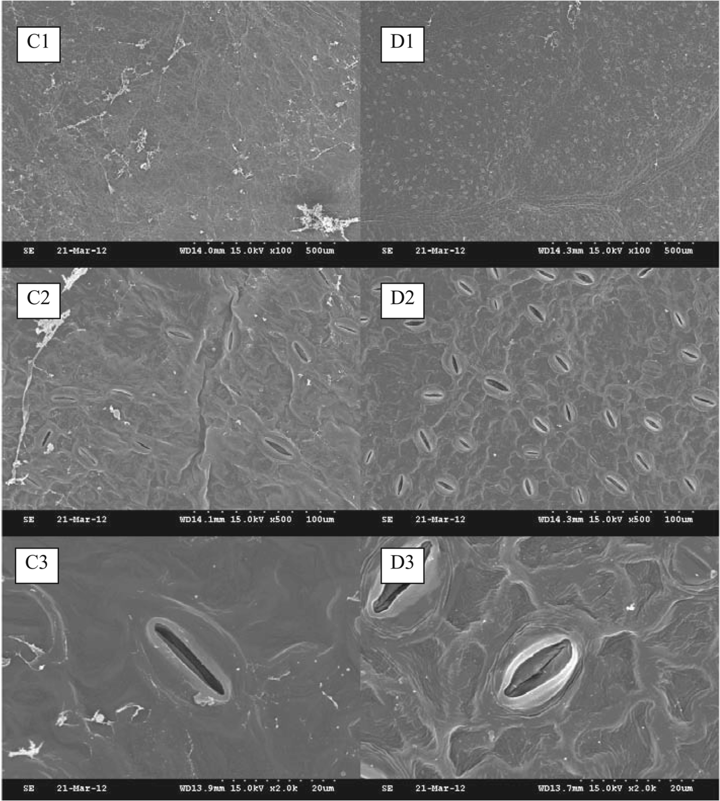
Following the salt treatment, the control plant leaf surfaces showed large changes from the salt stress. The majority of the stomata were closed and the number of stomata was significantly decreased in comparison with normal growth conditions (Fig. 5C1–C3). In contrast, transgenic plants also appeared to have lost water, but the leaf structure did not appear to be seriously damaged. To adapt to salinity, transgenic plants showed an increase in stomatal density, but stomatal size was not significantly altered (Fig. 5D1–D3).
Leaf surface structure of transgenic and control plants under NaCl stress. C1, the mycelia attached to the surface of control plants (×100); C2, stoma of control plants surface (×500); C3, stoma of control plants surface (×2·0 K). D1, the mycelia attached to the surface of transgenic plants (×100); D2, stoma of transgenic plants surface (×500); D3, stoma of transgenic plants surface (×2·0 K).
Effect of different concentrations of NaCl stress on leaves of transgenic apple
After 5 days in aqueous solution, there were no significant changes in the leaves of either transgenic or control apple plants, indicating that the detached leaves were not harmed under salt stress. However, at 0·5% NaCl, transgenic plants still showed no significant changes by the 5th day, whereas the petioles of control plants began to show browning, and the chlorophyll content was reduced (Fig. 6a). When the NaCl concentration was increased to 1·0%, the petioles and leaves of control plants showed browning and were less green by the 4th day, and the effects were increased under higher NaCl stress by the 5th day. Conversely, transgenic leaves still had no significant change. When the NaCl concentration was 1·5%, control leaves, petioles and leaf margins were less green by the 3rd, 4th and 5th day as the NaCl stress increased, whereas the transgenic leaves began to show a lack of green colour after 4 days (Fig. 6b).
(Colour online) Effect of different concentrations of NaCl on the leaves of apple in vitro. (a) Upper leaf surfaces; (b) lower leaf surfaces; WT, leaves of the control plants; T, leaves of the transgenic plants.
DISCUSSION
The TGA transcription factors comprise a recently identified family of plant-specific transcription factors. The TGA transcription factors contain a bZIP structural domain, belonging to the bZIP transcription factors D-class Asian family. The bZIP proteins have been identified in a variety of plants and play crucial roles in almost all biological processes (Jakoby et al. Reference Jakoby, Weisshaar, Dröge-Laser, Vicente-Carbajosa, Tiedemann, Kroj and Parcy2002). Plant bZIP transcription factors play very important roles in the resistance to adverse natural environmental processes, and their expression is induced by abiotic stresses such as high salinity, low temperature and drought (Stankovic et al. Reference Stankovic, Vian, Henry-Vian and Davies2000; Lee et al. Reference Lee, Choi, Hwang, du Choi and Hwang2006; Rodriguez-Uribe et al. Reference Rodriguez-Uribe and O'Connell2006; Nijhawan et al. Reference Nijhawan, Jain, Tyagi and Khurana2008; Seong et al. Reference Seong, Kwon, Ghimire, Yu, Cho, Lim, Kim, Heo, Lim, Chung, Kim and Lee2008; Zou et al. Reference Zou, Guan, Ren, Zhang and Chen2008; Wang et al. Reference Wang, Gao, Liang, Wang, Yang and Liu2010b). By regulating the activity of a plant-degrading enzyme, overexpression of the CAbZIP1 gene improved plant resistance (Karpinski et al. Reference Karpinski, Wingsle, Karpinska, Hällgren, Aro and Andersson2004; Lee et al. Reference Lee, Choi, Hwang, du Choi and Hwang2006), and the overexpression of exogenous bZIP proteins improved the resistance to a variety of stresses including salt, cold, methyl viologen oxidation, drought and heat (Lee et al. Reference Lee, Choi, Hwang, du Choi and Hwang2006; Liao et al. Reference Liao, Zou, Wei, Hao, Tian, Huang, Liu, Zhang and Chen2008; Zhang et al. Reference Zhang, Wollenweber, Jiang, Liu and Zhao2008). Furthermore, the overexpression of bZIP proteins could increase the photosynthetic capacity of plants under stress conditions to improve their resistance (Zhang et al. Reference Zhang, Wollenweber, Jiang, Liu and Zhao2008). In the present study, the MhTGA2 gene was transformed into Nagafu No. 6 using the A. tumefaciens-mediated method. Four transgenic apple lines were obtained, as confirmed by PCR and RT–PCR. The MhTGA2-transformed plants exhibited alterations of their physiological and ecological characteristics. With regard to the morphological characteristics, the roots of transgenic plants were more numerous, longer and stronger than the controls. Overall, the growth status of transgenic plants was better than control plants, probably due to their ability to adapt to the stress conditions and grow more rapidly. In contrast, the control plants were less able to adapt to the external environment: the growth rate was relatively slow and the plants were more susceptible to disease. With regard to physiology, the net photosynthetic rate, instantaneous carboxylation efficiency and stomatal conductance of transgenic plants was significantly greater than control plants, whereas the intercellular CO2 concentration was significantly lower than in control plants.
Different bZIP transcription factors have specific functions in the resistance of plants to adverse environmental conditions, and certain bZIP genes could increase the resistance to salt and cold, whereas others are related to abscisic acid (ABA) and could increase drought resistance (Kang et al. Reference Kang, Choi, Im and Kim2002; Fujita et al. Reference Fujita, Fujita, Satoh, Maruyama, Parvez, Seki, Hiratsu, Ohme-Takagi, Shinozaki and Yamaguchi-Shinozaki2005). Liao et al. (Reference Liao, Zou, Wei, Hao, Tian, Huang, Liu, Zhang and Chen2008) showed that salt tolerance conferred by different bZIP transcription factors varied with different developmental stages. In the seedling and bolting stages, the transgenic plants overexpressing soybean GmbZIP44, GmbZIP62, or GmbZIP78 showed increased resistance to salt and cold, whereas the overexpression of the GmbZIP78 gene failed to enhance the resistance to salt in the bud stage (Liao et al. Reference Liao, Zou, Wei, Hao, Tian, Huang, Liu, Zhang and Chen2008). In the present study, the MhTGA2 gene was transformed into Nagafu No. 6 to enhance the resistance of this variety to NaCl. Using different concentrations of a NaCl solution, the result showed that the overexpression of the MhTGA2 gene in the transgenic plants can enhance their salt-stress resistance. These results were also verified by scanning electron microscopy. Taken together, the present results suggest that the MhTGA2 gene may enhance the resistance to abiotic stress.
This work was supported by grants from the China Ministry of Science and Technology ‘863’ Programme No. 2011AA100204.