Introduction
Epilepsy is a chronic neurological disease that represents a public health problem and causes deterioration in the quality of life of patients who do not have guaranteed medical control of their condition and are marginalized and discriminated against in society. In Mexico, the estimated prevalence of patients with epilepsy is between ten and 20 per 1000 individuals, suggesting that at least one million individuals in the country have some form of epilepsy (Ávila, Reference Ávila2004). The relationship between epilepsy and visceral larval migration syndrome (VLM) caused by Toxocara canis has been elucidated previously (Nicoletti et al., Reference Nicoletti, Bartoloni and Regio2002, Reference Nicoletti, Bartoloni, Sofia, Mantella, Nsengiyumva, Frescaline and Preux2007, Reference Nicoletti, Sofia and Mantella2008), suggesting an association between the parasite and the incidence of epilepsy in the regions where the studies were conducted. However, Eraky et al. (Reference Eraky, Abdel-Hady and Abdallah2016) questioned the association between T. canis and cryptogenic epilepsy (epilepsy of unknown origin) in children, suggesting the need to clearly elucidate this relationship between epilepsy and T. canis larvae.
In Mexico, T. canis eggs have a high prevalence in public parks and playgrounds (Martínez-Barbabosa et al., Reference Martínez-Barbabosa, Fernandez-Presas, Vazquez-Tsuji and Ruíz-Hernández1998; Romero et al., Reference Romero, García, Mendoza, Torres and Ramírez2009; Encalada-Mena et al., Reference Encalada-Mena, Duarte-Ubaldo, Vargas-Magaña, García-Ramírez and Medina-Hernández2011) or a low prevalence but high viability in dog faeces in soil littering public parks (up to 97%) (Romero et al., Reference Romero, Mendoza, Bustamante, Crosby and Ramírez2011). In a previous study, the viability of T. canis eggs in the soil of public parks was reported to be 73%; furthermore, the viability of infective eggs found in other playgrounds was 86%, whereas that of eggs obtained directly from the dog faeces was 94% (Romero et al., Reference Romero, Yañez, Mendoza, Bustamante-Montes and Ramírez-Durán2013b). Exposure to canine faeces in parks and streets of urban areas is associated with a high risk of contamination for children, who can become infected with T. canis larvae through their habits such as pica or geophagy, by playing games on the contaminated soil and by lack of hygiene while living with infected dogs (Romero et al., Reference Romero, Mendoza, Yañez, Ponce and Bustamante2013a).
Toxocara canis is a parasite that affects various organs, particularly the liver, lungs, eyes and central nervous system. In humans, the parasite develops only until larval stage L2; the larvae migrate to various organs without reaching its adult stage (Beaver, Reference Beaver1969). After infection with larvated eggs of T. canis, a large number of these parasites can remain in the tissues for many years. In the brain, the larvae produce glycosylated proteins that induce a CD4 Th2 cell-type immune response with the production of IgE and interleukin-5, which promotes eosinophilic differentiation and vascular adhesion (Bächli et al., Reference Bächli, Minet and Gratzi2004). It has been suggested that the immune reaction triggered by the presence of the parasite induces generalized seizures, whereas the formation of granulomas causes focal seizures (Critchley et al., Reference Critchley, Vakil, Hutchinson and Taylor1982; Viovy et al., Reference Viovy, Jofré, Noemi, Erazo and Cerva1999).
There is currently no direct method to confirm the presence of T. canis larvae in humans, with indirect evidence being the only tool available to detect the presence of this parasite, specifically the detection of antibodies in human body fluids (Despommier, Reference Despommier2003). Several authors have reported that for the diagnosis of VLM, it is necessary to obtain evidence using enzyme-linked immunosorbent assay (ELISA) and Western blotting (Magnaval et al., Reference Magnaval, Fabre, Mauriéres, Charlet and Larrard1991; Iddawela et al., Reference Iddawela, Rajapakse, Perera and Agatsuma2007).
Considering the relevance of reported cases of the presence of T. canis in Mexico and that no studies have been performed in this country to determine the relationship between epilepsy and T. canis larvae, this study aimed to estimate the presence and specificity of anti-T. canis antibodies in the serum samples of Mexican children with epilepsy through serology using ELISA and Western blotting.
Material and methods
Study group
Between January 2010 and January 2013, 214 paediatric sera samples were collected from patients aged 1–18 years and divided into two groups: control group – children without epilepsy, comprising 103 clinically healthy individuals without personal medical or family history of epileptic seizures and no epidemiological antecedents of the presence of the parasite; and epileptic group – 111 children diagnosed with epilepsy. The children were treated at the Neurology Department of Hospital Infantil de México Federico Gómez; thus, their clinical records were available at the hospital, as was information on risk factors and demographic data associated with T. canis larvae infection. Cranial imaging studies and analyses were performed in the epileptic group to confirm neurological damage. The classification of the types of epileptic seizures was based on the 1993 guidelines of the International Epilepsy Association (Commission on Classification and Terminology of the International League against Epilepsy, 1993). All patients included in the present study signed a written informed consent, and the study was approved by the Ethical Committee of Hospital Infantil de México Federico Gómez.
L2 larvae of T. canis and Ascaris lumbricoides
Larvae were obtained from T. canis eggs by dissecting the gravid uteri of female worms, whereas the larvae of A. lumbricoides were collected from human faecal samples obtained from various populations in the state of Guerrero, Mexico. In both cases, the larvae were incubated for 30 days at 28°C in a solution containing 2% formaldehyde and 1% sodium hypochlorite for embryonation. The larvated eggs from both parasites were suspended separately in 0.26% sodium hypochlorite at room temperature (22°C) with constant stirring for 30 min and washed twice with saline solution (85% NaCl) under sterile conditions, and the larvated eggs from both parasites were suspended separately in 0.26% sodium hypochlorite at room temperature (22°C) with constant stirring for 30 min and washed twice with saline solution (85% NaCl) under sterile conditions and stirred vigorously until eclosion of the larvae was observed under the microscope. The content was then transferred to a conical tube containing a sterile culture medium. The larvae were collected using the modified Oaks and Kayes technique (Oaks & Kayes, Reference Oaks and Kayes1979) and maintained in RPMI 1640 medium with dextrose (Sigma R-0883) and antibiotics (100 mg/mL penicillin and streptomycin) at 37°C in an atmosphere of 5% CO2 (Queu® incubator West Virginia, USA no. 2710).
Excretion-secretion antigens (ES) of L2 larvae of T. canis and A. lumbricoides
The excretion-secretion (ES) antigens were obtained using the Savigny method (Savigny, Reference Savigny1975). The supernatant of the larvae cultivated in RPMI 1640 medium supplemented with 1% dextrose and glutamine (Sigma R-0883) was drawn off every week for 30 days and concentrated in Amicon UM-2 tubes by centrifugation (Sorvall® Centrifuge RT6000B Massachusetts, USA) at 5000 g for 30 min at 4°C and washed off with Tris buffer (pH 7.4; UltraPureTris, Invitrogen Scotland, UK® no. 15504-020) to remove impurities and concentrate ES from the larvae of T. canis (TcES). To inactivate the enzyme activity of the antigens obtained, a mixture of protease inhibitors was added: 10 mm Etiendiaminotetraacétic (Sigma E-9884), 20 mm Phenylmethylsulfonyl fluoride (Sigma P-76-26), 10 mm leupeptin (Sigma L-2884) and 1 mm pepstatin (Sigma P-7626). The concentration of the proteins contained in TcES was determined using the Bradford method (Bradford, Reference Bradford1976), and the material was stored at −70°C until use.
Determining the sera anti-T. canis antibodies of children with epilepsy and of controls without epilepsy by using TcES-based ELISA
Serum samples from the control and study groups were evaluated by using TcES-based ELISA. The method required 96-well polystyrene plates (Costar 3590) sensitized with 5 µg/well of TcES from T. canis overnight at 4°C; washing was performed with phosphate-buffered saline (PBS) Tween 20 0.05%, and blocking was performed with 5% milk-PBS for 2 h at 37°C. After washing, the diluted sera were added to 5% milk-PBS in a 1:500 dilution and incubated overnight at 4°C. The anti-human IgG horseradish peroxidase conjugate was then added (diluted to 1:10,000) (Sigma A-9044) and incubated for 2 h at 37°C. The plate was subsequently developed with 0.1 mg/ml of orthophenylendiamine (Sigma P-9029) diluted in a phosphate/citrate buffer with hydrogen peroxide (H2O2) as the substrate. The plates were then incubated for 15 min, and the reaction was terminated with sulfuric acid (2N). The absorbance of each well was determined by reading in a PR 2100 SANOFI spectrophotometer (Bio-RAD® Virginia USA) with a 495-nm filter. All reactions were performed in duplicate (Iddawela et al., Reference Iddawela, Rajapakse, Perera and Agatsuma2007).
Cross-reaction between anti-T. canis and anti-A. lumbricoides antibodies in the sera of children with epilepsy
The cross-reaction procedure was performed for the sera of epileptic children positive for anti-T. canis antibodies. The plates were sensitized with 5 µg/well of TcES and ES from the larvae of A. lumbricoides (AlES) using a previously described technique (Iddawela et al., Reference Iddawela, Rajapakse, Perera and Agatsuma2007).
Unidimensional electrophoresis of TcES and AlES
TcES (excretion-secretion T. canis and AlES (excretion-secretion A. lumbricoides)kept in vitro (5 µg/line) were analysed on 12% sodium dodecyl polyacrilamide gel electrophoresis (SDS-PAGE) polyacrylamide gels (San Louis, USA Sigma® no.79-06-1/bisacrylamide Sigma San Louis, USA® no.110-26-9 at 30%; UltraPureTris, Invitrogen®Scotland, UK no. 15504-020 pH 8.8; sodium dodecyl sulfate, San Louis, USA Sigma® no. 205-788-1 at 10%; distilled water; ammonium persulfate, San Louis, USA Sigma® no. 7727-54-0 and TEMED, San Louis, USA Sigma® no. 203-744-6) in an electrophoretic chamber at 100 V for 45 min. (Bio-RAD® Mini Protean) using the Laemmli and Favre method (Laemmli & Favre, Reference Laemmli and Favre1973) and a running buffer for protein electrophoresis (UltraPureTris Scotland, UK Invitrogen® no. 15504-020; sodium dodecyl sulfate San Louis, USA, Sigma® no. 205-788-1; glycine, Sigma® no. 56-40-6 and distilled water). The gels were incubated for 24 h in a fixative solution of silver (methanol; glacial acetic acid, Golden Bell® no. 20100; concentrated enhancer and distilled water) and stained with a silver stain (Silver Complex Solution, Bio-RAD® no. 161-0462; Image Development Reagent, Bio-RAD® no. 161-0464; Reduction Moderator Solution Bio-RAD® no. 161-0463, Development Accelerator Solution Bio-RAD® no. 161-0449).
Western blotting analysis for assessing cross-reactivity among the excretory–secretory antigens of T. canis and A. lumbricoides
For Western blotting analysis, 150 µg/ml of TcES and AlES were used, both of which were separated on acrylamide-bisacrylamide preparative gels (SDS-PAGE) at 12% under reducing conditions at 100 V (Towbin et al., Reference Towbin, Staehelin and Gordon1979) and transferred onto 0.2-μm nitrocellulose membranes (Bio-RAD® no. 162-0112) using a Bio-RAD® Transblot transference chamber. The Western blotting analysis was performed according to the technique described by Magnaval et al. (Reference Magnaval, Fabre, Mauriéres, Charlet and Larrard1991). The membranes of both gels were cut into strips of 3–4 mm and incubated with blocking solution (milk at 5% diluted in PBS) overnight at 4°C. The sera confirmed as positive for T. canis using ELISA were diluted to 1:250 in the blocking solution and incubated overnight at 4°C in the strips of nitrocellulose paper with T. canis and A. lumbricoides antigens, washed five times with PBS-Tween 20 0.05% and incubated with the anti-human IgG conjugate bound to peroxidase (Sigma® no. A-9040) at 1:10,000 for 2 h at 37°C. The strips were washed with PBS-Tween 20 0.05%. After washing, immunodetection was developed with 4-chloronaphthol (Sigma® no. C-8890) in a saline phosphate buffer with H2O2 as the substrate. The reaction was terminated with distilled water.
Statistical analyses
Descriptive data analysis was performed, including the estimation of relative frequencies stratified by clinical and demographic characteristics of the subjects (patients with and without epilepsy). Independent hypotheses between the diagnosis of epilepsy and each of the remaining variables were evaluated using two-way tables and Fisher's exact test. Binary logistic regression models were adjusted to evaluate the association between the vector of covariates and the diagnosis of epilepsy. The Hosmer–Lemeshow test was used to assess the goodness-of-fit of logistic regression models, whereas the Akaike and Bayesian information criteria were used for comparison between the models. All procedures were performed using the statistical programme STATA version 14.1 StataCorp Texas 77845, USA.
Results
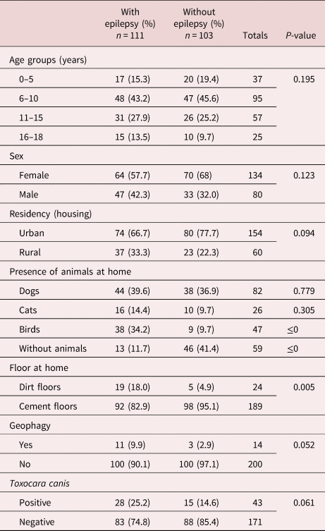
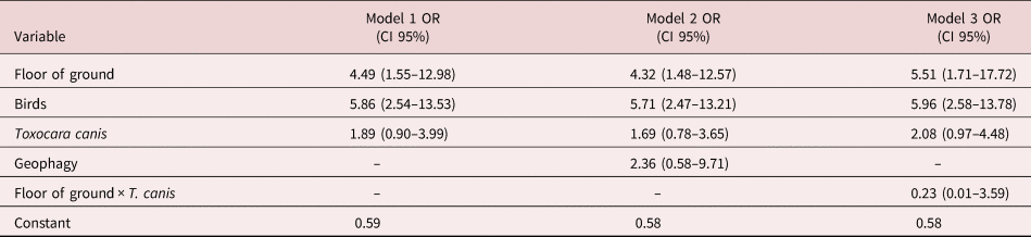
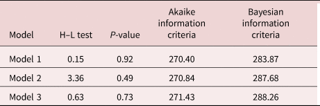
As shown in table 1, of the 103 control children, 70 were female and 33 were male. The epileptic group comprised 64 females and 47 males aged between one and 18 years, whereas the majority of children in the control group were aged 5–9 years. Table 1 also shows the epidemiological and demographic factors that were evaluated in both groups. There was a significant difference between the two groups in the positivity to T. canis detected in the tests, as the number of cases with positive results was higher in the study group than in the control group. This suggests a significant association between the parasites and the disease. The analysis of different risk factors was associated with the co-existence of animals in the two study groups; it was higher in the epileptic group, whose percentage of co-existence with dogs was 39.6% compared with 36.9% in the control group. The percentage of co-existence with pet and farm birds in the study group was 34.2% compared with just 9.7% in the control group. On interpreting the regression coefficients of the dirty floor factor, the presence of dirty floors was positively associated with having epilepsy, increasing the odds to 5.96 in the first model and to 5.71 and 4.49 in the second and third models, respectively. The presence of pets and farm birds on the dirty floors was also positively associated with the diagnosis of epilepsy, increasing the odds to 5.51 in the first model and to 4.32 and 5.86 in the second and third models, respectively (table 2). According to the Hosmer–Lemeshow tests, these three models were considered a good fit to the data, as the values obtained from these models and their respective P-values (not significant) may be interpreted as a result of the goodness-of-fit model (table 3).
The epidemiological and demographic factors in patients and healthy participants.

P ≤ 0.05.
Multivariate-adjusted odds ratios for various characteristics associated with epilepsy.

Dependent variable: epilepsy. OR, odds ratio; CI, confidence interval.
Results of the Hosmer–Lemeshow (H–L) tests.

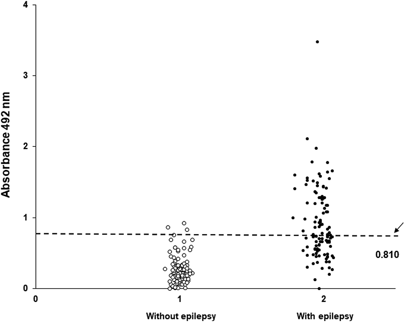
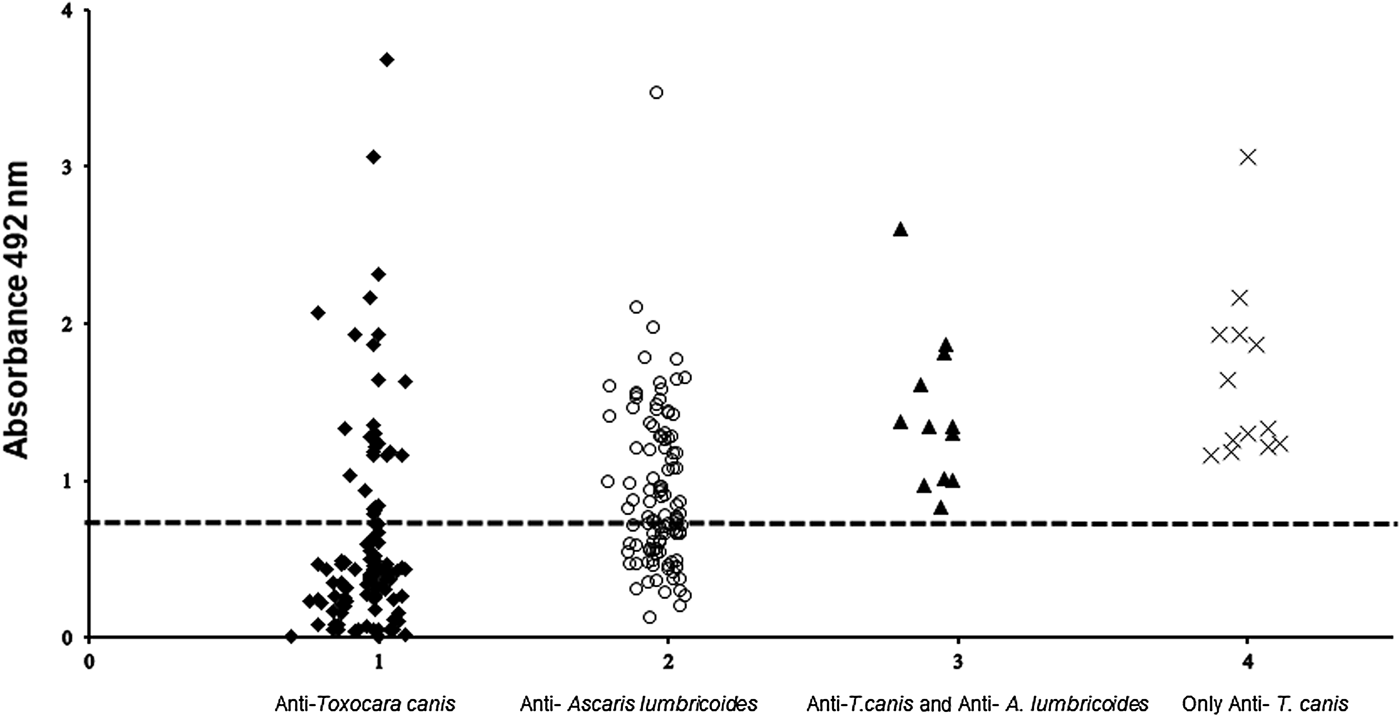
The levels of anti-TcES antibodies are shown in fig. 1, indicating seropositivity in 28/111 (25.2%) and seronegativity in 83/111 (74.8%) cases, in accordance with the cut-off point of optical density 0.80. The cut-off point was calculated as the average of negative sera ±2 standard deviations, whereas positive values greater were than 0.8 optical density(Espinoza et al., Reference Espinoza, Huapaya, Suárez, Chávez, Sevilla, Dávila, Huiza, Náquira and Alva2003; Cardona et al., Reference Cardona, Fabian and Gomez2005).
Serological analysis evaluated by ELISA: detection of antibodies anti-T. canis in groups of children with and without epilepsy (patients, n = 111; healthy individuals, n = 103). Samples with absorbance of less than 0.8 were defined as negative and those of more than 0.8 as positive. Dashed line represents cut-off value.

The ELISA results for both groups are presented in fig. 2. Data obtained showed a positive reaction to anti-AlES antibodies in 52/111 (46.8%) cases in the epileptic group and 3/103 (2.91%) cases in the control group, according to the cut-off point of D.O 0.80.
Cross-reaction antibody anti-T. canis and A. lumbricoides in sera of children with epilepsy (n = 111). Presented anti-A. lumbricoides antibodies did not give a cross-reaction of sera positive for T. canis. Samples with absorbance of less than 0.8 were defined as negative, and those more than 0.8 as positive. Dashed line represents cut-off value.

The cross-reaction between the serum samples of the children with epilepsy using the antigen of A. lumbricoides is shown in fig. 2, where the recognition percentage was 52/111 (46.8%). The number of serum samples recognizing antigens of TcES and AlES was 12/111 (10.8%), and that of serum samples not having cross-reaction with A. lumbricoides and exclusively recognizing antibody anti-T. canis was 13/111 (11.7%) (fig. 2).
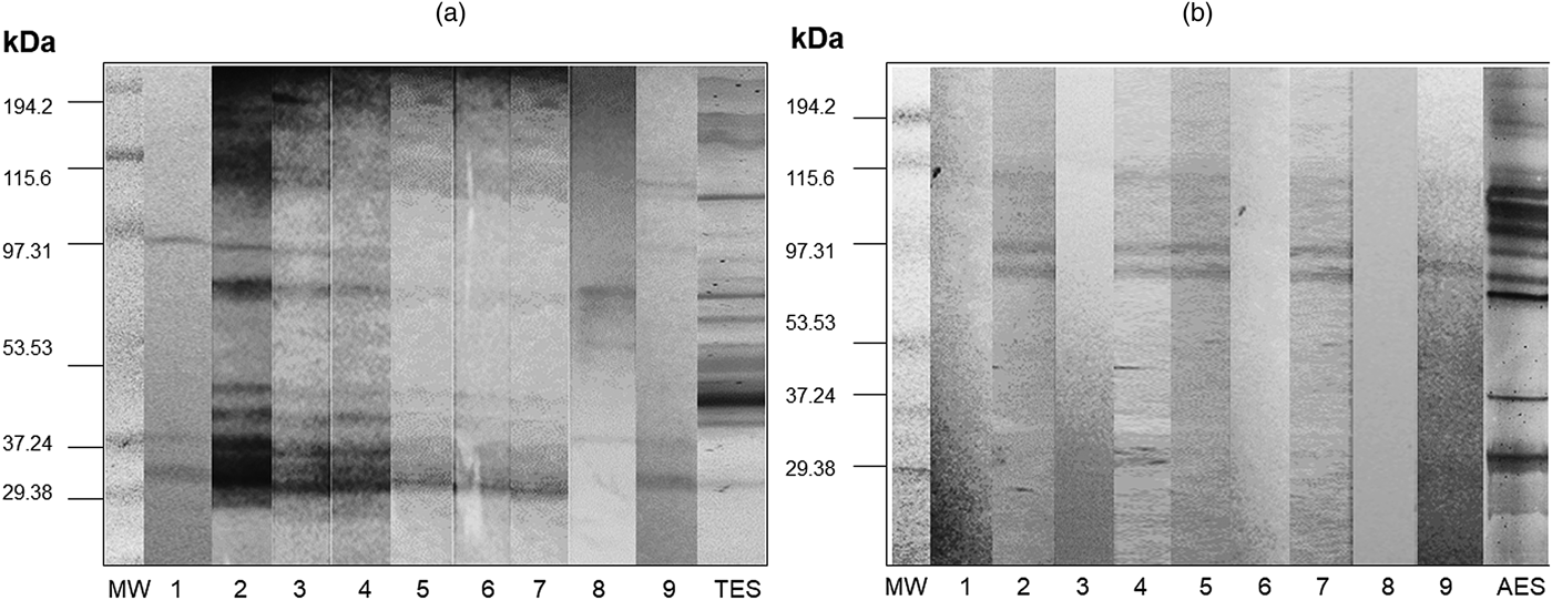
The comparison of the electrophoretic profiles of TcES and AlES on polyacrylamide gels is shown in fig. 3. The profiles showed differences in the tonality and number of proteins, with a 25–210-KDa complex in the TcES profile (fig. 3a, b).
Analysis of Western blotting of sera from children with epilepsy previously evaluated by ELISA. (A) Electrophoretic analysis in 12% SDS-PAGE of the TES. Sera from children with epilepsy and TcES. (B) Electrophoretic analysis in 12% SDS-PAGE of the AlES. Sera from children with epilepsy and AlES. Abbreviations: MW, molecular weights; 1–9, serologic specimens.

The results of the Western blotting analysis of the nine serum samples positive for anti-T. canis antibodies showed recognition of the 24-, 35-, 55-, 70-, 120- and 210-kDa proteins (fig. 3a). Figure 3b shows the results of the Western blotting analysis of the nine serum samples with anti-T. canis antibodies using AlES, showing recognition of the 70-, 80- and 110-kDa proteins.
Discussion
The analysis of epidemiological and demographic factors of the groups included in this study and their relationship with positive reactions to the antigens TcES of the larva of T. canis in ELISA revealed the probability that this pattern of association may be coincidental was only 6.1%, as the evidence obtained in this study supported a possible relationship between T. canis larvae and the prevalence of epilepsy. Remarkably, as the observed difference between the two groups in the positivity of the results was not marked, this may indicate a bias resulting from the expected results of the study. Moreover, controls were from the same hospital, but without clinical manifestations of epilepsy, and also came just for external therapy, and they usually have conditions and characteristics that differ from those of the rest of the population. Glickman et al. (Reference Glickman, Cypess, Crumrine and Gitlin1979) have also examined the presence of anti-T. canis antibodies in their serological study, comparing 51 children with epilepsy of unknown aetiology with 33 children with epilepsy of suspected aetiology. The results obtained for these two groups did not differ significantly, but when the seropositivity ranges were compared between these patients and a group of healthy children, the results revealed significantly higher antibody titres in the children with epilepsy of unknown aetiology. However, several studies have indicated that the rates of infection with Toxocara spp. are not high in children with symptomatic epilepsy. These findings have been related to the fact that children with epilepsy have a higher tendency to consume unhygienic foods, and it has been suggested that such behaviour patterns predispose these children to infection with Toxocara spp.
Models of multivariate logistic regression analyses indicated that contact with farm birds and interaction with dirt floors were factors associated with the presence of epilepsy in patients; the dispersing and infectivity of T. canis eggs has been associated with these variables. Fitz et al. (Reference Fitz, da Silva, Pelegi, de Lima, Pereira and Alvares2018) concluded that chickens play an important role in the dispersal of T. canis eggs in the environment, as well as dogs, who are definitive hosts and transmission vectors to other paratenic hosts. Our results suggest that co-existence with farm birds is a significant risk factor.
An association between epilepsy and toxocariasis was reported in a rural area of Bolivia by Nicoletti et al. (Reference Nicoletti, Bartoloni and Regio2002); they observed that correctly classifying epileptic seizures is a key element in interpreting the results of analytical studies. Their work confirmed the important roles played by parasites, including cysticercosis and toxocariasis, in the incidence of epilepsy within their study area. In studies previously performed in a Kiremba population in Burundi, and in Italy, a significant association was found between seropositivity to T. canis in patients and epilepsy, and it was suggested that T. canis may play a critical role in the incidence of epilepsy in endemic regions (Nicoletti et al., Reference Nicoletti, Bartoloni, Sofia, Mantella, Nsengiyumva, Frescaline and Preux2007; Eraky et al., Reference Eraky, Abdel-Hady and Abdallah2016).
These findings increase the value of the present study conducted in Mexico, as there are no registered cases of children with epilepsy caused by T. canis. The results of the present study examining serum samples from the children with cryptogenic epilepsy (n = 111) showed that 11.7% children were positive for anti-T. canis antibodies, suggesting that they had been in contact with this parasite and had generated immunological memory as a response. In addition, 46.8% of the study group was positive for anti-A. lumbricoides antibodies, a significant cross-reaction percentage similar to that reported by Badley et al. (Reference Badley, Grieve, Bowman, Glickman and Rockey1987) and Rosamanta et al. (Reference Rosamanta, Romero and Arias2003). These previous studies have reported that T. canis antigens may cross-react with other parasites (ascariasis, fasciolosis, strongyloidiasis, trichinosis and anisakiasis) due to their composition, as their walls contain glycosylates comprising 40% carbohydrates. Speiser & Gottstein (Reference Speiser and Gottstein1984) observed a moderate cross-reaction with other helminths on performing ELISA.
Few researchers have compared TcES and AlES; however, Kennedy et al. (Reference Kennedy, Qureshi, Fraser, Haswell-Elkins, Elkins and Smith1989) compared the secreted and somatic antigens of three parasites that affect humans: A. lumbricoides, A. suum and T. canis. They found an antigenic similarity among these nematodes: an internal 14-kDa protein that has its homologue in all three worms. This molecule activates an immune response of IgG in Ascaris infection, but does not activate a measurable response in VLM caused by toxocariasis. In this previous study, sera were used from previously infected rabbits.
Electrophoretic profiles of T. canis and A. lumbricoides were obtained to corroborate the presence of antigenic proteins of the larval stage of both parasites kept in vitro. Those profiles showed differences in the intensity of the proteins when run with 5 µg/μL of the protein. TcES was more evident than AlES. The 30-, 60- and 90-kDa proteins were present in both parasites; furthermore, low-molecular-weight (17–40 kDa) proteins were present in both profiles. The typical Western blotting VLM pattern for T. canis in humans was reported by Speiser & Gottstein (Reference Speiser and Gottstein1984) using positive sera from patients, which revealed 10–14 characteristic 25–200-kDa bands. Other studies have indicated that TES can be divided into two groups: one group with high-molecular-weight proteins (132, 147 and 200 kDa) and another one with low-molecular-weight proteins (24, 28, 30 and 53 kDa) (Magnaval et al., Reference Magnaval, Fabre, Mauriéres, Charlet and Larrard1991).
The proteins most frequently recognized using Western blotting of the sera of children with cryptogenic epilepsy using TcES had low molecular weights (20–40 kDa), whereas the Western blot analyses of AlES did not recognize low-molecular-weight proteins, detecting only proteins with molecular weights >95 kDa. The results showed that the sera of nine children with cryptogenic epilepsy were positive for anti-T. canis antibodies, but not for cross-reactivity against A. lumbricoides using ELISA. Our results also showed that 68.7% of samples were positive, as they presented with proteins of 20–40 kDa. Roldán & Espinoza (Reference Roldán and Espinoza2009) published the results of a Western blot study of several parasites, showing that the most specific proteins for VLM were those with a low molecular weight. Therefore, the absence of cross-reactivity with other helminths allows these proteins to be utilized for diagnostic purposes. The low-molecular-weight proteins of 24–35 kDa form a protein complex that has a high specificity regarding anti-T. canis IgG antibodies (Qualizza et al., Reference Qualizza, Incorvaia, Grande, Makri and Allegra2011). The analysis of the cross-reactivity with AES showed the presence of these antigens in 66% of the tested serum samples, indicating a significant percentage of cross-reactivity. This coincides with the results of other studies (Magnaval et al., Reference Magnaval, Fabre, Mauriéres, Charlet and Larrard1991; Roldán & Espinoza, Reference Roldán and Espinoza2009; Bellanger et al., Reference Bellanger, Humbert, Gavignet, Deschaseaux, Barisien, Roussel, Millon, Aubin and Piarroux2010; Santillán et al., Reference Santillán, Bastin, Céspedes and Monkiewicz2013) that identified three proteins of 55, 100 and 120 kDa responsible for cross-reactivity with A. lumbricoides and other etiological agents, including virus or parasites. Similarly, Santillán et al. (Reference Santillán, Bastin, Céspedes and Monkiewicz2013) and Magnaval et al. (Reference Magnaval, Fabre, Mauriéres, Charlet and Larrard1991) reported four bands specific to T. canis (24, 28, 30 and 35 kDa). Roldán & Espinoza (Reference Roldán and Espinoza2009) confirmed these results using Western blotting, whereas Robertson et al. (Reference Robertson, Bianco, McKerrow and Maizels1989) observed that the proteins specific to T. canis were of molecular weights 400, 120, 70, 55 and 32 kDa. In the present study, sera samples of 11.7% of the children with epilepsy contained anti-T. canis antibodies with no cross-reactivity against AES, and the results of Western blotting revealed specific recognition to TEA in the range of 20–40 kDa.
There are no previous reports on the relationship between this parasite and children with epilepsy in Mexico. For the first time, this relationship between epilepsy and T. canis larvae was demonstrated.
Acknowledgement
We thank Dr. Pilar Crisóstomo Vázquez of the Hospital Infantil de México Federico Gómez for the donation of adults worms of A. lumbricoides used in this work.
Financial support
This study was supported by federal funding from the Government of Mexico and by a grant from CONACyT Mexico (project SALUD-2009-COI-115346).
Conflicts of interest
None.
Ethical standards
All authors assert that all procedures contributing to this work comply with the ethical standards of the ethics committee from the Hospital Infantil of México Federico Gómez HIM/2010/029 and have, therefore, been performed in accordance with the ethical standards laid down in the 2017 Declaration of Helsinki.