Introduction
A pharyngocutaneous fistula (PCF) occurs when there is breakdown or inadequate closure of the suture line of the reconstructed pharynx following (pharyngo)laryngectomy. As with all fistulae, intra-luminal contents, in this case saliva, flows through the dehiscence and becomes ‘extra-luminal’, with resultant draining via an external wound.Reference van la Parra, Kon, Schellekens, Braunius and Pameijer1–Reference Krouse and Metson4 Typically, PCFs become clinically apparent between 7 and 11 days after surgery.Reference Mäkitie, Irish and Gullane5–Reference Virtaniemi, Kumpulainen, Hirvikoski, Johansson and Kosma7
A PCF is by far the most common serious complication after major laryngeal surgery with published incidences as high as 48 per cent in some centres.Reference Carsuzaa, Capitaine, Ferrié, Apert, Tonnerre and Frasca6–Reference Galli, De Corso, Volante, Almadori and Paludetti8 Interestingly, despite advances in techniques and equipment over the past 40 years, the incidence remains high. It is commonly reported in the literature as between 4 and 38 per cent in the 1970-80s and between 9 and 23 per cent in the past two decades.Reference Mäkitie, Irish and Gullane5, Reference Ahn, Lee and Sohn9–Reference Aydoğan, Kiroğlu, Tuncer and Soylu11 Multiple risk factors have been implicated in the development of PCFs, including advancing age, diabetes, hypertension, poor nutritional status, anaemia, advanced tumour staging, pre-operative tracheostomy and pre-operative or concurrent chemoradiotherapy.Reference Carsuzaa, Capitaine, Ferrié, Apert, Tonnerre and Frasca6, Reference Galli, De Corso, Volante, Almadori and Paludetti8, Reference Cavalot, Gervasio, Nazionale, Albera, Bussi and Staffieri12, Reference Qureshi, Chaturvedi, Pai, Chaukar, Deshpande and Pathak13 The increased use of pedicled and free tissue transfer in a salvage setting has helped reduce published PCF rates.Reference Patel, Moore, Wax, Rosenthal, Sweeny and Militsakh14, Reference Patel and Keni15
A clinically evident PCF has potential severe consequences for the patient that extend well beyond the local infective and wound management issues. Delaying the commencement of oral intake, voice rehabilitation and discharge from hospital can all be significant, as well as possibly impeding further adjuvant treatment which may impact recurrence rates.Reference van la Parra, Kon, Schellekens, Braunius and Pameijer1, Reference Friedman, Venkatesan, Yakovlev, Lim, Tanyeri and Caldarelli3, Reference Krouse and Metson4, Reference Carsuzaa, Capitaine, Ferrié, Apert, Tonnerre and Frasca6 A PCF also predisposes other serious acute complications such as flap necrosis and carotid artery rupture with their associated morbidity and mortality risks.Reference van la Parra, Kon, Schellekens, Braunius and Pameijer1, Reference Krouse and Metson4, Reference Ikiz, Uça, Güneri and Erdağ16, Reference Papazoglou, Doundoulakis and Terzakis17
There have been considerable efforts to identify and mitigate pre-, intra- and post-operative risk factors for PCF formation. There is a considerable ongoing debate as to the merits of early versus late initiation of oral intake post laryngectomy, but the most common post-operative practice involves the patient remaining nil by mouth for the first 7-10 days after surgery to allow for the suture line to heal without being bathed in proteolytic and bacteria-ladened saliva.Reference Boyce and Meyers18, Reference Milinis, Gaskell, Lau, Lancaster and Jones19 In this period, the patient is normally fed via a feeding tube with or without a salivary tube in place to bypass the suture line. The early radiological detection of any PCFs is therefore a key but poorly researched topic that has a key role in prompt intervention that can potentially (partially) mitigate the adverse effects.Reference Mäkitie, Irish and Gullane5, Reference Ahn, Lee and Sohn9, Reference Aydoğan, Kiroğlu, Tuncer and Soylu11 A barium or water-soluble contrast swallow study is typically performed to assess the integrity of the pharyngeal repair prior to the commencement of oral intake.Reference Ahn, Lee and Sohn9, Reference AM, Cohen and Adams10, Reference White, Golden, Sweeny, Carroll, Magnuson and Rosenthal20, Reference Morton, Mehanna, Hall and McIvor21
Despite the obvious importance of PCF detection, there is no definitive gold-standard investigative modality recommended in the literature.Reference Friedman, Venkatesan, Yakovlev, Lim, Tanyeri and Caldarelli3, Reference Narayan, Limbachiya, Balasubramanian, Subramaniam, Thankappan and Iyer22 Water soluble or barium contrast swallow studies are the most widely used investigation; however, their value is not universally accepted with publications highlighting limitations including low sensitivity, the provision of imprecise two-dimensional images and minimal value in detecting early clinically silent PCFs.Reference van la Parra, Kon, Schellekens, Braunius and Pameijer1, Reference Ahn, Lee and Sohn9, Reference Imaizumi, Suzuki and Murono23, Reference Moses, Eisele and Jones24 Other articles point to the lack of standardisation for performing the swallow studies themselves.Reference Narayan, Limbachiya, Balasubramanian, Subramaniam, Thankappan and Iyer22 Disparate views exist however, with some championing the merits of contrast swallow studies and their ability to detect blind-ending PCFs potentially mitigating the evolution into true PCFs.Reference Krouse and Metson4, Reference Carsuzaa, Capitaine, Ferrié, Apert, Tonnerre and Frasca6, Reference Chabrillac, Baudel, Vergez, Woisard, Farenc and Morisseau25 Clinical use internationally is therefore variable with a lack of consensus for appropriate use.Reference Krouse and Metson4, Reference Carsuzaa, Capitaine, Ferrié, Apert, Tonnerre and Frasca6, Reference Moses, Eisele and Jones24
This study aims to address this by evaluating the diagnostic accuracy of water-soluble contrast swallow studies in the detection of PCF and its role in informing post-operative management decisions in laryngectomy patients.
Methods
Study participants
A retrospective case note review was performed on all patients who underwent a laryngectomy under the care of the Liverpool Head and Neck Centre at Aintree University Hospital, Liverpool, between April 2018 and July 2023. All available electronic patient records, radiological images and reports and scanned paper notes were reviewed with primary outcome data recorded for the incidence of post-operative clinical PCF. The contrast swallow study is typically performed between post-operative days 7 and 14 using the water-soluble contrast material (e.g., gastrograffin). The images are contemporaneously reported by an appropriately head and neck trained member of the radiological team. The study was approved by the institutional clinical governance team.
Inclusion criteria
All patients who underwent a contrast swallow study after total (TL) or partial laryngectomy (PL) prior to commencing oral diet were included regardless of whether their operation was a primary or salvage procedure. Patients who underwent a reconstruction with a free-flap or regional pedicle flap were eligible for inclusion.
Exclusion criteria
Any patients with clinical evidence of PCF prior to a contrast swallow study being performed were excluded.
Outcomes
The primary outcome was the incidence of clinical PCF in patients with positive findings from water contrast swallow for a radiological fistula, compared with the patients who had no abnormal radiological study findings. The secondary outcome was the length of hospital stay. Patients were followed up for at least 30 days or until they were discharged from hospital, with data recorded and comparison made between those who had radiological evidence of a PCF and those who didn’t, and how they were managed and when oral diet was commenced post-operatively.
Statistical analysis
The accuracy of the contrast swallow study was assessed using sensitivity, specificity, negative predictive value and positive predictive value. Demographic and clinical variability were compared between the study groups using the chi-squared test for the categorical data and Mann–Whitney U test for continuous parametric data.
Results
A total of 94 patients were evaluated for inclusion into the study. Following exclusion of patients with pre-contrast study (n = 6) clinical PCF and those without contrast study performed (n = 6), 82 patients met inclusion criteria into the study. Salvage surgery was performed in 35 (43 per cent) patients. Clinical characteristics are summarised in Table 1. Contrast studies were performed in all cases between 6- and 16-days post-operatively (median at day 10). A total of 74 (90 per cent) of all the contrast studies were reported by head and neck trained radiographers. A radiological fistula was positively reported in 22 of the 82 eligible patients (27 per cent).
Demographic characteristics of the study participants

IQR = interquartile range; SD = standard deviation.
Accuracy of the contrast swallow study
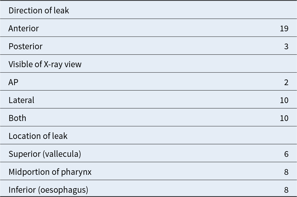
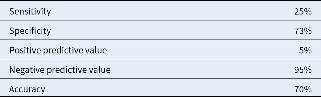
Radiological fistulae were suspected in 22 patients (27 per cent). The most frequently identified locations of the leak included inferior and midportions of the pharynx which were pinpointed in 16 out of the 22 radiological fistulas (73 per cent) (Table 2). In the 22-patient set where a radiological fistula was identified, only 1 patient went on to develop a true clinical PCF despite monitored reintroduction of feeding due to low clinical concern in 16 patients (see below). In the 60-patient set where no evidence of radiological fistula was found, oral feeding was commenced on the same day or the day after the swallow study. Three of these patients went on to develop a clinical PCF. These results represent modest diagnostic accuracy for the contrast swallow study of 70 per cent. In our cohort, the results suggest a sensitivity for the contrast swallow study of 25 per cent and a specificity of 73 per cent and give positive and negative predictive values of 5 and 95 per cent accordingly (Table 3).
X-Ray characteristics of the 22 patients with radiological PCFs

AP = anterior–posterior; IQR = interquartile range; PCF = pharyngocutaneous fistula; SD = standard deviation.
Statistical accuracy of the contrast swallow study

Primary and secondary outcomes
Of the total 94 patients evaluated, 10 went on to develop a PCF, giving an overall PCF incidence of 10.6 per cent for the Liverpool Head and Neck Centre for the five-year study period.
Looking at the 82 patients who met the inclusion criteria a total of 4 patients (5 per cent) developed clinical PCFs (Table 4). Three patients showed clinical signs of PCF despite normal contrast swallow study, while only 1 out of 22 patients with radiological fistula developed clinical PCF. The proportion of patients developing complications was comparable between the two groups (5 per cent vs. 5 per cent, p = 0.82). The time to initiation of oral feeding was also not significantly different between normal and positive fistula wall shear stress (WSS) (10 vs. 8 days, p = 0.82) (Figure 1).
Comparison of radiological PCFs to clinical PCFs after laryngectomy at AUH between April 2018 and July 2023. PCF = pharyngocutaneous fistula.

Primary and secondary outcomes

Management of radiological fistula
After careful clinical appraisal, 16 patients began oral feeding between 0 and 4 days after the swallow study. None of these patients went on to develop any evidence of a clinical PCF.
In the remaining six patients, the radiological fistula was more convincing and the clinical picture less reassuring. These patients, therefore, underwent repeat or serial contrast studies. In three of the six, the radiological fistula had completely resolved on subsequent imaging, and they began oral intake without issue. One patient made the informed decision to feed despite a persistent radiological fistula, they also remained well and did not develop a clinical PCF.
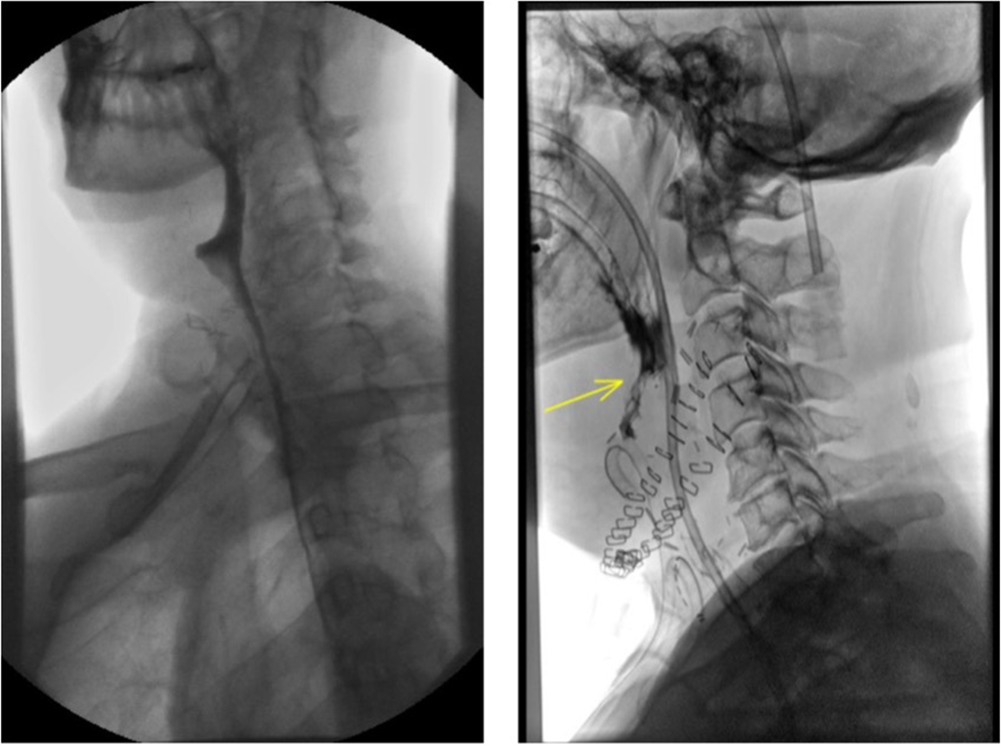
Due to patient factors and convincing radiological signs, one patient was discharged with a nasogastric feeding tube in situ. They re-commenced oral diet several weeks later, following outpatient clinic review, with no clinical sequalae (Figure 2).
Water soluble contrast images of two patients with radiological PCFs. The left-hand image reported as ‘A small anastomotic leak is noted anteriorly in the hypopharynx’, the right-hand image reported as ‘There is a left anterolateral contrast leak extending from the region of the pharyngeal suture line running inferiorly’. Only the patient on the right went on to develop a clinical PCF. PCF = pharyngocutaneous fistula.

Clinical PCF
Four patients developed a clinical PCF from this cohort. The records for these patients have been scrutinised to assess for any possible patterns that may warrant further analysis or investigation. At their time of surgery, the patients ranged in age from 54 through to 70, with an even gender mix. Two patients underwent laryngectomy (primary and salvage), and the other two underwent pharyngolaryngectomy (primary and salvage). Flap reconstruction was undertaken using an ALT flap in three out of the four patients. No primary speaking valves were placed. The four patients were under the care of and operated on by four different surgeons. From a thorough review, there does not appear to be a characteristic common to these four patients that may explain the subsequent negative clinical course.
Discussion
In this large cohort study, the utility of contrast swallow studies for identifying PCF after laryngectomy has been interrogated. Of the 82 patients eligible for inclusion, 22 were reported to have radiological features of a PCF. Of these, only one patient went on to develop a true clinically apparent fistula. Of the 60 patients with no radiological evidence of a PCF, 3 went on to develop a clinically discernible PCF. The present study therefore raises the hypothesis that contrast swallow is a sub-optimal and clinically unhelpful investigation. The moderately high specificity of 75 per cent and the very high negative predictive value of 95 per cent may serve to provide reassurance in situations where there is little to no clinical concern, but this comes at a cost of valuable radiological staff and facilities time, patient anxiety and potential delayed discharge. Of note, the low sensitivity of 25 per cent and very low positive predictive value of 5 per cent represents a significant over-reporting of radiological fistulae. As a point of reference, these values are considerably lower than those deemed acceptable for screening programmes.26–Reference van der Velde, Blanker, Stegmann, de Bock, Berger and Berendsen28 The pharyngeal wall outlined in this investigation post-operatively can be significantly altered by suture lines, mucosal inversion and oedema, making concurrent reporting of the live investigation very difficult. Indeed, subsequent surgeon review of positive reported findings resulted in a downgrading of suspicion in almost 75 per cent such that oral feeding was recommenced.
The clinical value of contrast swallow studies remains a contentious issue. Krouse et al. reviewed a similar size and aged cohort, identifying three independent predictors of PCF development: (1) previous radiotherapy, (2) presence of other post-operative complications (other than salivary leak) and (3) whether the radiologically identified sinus tract was larger than 2 cm. They concluded that the information gained from performing a contrast swallow study had a predictive value, in that blind-ending tracts that were longer than 2 cm were more likely to progress into true PCFs, but this knowledge could not be utilised to prevent an impending fistula.Reference Krouse and Metson4
In contrast, Naragan et al. recognised that contrast swallow studies had a high negative predictive value; in keeping with this study, however, they attributed this to early detection of sub-clinical/blind-ended fistula which were managed by returning the patient to a nil by mouth/tube fed status, thereby allowing the ‘impeding’ fistula to heal. Their conclusion was therefore to advocate for contrast swallow studies in all patients to prevent conversion of sub-clinical to frankly clinical PCFs,Reference Narayan, Limbachiya, Balasubramanian, Subramaniam, Thankappan and Iyer22 a clinical progression we failed to observe in our study, despite oral feeding provocation. Other studies have taken a more targeted approach, with for example Moses et al. observing clinical signs in all patients that progressed to PCF and therefore concluding that these clinical findings should prompt a contrast swallow study for radiological confirmation.Reference Moses, Eisele and Jones24 Certainly the prognostic value of abnormal radiological findings in the absence of clinical signs remains unclear,Reference van la Parra, Kon, Schellekens, Braunius and Pameijer1 and we support a decision-making process that is based primarily on clinical presentation rather than routine investigations.
It is important to recognise that, in terms of delayed discharge and patient anxiety, the doubt created by a false positive result can partially mirror the negative effects of a true PCF. The negative psychological impact of false positive results is well recognized,Reference Salz, Richman and Brewer27–Reference Tluczek, Mischler, Bowers, Peterson, Morris and Farrel29 particularly in this vulnerable post-operative group.Reference Semple, Parahoo, Norman, McCaughan, Humphris and Mills30, Reference Katz, Irish, Devins, Rodin and Gullane31
Avoiding reliance on low accuracy tests can also allow other clinical strategies, with the patient at the centre of the decision-making process. Several studies have reported comparable outcomes when reviewing early versus late feeding, with early oral feeding associated with significantly shorter inpatient hospital stays.Reference Milinis, Gaskell, Lau, Lancaster and Jones19, Reference Sousa, Porcaro-Salles, Soares, de Moraes, Silva and Sepulcri32–Reference Prasad, Sreedharan, Dannana, Prasad and Chandra34 Early feeding forms part of the evidence-based ERAS (Enhanced Recovery After Surgery) protocol which is widely accepted and used within general surgery following higher risk gastrointestinal anastomoses.Reference Ljungqvist, Scott and Fearon35 This strategy should be more widely considered in appropriately selected patients following laryngectomy.Reference Milinis, Gaskell, Lau, Lancaster and Jones19, Reference Kishikova and Fleming36
The limitations of this retrospective series are acknowledged; despite being performed at a regional head and neck centre over a five-year period, the number of positive PCF events are low. Although this represents very low complication rates in a mixed primary/salvage cohort at our institution with comparable published outcomes, it does limit the statistical validity of comparative analyses within the study as well as the ability to adequately match patient groups. However, with a large group of post-laryngectomy patients followed for adequate time, it remains clear that 73 per cent of positively reported fistulae on contrast swallow investigations were felt to be inaccurate or not clinically significant, and subsequent oral feeding in the ensuing few days had no adverse impact on the fistula event rate.
• A pharyngocutaneous fistula (PCF) is by far the most common serious complication following a laryngectomy, with an incidence of between 9 and 23 per cent
• Despite the clinical significance of PCFs and the importance of their detection there is no definitive gold-standard investigation recommended in the literature
• Contrast swallow study is the most widely reported modality in the literature, however its accuracy is not well defined
• In this cohort, the contrast swallow study was found to be a sub-optimal investigation for identifying PCFs following laryngectomy. The high negative predictive value of 95 per cent may provide reassurance in situations where there is little clinical concern; however, the very low positive predictive value of 5 per cent presented a significant over-reporting of radiological fistulas, which may serve to introduce doubt and cloud clinical judgement
• The findings call into question the merits of contrast swallow study in guiding the commencement of oral feeding and call for alternative management strategy
Conclusion
This retrospective series suggests that, in the post laryngectomy patient, contrast swallow studies are sub-optimal to identify those likely to develop a clinical PCF. The high negative predictive value may support clinicians in a reassurance context, but the very low positive predictive value may complicate recovery and delay return to function for the rehabilitating patient. It is our belief that clinical findings should dictate the use of these investigations and further efforts made to evaluate alternative imaging modalities such as contrast computed tomography or ultrasound.
Author contributions
Henry Lonsdale: Data curation + analysis; writing – original draft + review and editing. Kristijonas Milinis: conceptualization; analysis; writing – review and editing; supervision. Alexandra Bell: Data curation. Christopher Loh: Supervision. Katharine Davies: Supervision. Jason Fleming: Writing – review and editing; lead senior supervisor.
Funding statement
None.
Competing interests
None known.