Introduction
The field of colloidal assembly was driven for a long time by fundamental interest in understanding crystallization in simple model systems. More recently, colloids and their nanoscale analogs, nanoparticles (NPs), have emerged as new functional blocks for optical, energy, and biomedical applications. These developments have opened a new frontier of rational self-assembly of materials made from such particles. The structures of the assemblies are determined by the interplay among interaction forces, entropic effects, and kinetics. Unlike atoms, colloids and NPs can vary widely in their sizes, shapes, and interactions. Nevertheless, it has been challenging to establish a broadly applicable platform for “a la carte” assembly of ordered particle arrays and their dynamic control.Reference Klinkova, Choueiri and Kumacheva1–Reference Rogers, Shih and Manoharan3
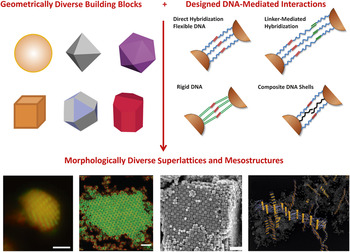
One of the most controllable assembly strategies relies on programming interparticle interactions using the complementarity of DNA strands.Reference Rogers, Shih and Manoharan3 This provides selective and reversible interactions between particles of different sizes and shapes (Figure 1). DNA strands can also act as tailored structural elements in particle crystals, smart assembly guides, and reconfigurable structural elements. In recent years, much progress has been made in DNA-guided assembly of particle superlattices. By tuning the interactions, sizes, and shapes of NPs, a wide variety of structures have been assembled. Here, we discuss the most significant achievements and challenges in this field and future directions.
Schematic representation of the concept of DNA-mediated programmable self-assembly of particles. Particles can be coated with DNA shells, thus, the particle cores carry materials function, and DNA shells provide programmable interactions. (Top left panel): the diversity of particle shapes that can be synthesized. (Top right panel): richness of DNA-shell designs for modulating DNA-driven interparticle interactions. (Bottom panel): from left to right, crystallization of colloid particles,Reference Casey, Scarlett, Rogers, Jenkins, Sinno and Crocker4 binary mixture of colloid particles,Reference Wang, Wang, Zheng, Ducrot, Yodh, Weck and Pine5 nanoscale cubes and spheres,Reference Lu, Yager, Zhang, Xin and Gang6 and a symmetry breaking for DNA-interacting rods due to the collective chain effect results in a ribbon formation.Reference Vial, Nykypanchuk, Yager, Tkachenko and Gang7 Note: Scale bars, from left to right, are 2 μm, 5 μm, and 200 nm. Reprinted with permission from References Reference Casey, Scarlett, Rogers, Jenkins, Sinno and Crocker4 and Reference Wang, Wang, Zheng, Ducrot, Yodh, Weck and Pine5. © 2012 and 2015 Macmillan Publishers Ltd.

Crystallization of superlattices
DNA was proposed as a programmable “glue” for the assembly of NPs about two decades ago.Reference Alivisatos, Johnsson, Peng, Wilson, Loweth, Bruchez and Schultz8,Reference Mirkin, Letsinger, Mucic and Storhoff9 The basic idea is that DNA-functionalized NPs with complementary strands can hybridize due to the Watson–Crick base pairing between the strands grafted to two different particles. One can consider two approaches to link particles with DNA: direct hybridization (DH) and linker-mediated hybridization (LH) (see Figure 1). In the DH approach, the particles are functionalized by DNA terminated with mutually complementary single-stranded segments (also known as a “sticky end”). In the LH approach, the grafted DNA pairs are not complementary, and appropriately encoded DNA linkers are required to connect them. In addition, the grafted DNA can vary in their rigidity, length, and grafting density, all of which lead to modification of the shell, formed by DNA around the particle core, and, consequently, it will affect particle interaction characteristics.
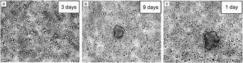
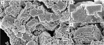
DNA-mediated particle binding without additional optimization or annealing would normally lead to amorphous aggregates due to unfavorable interactions and complex kinetic pathways to crystallization.Reference Valignat, Theodoly, Crocker, Russel and Chaikin10,Reference Maye, Nykypanchuk, van der Lelie and Gang11 Crocker’s group reported the first example of crystallization for DNA-coated particles for micrometer-scale colloidsReference Kim, Biancaniello and Crocker12 (see formation of small crystallites in Figure 2a–c). The GangReference Nykypanchuk, Maye, van der Lelie and Gang13,Reference Xiong, van der Lelie and Gang14 and MirkinReference Park, Lytton-Jean, Lee, Weigand, Schatz and Mirkin15 groups independently reported a major breakthrough, an assembly of crystalline 3D lattices from NPs using DNA.The key advances were (1) the use of a thermal annealing procedure combined with in situ small-angle x-ray scattering for sample structure monitoring; and (2) rational design of the DNA shell to optimize the interparticle interactions. The optimal temperature window for the onset of crystallization was shown to be about a few degrees below the DNA melting temperature.
Crystallization results for 1-μm diameter DNA-grafted colloidal particles after (a) three days (∼1500 DNA/particle), (b) nine days (∼1500 DNA/particle), and (c) one day (∼9500 DNA/particle). Particles are 1μm in diameter. Reprinted with permission from Reference Reference Kim, Biancaniello and Crocker12. © 2006 American Chemical Society.

TheoryReference Tkachenko16 suggested that order requires a particular balance of attraction and repulsion, which can be controlled by the design of the DNA shell, for example, through manipulating the DNA rigidityReference Nykypanchuk, Maye, van der Lelie and Gang13 and mixing different strand types.Reference Maye, Nykypanchuk, van der Lelie and Gang11,Reference Wu, Feng, Sha, Dreyfus, Grosberg, Seeman and Chaikin17 Utilizing these approaches, the crystallization of NPs into body-centered-cubic (bcc) or face-centered-cubic (fcc) phases was achieved.Reference Nykypanchuk, Maye, van der Lelie and Gang13–Reference Park, Lytton-Jean, Lee, Weigand, Schatz and Mirkin15 The fcc phase was formed from single-type particles (all particles in the system have the same DNA shell), while the bcc phase (isostructural to CsCl) (i.e., presence of two types of species in the lattice) was observed in binary particle systems (particles with two types of DNA) with mutual attraction, consistent with earlier theoretical predictions.Reference Tkachenko16 A systematic studyReference Xiong, van der Lelie and Gang14,Reference Xiong, van der Lelie and Gang18 of LH NP systems identified the minimum number of DNA linkers required for robust crystallization, which was later confirmed by Monte Carlo simulations in which DNA shells were modeled as “soft colloids.”Reference Angioletti-Uberti, Varilly, Mognetti, Tkachenko and Frenkel19 Experiments indicated that phase behavior was sensitive to the details of shell tailoring,Reference Casey, Scarlett, Rogers, Jenkins, Sinno and Crocker4 and the morphology of assembly could be kinetically tuned.Reference Di Michele, Varrato, Kotar, Nathan, Foffi and Eiser20,Reference Varrato, Di Michele, Belushkin, Dorsaz, Nathan, Eiser and Foffi21
Theory and modeling of DNA-driven particle systems
From a theoretical point of view, particles with DNA-mediated particle binding represent a unique model system with designed interactions and a significant methodological challenge due to its multiscale character. The theoretical and computational approaches for describing DNA-mediated particle interactions can be roughly divided into three groups: (1) models in which the DNA subsystem is replaced with effective pair potential; (2) simulations with explicit coarse-grained polymer models of DNA; and (3) hybrid approaches in which simulations of chains are used for the effective potential (EP).
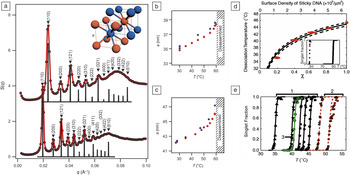
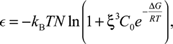
The EP models were among the first proposed.Reference Tkachenko16,Reference Biancaniello, Kim and Crocker22,Reference Licata and Tkachenko23 In general, the EP is well justified primarily for the colloids, which are larger than typical DNA shells. Fundamentally, these potentials can be calculated as a free energy of the DNA subsystem for a given interparticle separation. In fact, such pair potential was experimentally measuredReference Biancaniello, Kim and Crocker22 using optical tweezers, and the results were in agreement with a simple multichain binding model. Later, a conceptually similar model was used to successfully describe experimental data on reversible aggregation and melting in DNA-colloidal systemsReference Dreyfus, Leunissen, Sha, Tkachenko, Seeman, Pine and Chaikin24,Reference Dreyfus, Leunissen, Sha, Tkachenko, Seeman, Pine and Chaikin25 (see Figure 3d–e). Multiple refinements of this theory have been proposed over the years.Reference Tkachenko16,Reference Angioletti-Uberti, Varilly, Mognetti, Tkachenko and Frenkel19,Reference Biancaniello, Kim and Crocker22–Reference Rogers and Crocker27 The essential physics of most can be captured with a very basic expression for the two-particle binding free energy:
 $$\varepsilon = - k_B TN\ln \left( {1 + {\rm{\xi }}^3 C_0 e^{ - {{{\rm{\Delta }}G} \over {RT}}} } \right),$$
where N is the number of complementary single-stranded DNA (ssDNA) pairs that can form a double-stranded DNA (dsDNA) duplex, ∆G is the corresponding hybridization free energy,  ${\rm{\xi }}$ is a characteristic length scale that is of the order of the thickness of the DNA shell, C 0 is a reference concentration of 1M, k B is the Boltzmann constant, T is temperature, and R is the ideal gas constant. In this formula,
 ${{\rm{\xi }}^3}{C_0}{e^{ - {\rm{\Delta }}G/RT}}$ represents the hybridization probability. Interestingly, strong binding between two particles is possible even in the regime where this probability is small, given the large number, N, of sticky ssDNA per contact. In fact, this is a typical regime for colloids. Also, the shape of the EP is nearly irrelevant for colloids, since its range is much shorter than the particle size, and they can be viewed as sticky spheres with type-dependent mutual affinity.
Structural study of assembly and temperature behavior of nanoparticle (NP)–DNA lattices. (a) Small-angle x-ray scattering obtained structure factors for assembled DNA-coated 10-nm gold NP with different DNA shellsReference Nykypanchuk, Maye, van der Lelie and Gang13 (a) reveals the body-centered-cubic ordering, as shown in the inset, with (b–c) temperature-dependent lattice constant (a). (b–c) Correspond to the systems shown (d); red circles and blue squares are obtained for heating and cooling cycles, respectively. (d–e) Reversible aggregation of DNA-coated colloids for variable composition of DNA shells.Reference Dreyfus, Leunissen, Sha, Tkachenko, Seeman, Pine and Chaikin24 Experimental and theoretical dissociation temperature as a function of χ, the fraction of sticky DNA. The red error bars on χ reflect the estimated 10% uncertainty in the coverage. The solid line is the predicted dissociation temperature. Inset: experimental (squares) and theoretical dissociation curves (solid line) before the entropy correction. (e) Melting curves are well described by a simple model, Equation 1; groups 1–3 represent different designs of the DNA linkers. Note: S, structure factor; q, wave vector transfer; T, temperature.

In the case of NPs, however, the core size is comparable or even smaller than the size of a DNA shell. Therefore, the applicability of EP is not guaranteed. Nevertheless, this class of models has been shown to have reasonable predictive power. The best known example is the complementary contact model (CCM).Reference Macfarlane, Lee, Jones, Harris, Schatz and Mirkin28 It takes into account two key properties of the NP–DNA system: effective size of the particle (represented by the hydrodynamic radius) and DNA coverage. The model postulates that the self-assembled morphology maximizes the relative fraction of hybridized DNA linkers. This simple criterion adequately predicts a number of features of the observed phase diagram. In its core, CCM is closely related to the EP models and similarly favors structures that maximize contact between mutually attractive particles. A number of more recent studies propose a route toward building a full-scale theory further helped by generic algorithms.Reference Srinivasan, Vo, Zhang, Gang, Kumar and Venkatasubramanian29,Reference Vo, Venkatasubramanian, Kumar, Srinivasan, Pal, Zhang and Gang30
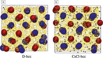
An alternative to the EP is to describe the DNA with a coarse-grain polymer model.Reference Mladek, Fornleitner, Martinez-Veracoechea, Dawid and Frenkel31–Reference Li, Sknepnek, Macfarlane, Mirkin and de la Cruz33 Because of the computational complexity, detailed modeling of DNA hybridization is typically not practical, and should be represented either by sticky monomers or other toy models. Even upon this simplification, the simulation of multiple DNA-functionalized particles remains a formidable task. Use of graphics processing unit (GPU)-based computing for molecular dynamics simulations of such NP–DNA systems proved to be remarkably efficient. Pioneered by Knorowski et al.Reference Knorowski, Burleigh and Travesset32 (see Figure 4a–b for illustration of phase formation), this approach was later used to numerically reproduce experimentally observed superlattices.Reference Li, Sknepnek, Macfarlane, Mirkin and de la Cruz33 A promising computational approach is construction of multiscale hybrid models.Reference Leunissen and Frenkel34–Reference Mladek, Fornleitner, Martinez-Veracoechea, Dawid and Frenkel36 The goal is to construct an analytic multiparticle Hamiltonian through simulation of the DNA-subsystem. Not only does this method produce well-justified pair potentials, it also allows one to take into account the multibody correction to the effective interparticle interactions,Reference Mladek, Fornleitner, Martinez-Veracoechea, Dawid and Frenkel36 essential for NP–DNA systems.
Superlattice self-assembly modeled via graphics processing unit (GPU)-based molecular dynamics simulations. Equilibrated snapshots of (a) “disordered”(body-centered-cubic)-bcc (D-bcc) and (b) CsCl-bcc, with blue and red type nanoparticles and hybridizations (black). Superlattice self-assembly modeled via GPU-based molecular dynamics simulations. Reprinted with permission from Reference Reference Knorowski, Burleigh and Travesset32. © 2011 American Physical Society.

Multicomponent lattices based on spherical particles
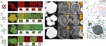
One of the most practical ways to achieve greater morphological diversity is to vary the size ratio of the constituent particles.Reference Leunissen, Christova, Hynninen, Royall, Campbell, Imhof, Dijkstra, van Roij and van Blaaderen37,Reference Shevchenko, Talapin, Kotov, O’Brien and Murray38 This led to the assembly of several types of NP–DNA superlattices. The observed phase behavior has been partially explained by the CCM approach,Reference Macfarlane, Jones, Senesi, Young, Lee, Wu and Mirkin39 and it has been further explored in recent studies.Reference Srinivasan, Vo, Zhang, Gang, Kumar and Venkatasubramanian29,Reference Vo, Venkatasubramanian, Kumar, Srinivasan, Pal, Zhang and Gang30 Pine et al.Reference Wang, Wang, Zheng, Ducrot, Yodh, Weck and Pine5 recently conducted similar studies for micrometer-scale colloids by revealing similarity with NP systems (shown in Figure 5a–d) and providing details of phase development.
Confocal fluorescent images (scale bar 5 μm) and corresponding drawings of various DNA-coated colloidal crystals: (a) face-centered-cubic (fcc); (b) body-centered-cubic (bcc) (isostructural to CsCl); (c) an AB2 crystal (isostructural to AlB2); and (d) an AB6 crystal lattice (isostructural to Cs6C60). Reprinted with permission from Reference Reference Wang, Wang, Zheng, Ducrot, Yodh, Weck and Pine5. © 2015 Nature. (e–i) Rhombic dodecahedron microcrystals from gold nanoparticles, (e–g) (left) transmission electron microscope and (middle and right) scanning electron microscope images, (h) small-angle x-ray scattering (SAXS) data, and (i) results of molecular dynamics simulations. Small x-ray scattering provides identification of assembled lattices, while the simulations reveal the assembly mechanism. Note: scale bars, left to right, are (e) 1 µm, 1 µm, and 2 µm; (f) 1 µm, 2 µm, and 4 µm; and (g) 0.5 µm, 1 µm, and 2 µm. Note: S, structure factor; q, wave vector transfer; q 0, wave vector transfer for the first SAXS peak; a, lattice parameter. Reprinted with permission from Reference Reference Auyeung, Li, Senesi, Schmucker, Pals, de la Cruz and Mirkin40. © 2014 Nature.

Successful formation of lattices typically results in a polycrystalline material. Well-defined microcrystalsReference Auyeung, Li, Senesi, Schmucker, Pals, de la Cruz and Mirkin40 shaped as Wulff polyhedra were successfully generated from DNA-functionalized NPs (Figure 5e–i) grown by a slow-cooling process. This thermodynamically favorable crystal shape is consistent with the crystal structure predicted from theoretical considerations. Due to the presence of the DNA shell, NPs often occupy only <10% by volume of the superlattice. Thus, it is possible to incorporate other components for generating more complex three-dimensional (3D) structures, for example, with optical functionalities.Reference Xiong, Sfeir and Gang41 Using a basic lattice of DNA-functionalized NPs as a “scaffold,” more complex assemblies were achieved by positioning molecular or nanoscale objects in the defined locations, as determined by the DNA encoding.Reference Xiong, Sfeir and Gang41,Reference Macfarlane, Jones, Lee, Auyeung and Mirkin42 In contrast to this incorporation method, a “deletion” of NP inorganic cores has been also demonstrated.Reference Auyeung, Cutler, Macfarlane, Jones, Wu, Liu, Zhang, Osberg and Mirkin43
Although gold NPs are often used in the studies, it is clear that the DNA-based platform can facilitate programmable assemblies of different types of particles, so long as they can be coated with DNA. Several approaches were recently demonstrated for generating binary mixtures of NPs with different inorganic cores. For example, bcc superlattices were assembled from an amino-DNA functionalized quantum dot and thiolated-DNA capped gold NP,Reference Sun and Gang44 in which the gold NPs were positioned in a simple cubic manner. Generally applicable carboxylic-based conjugationReference Sun and Gang44,Reference Zhang, Lu, Yager, van der Lelie and Gang45 and click-chemistryReference Zhang, Macfarlane, Young, Choi, Hao, Auyeung, Liu, Zhou and Mirkin46–49 approaches to functionalize NPs with DNA have been developed.
Using these methods, a broad range of functional NPs, including plasmonic, magnetic (iron oxide), catalytic (palladium), and luminescent (cadmium selenite, cadmium telluride) NPs were assembled into heterogeneous superlattices. It was found that the particle shape difference could suppress structural ordering, but the effect is minimized for larger ssDNA shells.Reference Zhang, Lu, Yager, van der Lelie and Gang45 Interestingly, the multicomponent lattices can exhibit compositional disorder for soft DNA shells; a progressive decrease of structural order and transition to clustering was also observed for small grafting numbers.Reference Zhang, Lu, Yager, van der Lelie and Gang45 Novel optical and field-responsive properties of the DNA-assembled multicomponent materials were demonstrated as well.
Assembly of anisotropic particles
Particle shapes have profound effects on the type of formed structures, even for solid objects.Reference Damasceno, Engel and Glotzer50 Assembly of DNA-functionalized NPs of different shapes, including rods, triangular prisms, octahedra, and rhombic dodecahedra, can result in the formation of dense phases in which particles maximize interactions of complementary rigid chains.Reference Jones, Macfarlane, Lee, Zhang, Young, Senesi and Mirkin51 For example, prisms form linear arrays, while rods form hexagonally packed membranes. Such organizations resemble the phases observed for solid objects. However, the polymeric nature of DNA chains can also induce unexpected assembly pathways. For example, it was observed that one-dimensional ladder-like ribbon structures emerge due to the spontaneous symmetry breaking of interparticle interactions during assembly of radially isotropic DNA-decorated nanoscale rods.Reference Vial, Nykypanchuk, Yager, Tkachenko and Gang7 Similar linear structures of alternating rods and spheres were also observed, indicating that collective effects of flexible linkers might promote new structural organizations.
Recent studies also demonstrated that both shape complementary and noncomplementary scenarios can be realized for assembly of dissimilar shapes into unified lattices with the help of DNA in binary mixtures (Figure 6a–c).Reference Lu, Yager, Zhang, Xin and Gang6,Reference O’Brien, Jones, Lee and Mirkin52 Although traditionally the shape difference would be expected to lead to phase separation, the complementarity between DNA on particles of different shapes allows for co-assembly. If the shapes have overall similar symmetry, the original phase is preserved even if the shapes are somewhat modulated, as was shown for nanocubes.Reference O’Brien, Jones, Lee and Mirkin52 However, for mixtures of anisotropic (nanocubes, octahedra) and spherical NPs, the anisotropic particle dictates the phase behavior,Reference Lu, Yager, Zhang, Xin and Gang6 while a certain softness of shells is required to accommodate the shape disparity. Such an approach permits designing lattice formation by the introduction of well-defined geometrical elements that impose specific binding coordination between particles, and where local symmetry correlates with the resulting self-assembled lattice. This idea was further demonstrated for the creation of designed low-dimensional lattices using DNA origami octahedra, 3D frame made out of folded DNA, as shaped interparticle linkers.Reference Tian, Wang, Liu, Xin, Li, Ke, Shih and Gang53
(a–c) Self-assembly of cubic lattice isostructural to NaCl for DNA-functionalized nanocubes and nanospheres.Reference Lu, Yager, Zhang, Xin and Gang6

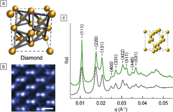
Recently, a new lattice assembly strategy was introducedReference Liu, Tagawa, Xin, Wang, Emamy, Li, Yager, Starr, Tkachenko and Gang54,Reference Tian, Zhang, Wang, Xin, Li and Gang55 based on the use of DNA frames, which are complex 3D DNA constructsReference Douglas, Dietz, Liedl, Hogberg, Graf and Shih56 that can prescribe the topology of interparticle connections.Reference Tian, Wang, Liu, Xin, Li, Ke, Shih and Gang53 These nontrivial building blocks, specifically designed DNA frames of different shapes, can be used as multiparticle connectors and as particle cages;Reference Licata and Tkachenko57,Reference Li, Liu, Yu, Jiang and Mao58 thus, they can dictate the desired anisotropic interactions without the complexity of particle design and without the necessity of changing particles for obtaining the targeted lattices. The diverse range of structures assembled using this strategy includes the diamond family of superlatticesReference Liu, Tagawa, Xin, Wang, Emamy, Li, Yager, Starr, Tkachenko and Gang54 (Figure 7a–c), traditionally considered an iconic challenge in colloidal self-assembly, as well as other rationally assembled lattices based on the polyhedral frames.Reference Tian, Zhang, Wang, Xin, Li and Gang55 Such an approach opens a new frontier in the creation of designed NP lattices with DNA.
Assembly of a diamond lattice from DNA-coated gold nanoparticles and tetrahedral DNA origami frames. (a) Schematic of the structure; (b) scanning transmission electron microscope image, scale bar is 50 nm; and (c) small-angle x-ray scattering (SAXS) obtained structure factor S(q) and (inset) a model of diamond lattice are shown.Reference Liu, Tagawa, Xin, Wang, Emamy, Li, Yager, Starr, Tkachenko and Gang54 Green and black curves correspond to experimental and modeled structure factors, respectively. Excellent agreement between them confirm the assembly of the diamond structure. Note: A and B, particles coated with respectively complementary DNA; S, structure factor; q, wave vector transfer; q 0, wave vector transfer for the first SAXS peak.

Stimuli responsive superlattices
Dynamically reconfigurable superlattices can be implemented by using flexible ssDNA as a linker. In one study, osmotic pressure was shown to compress the NP–DNA lattice by as much as six times in volume, without destroying its order.Reference Srivastava, Nykypanchuk, Maye, Tkachenko and Gang59 Salt concentrationReference Xiong, Sfeir and Gang41,Reference Tan, Kahn, Derrien, Campolongo, Zhao, Smilgies and Luo60,Reference Srivastava, Nykypanchuk, Fukuto and Gang61 and hydrationReference Radha, Senesi, O’Brien, Wang, Auyeung, Lee and Mirkin62 are environmental parameters for controlling the crystallization and lattice constants of superlattices. Moreover, triggering a transformation of the two-dimensional NP system from a hexagonal lattice to string-like clusters and to a weakly ordered network has been observed and correlated with changed rheological properties of the monolayer.Reference Srivastava, Nykypanchuk, Fukuto, Halverson, Tkachenko, Yager and Gang63
Highly specific responses to external stimuli have been explored in the case where NPs in a lattice were linked with a switchable DNA “device.”Reference Maye, Kumara, Nykypanchuk, Sherman and Gang64 The lattice constant of a bcc crystal was modulated by about 25% by adding specific DNA strands that alter the device state between two crystalline configurations.Reference Maye, Kumara, Nykypanchuk, Sherman and Gang64,Reference Kim, Macfarlane and Mirkin65 In a recent study, a molecular intercalation-based strategy was demonstrated for the reversible extension of dsDNA-linked NP lattices by ∼30%, using ethedium bromide as the intercalator molecule.Reference Pal, Zhang, Kumar and Gang66 Such regulation of interparticle distances can be used to modulate coupling between NPs for applications in plasmonic, light-emitting, and magnetic materials.
There is significant interest in creating dynamic lattices that can change their global organizational properties, such as for creating materials with switchable functions. Colloidal particles interacting through DNA linkers can be designed to form solids that melt when either heated or cooled;Reference Angioletti-Uberti, Mognetti and Frenkel67 also, reentrant melting and arbitrarily wide gas–solid transition were demonstrated experimentally.Reference Rogers and Manoharan68 These advances will permit, for example, creating materials with unique mechanical responses to temperature.
Recently, an intriguing concept of programmable switching of the crystallographic symmetry of an entire NP lattice into different phases by tailoring in situ interparticle interactions was proposed and implemented.Reference Zhang, Srinivasan, Vo, Pal, Kumar and Gang69 Reversible transformations can also be achieved by designing DNA shell modifications.Reference Kim, Macfarlane, Jones and Mirkin70 These studies demonstrate that targeted modification of NP shells allows for reprogramming of interactions in a dynamical manner, resulting in a series of phase transitions. This result opens up opportunities for creating systems whose structures and functions can be changed on demand. A combined diversity of particle types can be integrated using DNA methods, Reference Vial, Nykypanchuk, Yager, Tkachenko and Gang7,Reference Zhang, Lu, Yager, van der Lelie and Gang45,Reference Zhang, Macfarlane, Young, Choi, Hao, Auyeung, Liu, Zhou and Mirkin46,Reference Jones, Macfarlane, Lee, Zhang, Young, Senesi and Mirkin51 advances in DNA functionalization of particles,Reference Zhang, Lu, Yager, van der Lelie and Gang45–Reference Heuer-Jungemann, Kirkwood, El-Sagheer, Brown and Kanaras48,Reference Sun and Gang71 and to potentially generate novel strategies for the lattice designReference Wang, Wang, Zheng, Ducrot, Yodh, Weck and Pine5,Reference Lu, Yager, Zhang, Xin and Gang6,Reference Macfarlane, Lee, Jones, Harris, Schatz and Mirkin28,Reference Vo, Venkatasubramanian, Kumar, Srinivasan, Pal, Zhang and Gang30,Reference Liu, Tagawa, Xin, Wang, Emamy, Li, Yager, Starr, Tkachenko and Gang54,Reference Tian, Zhang, Wang, Xin, Li and Gang55 a broad range of materials with desired performances.
Conclusions
In recent years, a new powerful approach based on DNA-driven particle interactions was demonstrated for and applied to the creation of ordered particle arrays. Programmable interactions provided by complementary DNA strands offer incorporation of multiple particle types, tunability of binding characteristics through the tailored design of DNA shells, and ample capabilities for structure reconfigurations. Significant progress was achieved in DNA-guided assembly of superlattices built from micrometer and nanoscale particles of different sizes, shapes, and chemical compositions. Moreover, by modulating the structure of DNA shells, nanoparticle shapes, and the topology of linking constructs, diverse lattice types were observed. While we are far from completely understanding these systems and predicting their phases, both experimental and theoretical efforts demonstrate enormous progress in revealing the leading parameters determining structure formation in these systems. Recent success in coupling complex DNA constructs and DNA-coated NPs into integral architectures reassures that in the near future, self-assembly for NP–DNA lattices might reach a new level of control. DNA-based strategies will allow for the assembly of desired targeted architectures. These advances in assembly of 3D particle arrays can be directly applied to fabrication of new types of materials from functional particles, thus enabling the full-scale engineering of desired optical, magnetic, and bio-related functions.
Acknowledgment
Research was carried out at the Center for Functional Nanomaterials, Brookhaven National Laboratory, and is supported by the US Department of Energy, Office of Basic Energy Sciences, under Contract No. DE-SC0012704.