Introduction
Laryngeal asymmetry is not uncommon. Like many other paired organs in the body, the laryngeal structures may have different sizes, positions and shapes. An anatomical study conducted by Hirano et al. on 50 excised cadaveric larynges found asymmetry of both the thyroid and cricoid cartilages.Reference Hirano, Kurita, Yukizane and Hibi1 Asymmetry was more accentuated in older specimens, but did not differ significantly within age groups or genders. There were differences in all angles and dimensions between the right and left side of the larynges. The thyroid cartilage as a whole tended to tilt to the right against the cricoid cartilage. Specifically, the right thyroid lamina tended to tilt laterally whereas the left one tended to tilt medially; as a result, the right tip of the superior thyroid notch was positioned more anteriorly compared with the left. Many authors have also described the anatomy of the crico-arytenoid joint as asymmetrical, alluding to the fact that even the shape and position of the corniculate and cuneiform cartilages can lack symmetry.Reference Sellars and Sellars2, Reference Sellars and Keen3
In our clinical experience, more often than not we observed asymmetry of the posterior laryngeal inlet, in keeping with the above anatomical findings. Few studies have elaborated on the incidence and clinical significance of such asymmetry.Reference Lindestad, Hertegård and Björck4, Reference Lacina5 Lindestad et al. reported a 70 per cent prevalence of arytenoid asymmetry in 109 subjects. This arytenoid asymmetry was more common in elderly subjects, with no gender preference. Differences in the antero-posterior position of the corniculate and/or cuneiform tubercles were the most frequent types of asymmetry, occurring in 66 per cent of total subjects and in 92 per cent of asymmetrical cases.Reference Lindestad, Hertegård and Björck4 Lacina et al. reported a higher incidence of adduction asymmetry in singers who had no voice problems, compared with those who had no voice problems. This was attributed hypothetically to the laryngeal adjustment needed by professional voice users during performance.Reference Lacina5
The clinical significance of arytenoid adduction asymmetry has not been elucidated in the literature. Hitherto, it has not been known whether patients with vocal symptoms are more likely to have arytenoid asymmetry, compared with asymptomatic individuals. The vocal effect of functional arytenoid asymmetry has also not been investigated. The present study thus aimed to report the prevalence of arytenoid asymmetry during adduction in non-professional voice users, and to assess the correlation between arytenoid asymmetry and vocal symptoms where present.
Materials and methods
Approval for the study was first obtained from the institutional review board of the American University of Beirut.
We reviewed the medical records and video recordings of 116 patients who had presented to the voice clinic for assessment and treatment.
Demographic data retrieved from the medical records included age, gender, and history of smoking, reflux and allergy. All the vocal symptoms described by the patients at the time of evaluation were recorded. These included hoarseness, breathiness, vocal fatigue, straining and inability to project the voice.
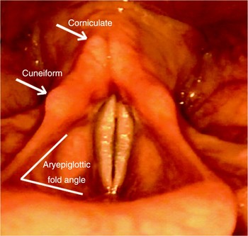
All patients had been examined using a 70° rigid laryngoscope (Karl Storz, Tuttlingen, Germany) coupled to a single chip camera. The video recordings were reviewed by two independent otolaryngologists who had no prior knowledge of the patients. The reviewers assessed the presence or absence of arytenoid asymmetry (defined in relation to the position of the corniculate cartilages), the cuneiform cartilages, and the angle between the aryepiglottic fold and the epiglottis (termed the aryepiglottic angle) (Figure 1). Asymmetry in the position of the cuneiform cartilages was reported when one side was anteriorly positioned compared with the other. Corniculate cartilage asymmetry was reported when one corniculate was seen to override the other. Asymmetry of the aryepiglottic angle was reported when the angle on one side was sharper than that on the opposite side. The degree of asymmetry was graded as mild, moderate or severe, and the site of asymmetry (right or left) was noted. However, the aryepiglottic fold angle was not objectively measured or subjectively categorised.
Anatomical structures of the laryngeal inlet during adduction.

The sample was described in terms of frequencies (for categorical variables) and means ± standard deviations (for continuous variables). The association between vocal symptoms and arytenoid asymmetry (treated as a dichotomous variable) was examined using the chi-square test or t-test, as appropriate. All analyses were conducted using the Statistical Package for the Social Sciences software program. A p value of <0.05 was considered statistically significant.
Results
Demographic data
There were 61 males and 55 females, with a mean age of 39 years and a standard deviation of 15 years. Almost one-third of patients had a history of reflux, 25 per cent had a history of smoking and 9.6 per cent had a history of allergy. Hoarseness was the most common symptom, occurring in 42.2 per cent of patients, followed by vocal fatigue (25 per cent) and inability to project the voice (12.9 per cent). Data are given in Table I.
Baseline patient characteristics

*n = 116. SD = standard deviation
Arytenoid asymmetry
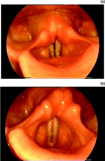
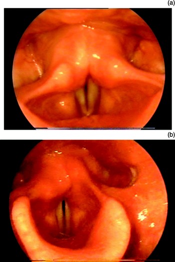
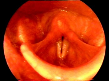
The total prevalence of arytenoid asymmetry was 37.1 per cent. The most common type of asymmetry was corniculate asymmetry, present in 27.6 per cent of patients and accounting for 74.39 per cent of total asymmetry cases (Figure 2). This was followed by cuneiform cartilage asymmetry, present in 15.5 per cent of patients and accounting for 41.77 per cent of total asymmetry cases (Figure 3). Aryepiglottic fold asymmetry was present in 8.6 per cent of patients (Figure 4). Data on arytenoid asymmetry are given in Table II.
(a) Mild corniculate cartilage asymmetry, with the right corniculate mildly overriding the left. (b) Severe corniculate asymmetry, with the right corniculate overriding the left.

(a) Mild cuneiform cartilage asymmetry, with mild anterior positioning of the right cuneiform cartilage compared with the left. (b) Severe anterior positioning of the right cuneiform cartilage compared with the left.

Aryepiglottic fold asymmetry.

Arytenoid asymmetry: types, laterality and severity

Data represent patient numbers (percentages). Prevalence of asymmetry types = *32 (27.6%); †18 (15.5%); ‡10 (8.6%).
Correlation between arytenoid asymmetry and vocal symptoms
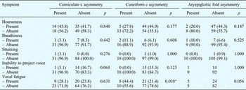
There was no correlation between arytenoid asymmetry and the occurrence of vocal symptoms, except for vocal fatigue (p = 0.038). Patients with vocal fatigue were more likely to have cuneiform asymmetry than those with no vocal fatigue or other vocal symptoms. Data are given in Table III.
Correlation between arytenoid asymmetry types and vocal symptoms

Data represent patient numbers (percentages), unless otherwise specified. *Significant at p < 0.05. C = cartilage
Discussion
The positioning of the arytenoids in the posterior laryngeal inlet is important during phonation, breathing and effort closure.Reference Sellars and Sellars2, Reference Sellars and Keen3 During phonation, the sliding movement of the arytenoids from the antero-latero-inferior position to a postero-medio-superior position is crucial. More often than not, otolaryngologists observe arytenoid asymmetry during adduction with no true understanding of its clinical significance. The need for medial approximation of the vocal folds during phonation is undisputed. However, it seems that adequate voice production requires more than just medial approximation but, rather, proper alignment of the vocal folds in both the vertical and horizontal dimensions. It is during this phase of alignment that asymmetry of the posterior larynx is invariably witnessed. Arytenoid asymmetry seems to be more prevalent during adduction than abduction. There is little published information on the prevalence of such asymmetry and its clinical significance.
Many anatomical studies have demonstrated asymmetry in the laryngeal framework.Reference Hirano, Kurita, Yukizane and Hibi1–Reference Friedrich and Kainz6 The presence of this laryngeal asymmetry is crucial in understanding the biomechanical behaviour of the arytenoids. Arytenoid asymmetry may involve the anterior positioning of the cuneiform cartilages, covering or overlapping of the corniculate cartilages by one another, and/or sharpening of the angle between the epiglottis and the aryepiglottic fold. We know that the corniculate tubercles form part of the posterior laryngeal inlet and are made up of cartilage attached to the top of the arytenoids by fibrous connections. In contrast, the cuneiform cartilages are more embedded in the connective tissue of the aryepiglottic folds anterior and superior to the corniculate cartilages. Asymmetry in the movement of the arytenoids is invariably translated into asymmetry in the position of these cartilages. This movement has been characterised as rocking of the arytenoids over the long axis of the cricoid facet, gliding parallel to the long axis of the cricoid facet, and a small component of axial rotation pivoting on the crico-arytenoid joint.Reference Fable7, Reference Von Leden and Moore8
Lindestad et al. have reported a relatively high percentage of arytenoid asymmetry (70%) in a group of healthy, non-dysphonic subjects.Reference Lindestad, Hertegård and Björck4 In normal individuals, arytenoid movement asymmetry seems to be a natural compensatory behaviour to adjust the position of the vocal folds and achieve symmetry during phonation. In our study, the total prevalence of arytenoid asymmetry was 37.1 per cent. This lower prevalence, compared with Lindestad and colleagues' findings, can be attributed to the relatively younger age group of our sample, and to the fact that none of our patients was a singer. Previous studies have reported a higher prevalence of arytenoid asymmetry in singers, a finding that has been attributed to non-specific functional factors, and to the fact that singers may require more perfect symmetry at the glottal plane compared with non-singers. Lacina has reported a higher percentage of laryngeal Arytenoid Adduction Asymmetry in singers who had no voice problems compared to those who had problems.Reference Lindestad, Hertegård and Björck4, Reference Lacina5
Friedrich and colleagues have demonstrated, in numerous radiological investigations of subjects with voice problems, that laryngeal asymmetry is common in patients with dysphonia, and is thus relevant to vocal function.Reference Friedrich, Kainz and Freidl9–Reference Friedrich and Kainz11 These findings corroborate those of Hirano et al., namely, that the thyroid cartilage is longer in the antero-posterior dimension on the left side compared with the right, with subsequent positioning of the thyroid notch more often on the right.Reference Hirano, Kurita, Yukizane and Hibi1
In our study, the prevalence of arytenoid asymmetry did not correlate with most of the vocal symptoms present; that is, patients with hoarseness, breathiness, inability to project the voice and/or straining were not more likely to have arytenoid asymmetry. Likewise, patients with arytenoid asymmetry were not more likely to have vocal symptoms. However, patients with vocal fatigue were more likely to have cuneiform cartilage asymmetry (p = 0.038).
The pathophysiology behind this correlation is unclear. We know that vocal fatigue often correlates with impaired breathing support and control, which is more commonly associated with a high-positioned larynx, and also frequently associated with low lung volume (rather than high lung volume). In this condition, more adjustment in the position of the arytenoids might be required. Of course, this remains a hypothetical explanation, as we lack objective data on the aerodynamic parameters and true position of the laryngeal framework structures during phonation. Another plausible explanation is hyperfunction of the intrinsic laryngeal muscles in an attempt to compensate for vocal fatigue. Patients with vocal fatigue often tend to use various laryngeal muscles excessively and inadvertently in an attempt to compensate for the loss of volume. This possible compensatory mechanism may lead to hyperfunctional behaviour, which may result in asymmetrical arytenoid movement during adduction. This speculative hypothesis requires further confirmation.
• Arytenoid asymmetry during adduction is common
• The presence of vocal symptoms, such as hoarseness, breathiness, inability to project the voice and straining, does not seem to correlate with the prevalence of arytenoid asymmetry
• Subjects with vocal fatigue are more likely to have cuneiform cartilage asymmetry
Our study had two limitations. The first was the lack of objective data on the voice, to enable better association of dysphonic symptoms with the position of the arytenoids. The second was the lack of any electromyographic measurement of the intrinsic laryngeal muscles, in order to substantiate the true activity of the arytenoids during adduction.
Conclusion
Arytenoid asymmetry during adduction is common. The presence of vocal symptoms such as hoarseness, breathiness, inability to project the voice and straining does not seem to correlate with the prevalence of arytenoid asymmetry. However, patients with vocal fatigue are more likely to have cuneiform cartilage asymmetry than other types of arytenoid asymmetry. Further studies are needed to better understand the biomechanical behaviour of asymmetrical arytenoid movement.