INTRODUCTION
The “two visual systems” theory differentiates between a visual–cognitive system for tasks such as object identification (perception or “what”) and a visuomotor system for tasks such as visually guided grasping (action or “how”; Milner & Goodale, 1995). These systems can operate independently but also interact when criteria for independence is not met (Creem & Proffitt, 1998, 2001b). Representations of tools may provide one example of an interaction between the two systems, as tools can be characterized by their identity and associated functions (what they are) as well as by their graspable structure (how to grasp). For example, a hammer has a known functional identity as something that hits a nail, but also can be processed on a perceptual level for the graspability of its structure. In this study, we define tools as objects that have graspable handles and have an identifiable way to be used. Research suggests that tools automatically activate representations for actions as demonstrated by both behavioral priming effects and neural activation of motor-related structures. However, it is difficult to determine whether this motor representation results from (1) a tool's inherent graspable structure or (2) the action plans associated with the tool, because these components are confounded in the presentation of familiar tools. Our goal was to use novel objects to examine the influence of observers' experience with associated skilled action plans on cognitive and neural representations for action. We examined patterns of real actions directed toward novel objects as well as the neural substrates underlying viewing objects and imagining actions revealed through functional magnetic resonance imaging (fMRI).
All objects in an environment have affordances, defined by Gibson (1979) as properties for action that are defined by an organism's goals. By this definition, objects have multiple ways in which to receive interaction. For example, a chair may afford sitting if one's goal is to rest, or it may afford standing if one's goal is to obtain something out of reach. With this view, even familiar tools have multiple affordances. While the dominant interaction with a toothbrush might be to hold it by its handle and use the bristles, it could also be held by its bristles if one's goal was to poke a small hole in a surface. One distinction between novel graspable objects and familiar tools is that representations for actions with novel objects are likely less influenced by knowledge of the object's function. Whereas viewing a toothbrush may evoke associations with brushing teeth, a view of a novel object can suggest action, but not a specific interaction. Second, familiar tools are associated with recipient objects (e.g., a toothbrush with teeth) and exert an influence or change of state of the environment. Third, tools' action goals are also potentially associated with a linguistic representation of a verb.
Recent research has suggested close links between viewing graspable objects and affordances. Behavioral paradigms have found facilitation effects when responses are compatible with an action plan congruent with the object, even when the response itself is irrelevant to the decision (Tucker & Ellis, 1998, 2001, 2004). These results suggest that objects may be automatically perceived for their potential actions. Functional neuroimaging paradigms have focused on object viewing and naming, imagined actions, and decisions about object function (Chao & Martin, 2000; Grezes & Decety, 2002; Grezes et al., 2003; Johnson-Frey et al., 2005). These studies have defined a network of regions involved in tool-related tasks, including the dorsal and ventral premotor cortex (DPM, VPM), supplementary motor area (SMA), inferior parietal lobule (IPL) including the intraparietal sulcus, posterior middle temporal gyrus (MTG), and the cerebellum.
A missing component of these studies is the differentiation between the motor processes evoked by an object's potential for action versus the action plans that rely on previous knowledge about the use of objects. Neuropsychological work has suggested that the latter may rely on processes mediated by the left IPL [Brodmann area (BA) 40, including or inferior to the anterior intraparietal sulcus]. Patients with ideomotor apraxia, usually a result of left IPL damage, show characteristic deficits in skilled action tasks such as production of gesture pantomime, imitation of postures, knowledge of appropriate manipulation of objects, and motor planning (Buxbaum, 2001; Buxbaum et al., 2005). Consistent with this is the proposed distinction between “purposeless” object-directed actions such as reaching and grasping mediated by the superior parietal cortex and “purposeful” actions involving conceptual knowledge and tool use mediated by the IPL (Johnson-Frey, 2003; Johnson & Grafton, 2003). Others have also proposed related dissociations between superior and inferior portions of the parietal cortex (Buxbaum et al., 2006; Creem & Proffitt, 2001a; Glover, 2004; Rizzolatti & Matelli, 2003). In addition, tasks of imagined and overt tool-use pantomimes suggest a particular role of left inferior parietal cortex in representing skilled actions (Choi et al., 2001; Moll et al., 2000; Rumiati et al., 2005)
Human and monkey studies show that the VPM (human BA 6, including the precentral and inferior frontal gyrus) serves both motor and cognitive functions (Picard & Strick, 2001; Rizzolatti et al., 2002) and has been implicated in the processing of tools and their associated actions. It is thought to be involved in object manipulation, coding of specific types of actions, and recognizing and understanding actions (Binkofski et al., 1999; Rizzolatti et al., 2002). It is also implicated in semantic or conceptual representations of tools, supported by findings of tool pantomime, imagined grasping, and viewing and naming tasks (Chao & Martin, 2000; Lewis, 2006). The VPM also forms a direct circuit with the anterior intraparietal cortex known to be involved in visually guided grasping (Culham et al., 2003). In addition, the pre-SMA (rostral–medial BA 6), interconnected with the prefrontal cortex, is involved in cognitive aspects of movement, including visuomotor associations and motor imagery (Picard & Strick, 2001) and its role may vary as a function of the meaningfulness of object-directed actions.
Our aim was to create tools in a controlled way by manipulating observers' experience with grasping and using novel objects. In this way, we could compare the objects' representations for action (both behavioral and neural) given manipulation versus skilled action training, modulated by different processing tasks during testing. Participants were trained on how to use half of a group of novel objects (tools) and had experience grasping and manipulating the other objects (shapes), without learning a specific use. During the fMRI session, participants viewed images of the three-dimensional (3D) objects and performed three different tasks: (1) viewing objects during a one-back memory paradigm, (2) imagining grasping the objects, and (3) imagining using the objects. Our primary goal was to compare neural activation in premotor and parietal cortex associated with manipulation versus complex action training. As described above, previous studies have implicated specific frontal, parietal, and temporal regions in tool and object manipulation/prehension and planning tasks, but it has not been clear whether they have a dominant role in processing graspable structure, goals for use, or a combination of both. Across three tasks and two types of stimuli, we predicted a shared network of activation in premotor and parietal cortex as well as ventral/lateral temporal regions involved in object recognition. Among the three task types, we predicted more activation in left VPM and IPL in the viewing task (which involved short-term memory for objects) and the using tasks (which involved imagined use of the object) when comparing tool versus shape stimuli. Because the grasping task was left unspecified in its goal, we left open the prediction about whether the two stimuli would show similar or distinct activation.
METHODS
Participants
Twenty-four, right-handed subjects (12 men; 18–38 years of age; mean, 26 years) participated in the experiment. Handedness was determined based on questions from the Edinburgh Handedness Inventory (Oldfield, 1971). One participant was excluded for excessive movement in the scanner, and twenty-three subjects were analyzed. Participants were naive as to the purpose of the experiment. The experimental procedures were approved by the University of Utah Institutional Review Board, and participants gave their informed consent before beginning the study.
MRI Acquisition
The fMRI tasks were performed on a Philips Eclipse 1.5 Tesla scanner. EPI images were acquired in a quadrature head coil with slice thickness 5 mm; field of view (FOV), 55.4 × 25.6 cm; data matrix, 128 × 64; TR, 2.2 seconds; echo time, 35 ms; and flip angle, 90 degrees. Twenty-five images were acquired during each repetition time. Anatomical images were acquired using a 3D RF-FAST sequence with TE, 4.47 ms; TR, 15 ms; flip angle, 25 degrees; bandwidth, 25 kHz; FOV, 25.6 cm; image matrix, 256 × 256; and slice thickness, 2 mm.
Objects and Design
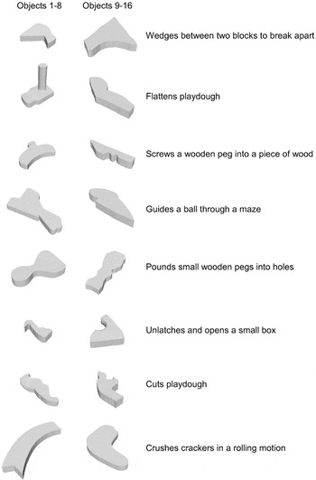
Sixteen different novel objects were created out of wood and painted a stone color (Krylon “Make It Stone” paint, see Figure 1). They were purposely created to have ambiguity about which part was the handle. The real novel objects were used in both behavioral pilot testing and training and testing sessions before the functional scans. Behavioral pilot testing indicated that, on average, there was not one preferred location to grasp the handle and that specific uses of the objects could not be identified. The objects were scanned for size and texture on a flatbed scanner to create 3D models of the objects at 30-degree intervals, to be used during the fMRI session. The images were rendered with Alias/Wavefront's Maya Unlimited. The novel objects were divided in two groups that we will refer to as 1–8 and 9–16. One complex action plan was assigned to each of the objects (see Figure 1). One group of 12 participants was trained on the use of objects 1–8 but not objects 9–16. The second group of 12 participants was trained on the action plans of objects 9–16 (using the same general action plans and recipient objects) but not objects 1–8. The half of the objects that did not have explicitly associated action plans were handled without knowledge of a goal-directed action.

Images and functions of 16 novel objects presented at a single orientation.
PROCEDURE
Behavioral Training
Training consisted of a one half-hour session, each of 2 days before the fMRI scanning session. The novel objects were never explicitly named based on their function. On day 1, the subjects watched the experimenter use each tool (objects 1–8 for one group and objects 9–16 for the second group) and then used it themselves. This procedure was repeated once. Then the subjects pantomimed the use of each tool, and they were corrected if they used the object incorrectly. Subjects then watched the experimenter handle each shape and then handled each object themselves. This procedure was repeated once. Then subjects were allowed to manipulate each shape. This typically involved rotating each shape within their hands to look at all sides and to grasp in different places. On day 2, subjects pantomimed the use of each tool at least twice until they reached 100% accuracy on the use. They also manipulated each shape at least two times. The goal was to give each participant equivalent time manipulating all of the objects over the 2 days, but to vary the extent to which they learned specific goal-directed actions associated with the objects.
Behavioral Testing
On the third day (the day of the scan), subjects completed a testing session at the location of the MRI scanner. Before the scan, the tools were set individually in front of the subject in a pseudo random order with the handle facing away from them at one of two oblique angles (45 and 135 degrees), while the subject's eyes were closed. After the object was placed on the table, the subject was given a signal to open their eyes and pick up the object with their right hand. Then subjects were asked to pantomime the use for each object. Following the grasping and using of the tools, subjects were allowed to handle each shape (to equate for motor experience immediately before the scanning session). Postscanning, subjects were presented with each shape individually and asked to grasp them twice. They were then asked to create a use for each shape. Shape grasping and using was performed after the scan to avoid the potential influence of thinking about a specific use before the imagined tasks in the scanner. All behavior was videotaped and coded at a later time.
fMRI Testing
During the scanning session, subjects performed three different tasks in the same order: view, imagine grasp, imagine use. This task order from no-action (view) to complex action (use) was held constant to avoid the possibility that explicit instructions to act in one task would influence strategies used on the less complex task. For example, imagining using the objects in one run could potentially lead the subject to explicitly imagine the use in the view or grasping task. The three fMRI tasks used the same standard boxcar block design with 16-second epochs. Eight images were presented in each block for 2 seconds each. The order of blocks alternated between scrambled images and novel objects (separate blocks for tools and shapes) leading to a four-block sequence (64 seconds) repeated five times. In these tasks, subjects viewed grayscale 3D images of the novel objects presented at different orientations. Each object was presented five times, always at a new orientation (rotated in depth with the handle at four oblique angles and one horizontal angle). Scrambled images, which served as baseline images, were created by placing a 10 × 10 grid over the intact images and randomly mixing the squares. The images were presented on a screen (37 × 27 in) at the foot of scanner using an LCD projector (Sharp XG-E12004). Participants viewed the screen through a mirror placed above their eyes. Subjects were told to keep their hands still, resting on their legs; no overt motor responses were required, and subjects were visually monitored for any hand movement.
Visual one-back task.
Subjects attended to all of the images and noticed when two of the same images were presented in succession.
Imagined grasping task.
Subjects imagined grasping and picking up each object with their right hand and fixated on the scrambled images.
Imagined using task.
Subjects imagined using each object with their right hand and fixated on the scrambled images.
Behavioral Analysis.
Grasping and using behavior was coded by two independent observers from the videotapes. For each grasping trial, it was determined whether the subject grasped the object by its “handle.” For each using trial, it was determined (1) whether the subject held the object by its “handle” and (2) whether the subject pantomimed the correct motion/function with the object. Grasping of shapes was coded using pictures of each of the objects. For coding, each image of the object was segregated into three graspable areas. For each trial, the location of where the subject grasped the object was highlighted on the picture. It was also coded whether this grasp was in the location of the “handle” (defined by its function as a tool for the other group). Using of the shapes was coded with pictures, as in the grasping task, as well as by a written description of how the subject used the object (e.g., as a hammer) and whether it was appropriate to the use of the object (defined by its function as a tool for the other group).
Imaging analysis.
Raw EPI data were ghost-corrected, distortion corrected, and reconstructed with in-house MATLAB routines to a 64 × 64 matrix with square 25.6-cm field of view and in-plane resolution of 4 mm. Statistical analysis were performed using MATLAB (Mathworks, Inc., Natick, MA) and statistical parametric mapping (SPM2, Wellcome Department of Cognitive Neurology, London, UK). The first five images of each task were discarded to ensure that the signal had reached equilibrium. EPI images were aligned to correct for head motion, and anatomical images were co-registered with the EPI images. All images were spatially normalized to the standard Montreal Neurological Institute (MNI) template and smoothed using isotropic Gaussian kernels of 8 mm. Individual and group analyses were performed. For each run (visual one-back, imagined grasping, imagined using), we applied a boxcar model convolved with the hemodynamic response function using a general linear model with four stimulus conditions for each participant (Friston et al., 1995). Two linear contrasts were defined to test for specific condition effects for Tools (Tool-ScrTool) and Shapes (Shape-ScrShape). The individual subject contrasts were used in subsequent group random effects analyses with the 23 total subjects, using one-sample t tests to assess the Tool and Shape effects relative to the scrambled images [threshold p = .05 corrected for family wise error (FWE), cluster size 10 voxels] and paired t tests to assess the difference between Tool and Shape effects (uncorrected p < .001, corrected to p < .05 at the cluster level of 60 voxels).
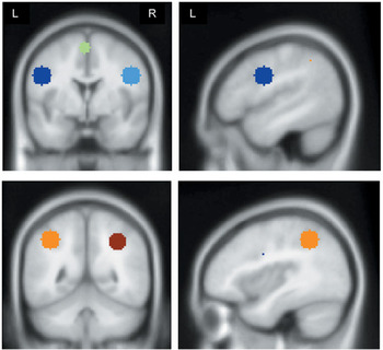
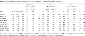
A second group random effect analysis was conducted across the three tasks for the combined tool and shape conditions (Tool − ScrTool + Shape − ScrShape), thresholded at p = .05 FWE. This analysis allowed us to choose regions of interest using Marsbar (Brett et al., 2002) present with both stimuli and to calculate effect sizes in individual subjects for those regions. The selection of the regions of interest (ROIs) was determined by a priori hypotheses about the role of premotor and parietal cortex in representing skilled actions. Local maxima coordinates resulting from the combined tool and shape conditions were used to identify the ROIs listed in Table 5 and a 10-mm sphere surrounding each coordinate was used as the ROI (see Figure 4).
RESULTS
Behavioral Results
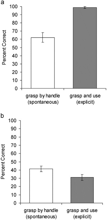
Grasping tools outside of the scanner showed that, when subjects were trained on the functions of novel objects, they grasped and used the objects in the intended way. Subjects grasped the objects by their handles on 62.1% of the spontaneous grasping trials and grasped and used the objects appropriately on 98.9% of the explicit use trials (see Figure 2a). Their using behavior indicated that subjects were successful at using the novel objects for their trained functions when explicitly given the instructions to do so. The grasping behavior is consistent with the findings of Creem and Proffitt's (2001b) real-tool study finding that, although all subjects do not spontaneously grasp all tools by their handles given neutral instructions that do not specify a use, on average, there is a tendency to reach for the handle. Grasping shapes outside of the scanner showed that, without complex action training, subjects grasped objects by their handles on 41.5% of the spontaneous grasp trials and grasped and used the objects appropriately on 31.1% of the explicit use trials (see Figure 2b). Paired t tests confirmed differences between tool and shape grasping [t(23) = 3.05, p < .01] and tool and shape using [t(23) = 18.89, p < .01]. (The percentage of shapes grasped by the handle 41%, did significantly differ from chance, 33.3%, given three locations.) These results suggest that shapes are less meaningful than tools with respect to a function-specific action plan.

a: Grasping and using performance for the eight tools outside of the scanner. b: Grasping and using performance for the eight shapes outside of the scanner
Imaging Results: Whole Brain Analyses
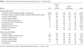
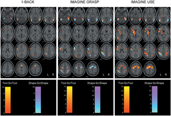
While viewing tools in the one-back task, there was significant activation bilaterally in large clusters of ventral occipital/temporal cortex and middle IPL, including the intraparietal area (although to a much smaller extent on the right) and SMA. DPM and VPM extending to the insula were found on the left. Right cerebellum was also found. The shape trials showed similar but smaller clusters of activation (see Table 1 for coordinates and Brodmann areas, and Figure 3). Notably, the premotor cortex activation found with the tool stimuli did not survive the threshold for shape stimuli. This is weak evidence consistent with our hypothesis that VPM function is influenced by knowledge of actions associated with tools, and not only their graspable structure. (The paired t test between the two contrasts revealed a 12-voxel cluster at the VPM (−54 4 30) when thresholded at p < .001 uncorrected. There were no other clusters above the threshold.)
Clusters of activation for the one-back viewing task (p < .05 corrected)


Activation for the three tasks resulting from the group random-effects t tests, superimposed on a single subject's anatomical structure (Tool-ScrTool contrast depicted in red-yellow; Shape-ScrShape depicted in cyan–purple; minimum t = 6.0).

A priori–defined frontal (ventral premotor cortex and presupplementary motor cortex, top) and parietal (inferior parietal lobule, bottom) regions of interest (10-mm sphere, see Table 5 for center coordinates).
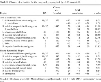
For the imagined grasping task, there was a similar left-lateralized premotor–parietal–temporal network of activation for both the tool and shape stimuli (See Table 2 and Figure 3). (The paired t test between the two contrasts revealed no clusters above the threshold. It is notable that this lack of difference is likely attributed to an increase in activation for shapes, and not a decrease in activation for tools. See Figure 3). This network of regions is consistent with the growing number of studies involving imagined grasping (Johnson-Frey et al., 2005) and appears from the present results to differ little, depending on the extent of knowledge about functional action plans. One explanation is that, given the lack of explicit instruction about how to grasp the objects, there may have been less consistency both within and between subjects about their imagined behavior or a tendency to attribute handles to the shapes (see General Discussion for further discussion of this result).
Clusters of activation for the imagined grasping task (p < .05 corrected)

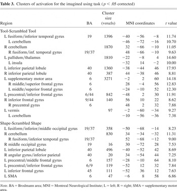
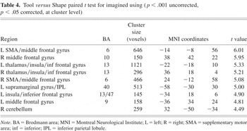
In imagined using, although there was some overlap between the two stimuli in posterior visual cortex, DPM, VPM, and IPL, Table 3 and Figure 3 illustrate the overall greater activation with tools versus shapes specifically in the left IPL, DPM including SMA, VPM extending to the IFG, the left inferior temporal gyrus, and cerebellum. The VPM activation was mostly more inferior than the VPM ROI, which helps to explain the marginal significance for the effect size analysis reported below. There was also a large cluster of activation in the basal ganglia, thalamus, and insula given the tool stimuli. The Tool versus Shape paired t test confirmed these differences and also showed additional clusters in bilateral middle frontal gyrus and inferior frontal gyrus, not overlapping the VPM ROI (see Table 4).
Clusters of activation for the imagined using task (p < .05 corrected)

Tool versus Shape paired t test for imagined using (p < .001 uncorrected, p < .05 corrected, at cluster level)

Imaging Results: ROI Analyses
Paired t tests compared the mean effect sizes for the two contrasts (Tool-ScrTool and Shape-ScrShape) resulting from the individual ROI analyses. The results are presented in Table 5. Because of the use of a simple t-contrast, the effect size values are equal to the parameter estimate. For the one-back task, effect sizes were significantly greater in the left IPL and VPM. For the grasping task, there were no significant differences. For the imagined using task, effect sizes were greater in the left IPL and pre-SMA, and marginally in the left VPM.
ROI coordinates, mean contrast (effect size), and paired t test statistics for the three tasks

DISCUSSION
There is prior evidence of a distributed frontal–parietal–temporal network involved in action planning and the objects associated with those actions (Grezes & Decety, 2002; Johnson-Frey et al., 2005). Our goal was to examine whether behavioral and neural distinctions exist between motor representations that are based on visual and motor experience with grasping objects (exclusive of use) versus those that are explicitly associated with skilled use of objects. Behaviorally, spontaneous and explicitly directed tool behavior was influenced by experience with object use. Neuroanatomically, we focused on regions that are part of the reach/grasp circuit, but that were predicted to be specifically involved in planning for use, the left VPM and IPL, as well as pre-SMA, predicted to be involved in cognitive representations of actions.
Primarily the left IPL and to a lesser extent the left lateral and medial premotor cortex were differentially recruited as a function of skilled action knowledge versus manipulation experience with novel objects, also modulated by the nature of the task required. In the viewing and using tasks, left IPL, and VPM/IFG were influenced by the object group with an additional effect of the pre-SMA for imagined using. Notably, the activation in the left IPL and VPM did not differ between the two object types in the imagined grasping task. One distinction of this task versus the others is that processing of complex action knowledge was not explicitly required in instructions to grasp. In viewing objects to remember them, observers may likely think about the meaning of the objects; and imagining using requires necessary retrieval of the skilled action representation. The ambiguity of the imagined grasping task, in hindsight, likely led to variable performance both within and between subjects. The nature of the task is such that it is difficult to conclude whether the lack of effect is a result of imagined versus overt grasping, variability in imagining grasping the object by the handle, a tendency to assign a “handle” to the shapes, or other accounts. Future work may examine the influence of task constraints on related tool representations.
Broadly, the present research poses the fundamental question, are all graspable objects tools? Gibson's theory of affordances would argue yes; both environments and objects are processed for the intent of an observer's actions. However, a recent study by Creem-Regehr and Lee (2005) demonstrated that simply viewing 3D shapes, although potentially graspable, did not activate the action system as the observation of tools has been shown to do (Chao & Martin, 2000; Creem-Regehr & Lee, 2005). Thus, they suggested that the left-hemisphere parietal and premotor network seen in tool-related tasks likely relies on knowledge of tool use as well as perception of potential graspability. Our current findings support this claim. Weisberg et al. (2007) recently conducted a related study involving an object-matching task after tool-like training of novel objects. Consistent with our findings, they found activation modulated by training in the left IPL and premotor cortex and middle temporal gyrus.
There were two critical manipulations in the present experiment, the presence/absence of skilled action training and the behavioral tasks both inside (viewing, imagined grasping, imagined using) and outside (grasping and using) the scanner. We differentiated between grasping and using in the behavioral tasks but presented objects that were grasped in a way that was consistent with the way they were used. Recent fMRI work has aimed to dissociate grasp and use processes with objects that conflict in the way they are grasped and used, finding greater activation in the left IFG, IPL, and superior temporal gyrus for use versus grasp trials (Buxbaum et al., 2006). These findings, along with our significant effects for the use but not grasp task, contribute to a similar conclusion that particularly the left IPL and second, the VPM/IFG is involved in motor planning for skilled object use. This claim is also supported by the deficits seen in left-IPL–damaged patients involving skillful tool use (Rothi & Heilman, 1997; Sirigu et al., 1995), and the mental representations of hand actions (Buxbaum et al., 2003; Sirigu et al., 1996). Using functional neuroimaging techniques, the middle (supramarginal gyrus and intraparietal sulcus) IPL has been defined in both motor execution and imagery tasks (Grezes & Decety, 2001; Hanakawa et al., 2003; Johnson-Frey et al., 2005), as well as some, but not all, tool-judgment tasks. It has been shown that the anterior IPL subserves visually guided grasping functions in both humans (Culham et al., 2003) and monkeys (Sakata & Taira, 1994) and that subdivisions defined in the monkey IPL form direct prehension circuits with subdivisions in the inferior frontal premotor cortex (Rizzolatti & Luppino, 2001).
It is important to recognize that the grasping and using tasks performed in the scanner in the present study involved only imagination and not execution. Previous work (Johnson-Frey et al., 2005; Moll et al., 2000) has investigated distinctions between motor preparation/imagery and execution in the context of meaningful hand movements. Although there is a good amount of overlap in the left hemisphere frontal–parietal network found across real and imagined skilled hand actions, the present work can only make neuroanatomical claims about the motor imagery tasks. In this context, the activation in pre-SMA is especially interesting. While the recruitment of pre-SMA in the using task may be a result of accessing a plan for complex use of an object, it also may have been differentially involved because of the cognitive nature of motor imagery. Future work could aim to test whether processes mediating complex action knowledge can be dissociated from those involved in motor imagery.
Although not the primary focus, the extensive activation found in both lateral and ventral temporal cortex in the present work has implications for theories of object recognition and semantic knowledge. Neuropsychological evidence supports distributed accounts of the organization of semantic knowledge that differentiate objects on dimensions of functional and visual/sensory information (Warrington & McCarthy, 1987; Warrington & Shallice, 1984). Category-specific deficits have been accounted for by a model that posits primarily functional representations for artifacts versus sensory representations for living things. Extensive neuroimaging work has supported category specificity in defining functional regions of the ventral and lateral temporal cortex (Chao et al., 1999). Sensory motor accounts extend beyond this dichotomy to propose that attributes are differentially associated with objects as a function of the experience with the objects (Allport, 1985). Following this model, small artifacts such as tools may specifically have motor-related representations because of their nature to be manipulated and used.
In addition to the frontal and parietal regions outlined above, neuroimaging findings suggest a role of the motion area MT and the MTG associated with motion properties of small artifacts (Beauchamp et al., 2002; Weisberg et al., 2007). We found activation centered on the inferior temporal gyrus, extending both to the MTG and fusiform gyrus in all three tasks for both the tool and shape stimuli. To examine this finding further, we defined two ROIs from the combined task random effects analysis at the middle and inferior temporal/fusiform gyri (see Table 5). There were no significant differences between tools and shapes that resulted from the direct comparisons of contrasts or the effect size analysis. One explanation for this is that both tools and shapes share similar appearance in size, color, and structural parts. Furthermore, because all stimuli were manipulated and grasped as real objects outside of the scanner, they likely shared some level of motor-related representations.
Additional brain regions emerged with significant effects, particularly in the imagined using task, including the prefrontal cortex (middle and inferior frontal gyri) and the cerebellum. The prefrontal regions likely play a role in representing objects for how they are manipulated based on task goals or meaning (Grezes et al., 2003; Johnson-Frey et al., 2003), consistent with similar activation found in tool-use planning, pantomime, and semantic tasks such as tool naming. The cerebellum is an integrated part of the frontal–parietal circuitry for action and is often found in motor imagery and tool-related tasks. Its greater role for tool versus shape stimuli in the present tasks may be a result of its representation of internal models for specific action schema (Imamizu et al., 2003; Lewis, 2006).
Some final considerations influencing the interpretation of the present data involve a distinction between function versus manipulation knowledge with respect to objects (Boronat et al., 2005; Buxbaum & Saffran, 2002). Boronat et al. (2005) found increased activation in the IPL for manipulation versus function decisions about objects, suggesting that representations of manipulable artifacts rely more on knowledge of object manipulation than on object function. Our results support the work of Boronat et al., as we did see increased IPL activation with the specificity of tool knowledge in the using task. However, we cannot determine whether it was the specificity of function or manipulation that relies most on this region. Future research could investigate the dissociation by providing functional knowledge about the use of novel objects without manipulation knowledge, or vice versa.
ACKNOWLEDGMENTS
We thank Bart Lee for help with the design of the novel objects, Amy Gooch for making the models of the novel objects, Shawn Yeh for help with statistical processing, and Dr. Greg Katzman for reviewing the MRI scans. This work was supported by a University of Utah Funding Incentive Seed Grant awarded to the first author.