Introduction
The recovery of bovine ovarian follicles in the preantral phase has become an important resource for human reproduction, being applied to patients with changes in fertility or reproductive disorders (Frydman and Grynberg, Reference Frydman and Grynberg2016). It is estimated that during the fetal phase, approximately 235,000 follicles are formed per ovary in bovine species (Silva-Santos et al., Reference Silva-Santos, Santos, Siloto, Hertel, Andrade, Rubin, Sturion, Melo-Sterza and Seneda2011). However, the majority (99.9%) of this stock of follicles culminates in atresia (Matsuda et al., Reference Matsuda, Inoue, Manabe and Ohkura2012). The interest in minimizing the atresia process and its deleterious effects, has led to the goal of developing an in vitro culture system that maintains follicular growth and allows the best use of the follicular reserve (Araújo et al., Reference Araújo, Gastal, Figueiredo and Gastal2014; Hatzirodos et al., Reference Hatzirodos, Hummitzsch, Irving-Rodgers, Harland, Morris and Rodgers2014).
The development of in vitro culture conditions is essential for a better understanding of folliculogenesis in animals such as cattle, goats, and murines, as a model for biotechniques applied to human reproduction (Langbeen et al., Reference Langbeen, De Porte, Bartholomeus, Leroy and Bols2015; Green and Shikanov, Reference Green and Shikanov2016; de Sá et al., Reference de Sá, Ferreira, Sousa, Duarte, Paes and Cadenas2020). In the search for the best conditions for in vitro culture of preantral ovarian follicles, several factors have been investigated, such as growth factors, antioxidants, and hormones, which can assist in successful follicular development and growth (Araújo et al., Reference Araújo, Gastal, Figueiredo and Gastal2014; Gomes et al., Reference Gomes, Lisboa, Silva, Max, Marino, Oliveira, González, Barreiros, Marinho and Seneda2015, Reference Gomes, Silva, González, Oliveira, Max, Lisboa, Barreiros, Santos, Sarapião, Gastal and Seneda2018; Silva et al., Reference Silva, Ribeiro, Menezes, Barberino, Passos, Dau, Costa, Melo, Bezerra, Donato, Peixoto, Matos, Gonçalves, van den Hurk and Silva2017; Bizarro-Silva et al., Reference Bizarro-Silva, Santos, Gerez, González, Lisboa and Seneda2018; Max et al., Reference Max, Bizarro-Silva, Búfalo, González, Lindquist, Gomes, Barreiros, Lisboa, Morotti and Seneda2018).
Another relevant factor related to the biotechnology of in vitro culture of preantral ovarian follicles refers to the production and release of excess free radicals, reflecting oxidative stress, cell damage, and deleterious effects on the female reproductive system (Andrade et al., Reference Andrade, Melo-Sterza, Seneda and Alfieri2010, Reference Andrade, van den Hurk, Lisboa, Hertel, Melo-Sterza, Moreno, Bracarense, Landim-Alvarenga, Seneda and Alfieri2012). In this context, the excessive production of reactive oxygen species (ROS) resulting from oxidative stress, generates a large volume of free radicals present in cells (Andrade et al., Reference Andrade, Melo-Sterza, Seneda and Alfieri2010, Reference Andrade, van den Hurk, Lisboa, Hertel, Melo-Sterza, Moreno, Bracarense, Landim-Alvarenga, Seneda and Alfieri2012; Gomes et al., Reference Gomes, Silva, González, Oliveira, Max, Lisboa, Barreiros, Santos, Sarapião, Gastal and Seneda2018). Therefore, supplementation with alpha-lipoic acid in an in vitro culture medium could promote an ideal developmental environment, considering that this antioxidant is known for its action in biological processes, acting directly in the elimination of ROS and metal chelation. In addition, it is an essential cofactor in multi-enzyme complexes, which catalyze the decarboxylation of α-keto acids in the Krebs cycle (Packer et al., Reference Packer, Witt and Tritschler1995).
There are still no reports of studies using alpha-lipoic acid supplementation with an extracellular matrix of agarose gel in the in vitro culture of bovine preantral ovarian follicles to evaluate follicular development, morphological integrity, and antioxidant potential. Therefore, the aim of this study was to evaluate these three factors in preantral ovarian follicles from Bos taurus indicus females grown in vitro and supplemented with the antioxidant alpha-lipoic acid in the culture medium.
Materials and methods
Unless otherwise noted, all chemicals used in this experiment were purchased from Sigma-Aldrich (St. Louis, MO, USA).
Obtaining and processing of ovaries
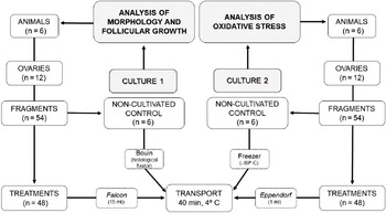
Ovaries (n = 24) of adult bovine females (n = 12) of the Nelore breed of Bos taurus indicus, cyclical, with a body condition score between 3 and 4 (scale from 0 to 5) (Ayres et al., Reference Ayres, Ferreira, de Souza Torres-Júnior, Demétrio, de Lima and Baruselli2009), were collected and processed in a slaughterhouse, 48 km (40 min) away from the Laboratory of Biotechnology of Animal Reproduction (Fig. 1). Immediately after slaughter, each pair of ovaries was washed separately three times with 0.9% saline solution (JP Farma, São Paulo, Brazil) and twice with 70% alcohol. The pairs of ovaries were fragmented with the aid of a sterile and disposable dermatological punch (6 mm; Kolplast, São Paulo, Brazil), resulting in fragments of approximately 9 mm2. Nine fragments were obtained from each pair of ovaries from random regions of the ovarian cortex, totalling 108 fragments. Afterwards, the obtained fragments were washed three times with 0.9% saline solution (JP Farma, São Paulo, Brazil) and twice with minimum essential medium (MEM; Gibco BRL, Rockville, MD, USA; osmolarity 300 mOsm/l, pH 7.2) contained in a 24-well plate. Fragments of the non-cultivated control (D0) were placed in a Falcon tube (15 ml) containing a Bouin fixator. Fragments were intended for analysis of morphology and follicular growth, and a fragment from each pair of ovaries was chosen randomly and placed in an Eppendorf microtube (1 ml), kept in a thermal container at 4°C, characterizing the non-cultivated control treatment (day 0; D0), for the analysis of oxidative stress. All fragments were transported to the laboratory under a constant temperature of 4°C.
Demonstrative scheme of obtaining the ovaries at the slaughterhouse.

In vitro culture of preantral ovarian follicles
In vitro cultures were performed separately to analyze follicular growth and morphology, and oxidative stress. The fragments (n = 96) of the ovarian cortex were cultured individually in 24-well plates containing only MEM or MEM supplemented with alpha-lipoic acid, comprising the following treatments: MEM, 50, 100, or 250 ng/ml, on agarose gel (Molecular Biology Grade Agarose; Kasvi, Brazil), in an oven at 38.5°C, in an atmosphere of 5% CO2 in air and saturated humidity. The base medium of the culture consisted of MEM supplemented with ITS (6.25 mg/ml of insulin + 6.25 mg/ml of transferrin + 6.25 mg/ml of selenium), 0.23 mM of pyruvate, 2 mM of glutamine, 2 mM of hypoxanthine, 1.25 mg/ml of bovine serum albumin (BSA; Gibco BRL, Rockville, MD, USA), 20 IU/ml of penicillin and 200 mg/ml of streptomycin. To prepare the extracellular matrix of the agarose gel, 3 g of powdered agarose (Agarose Molecular Biology Grade; Kasvi, Brazil) in 200 ml of distilled water was used to obtain a 1.5% agarose solution. After complete dilution, the solution was sterilized in an autoclave for 15 min. Subsequently, the solution was placed in a sterile glass Petri dish for gel solidification. Upon obtaining a gelatinous structure, the plate was kept in a 37ºC oven for 24 h to prove sterility, after which time the gel was segregated into fragments of approximately 1 cm3 (Silva et al., Reference Silva, Ribeiro, Menezes, Barberino, Passos, Dau, Costa, Melo, Bezerra, Donato, Peixoto, Matos, Gonçalves, van den Hurk and Silva2017), and each fragment was placed in a 24-well plate.
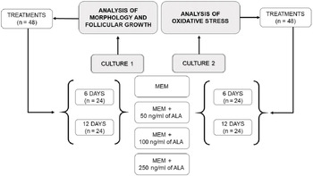
For each concentration of alpha-lipoic acid, fragments of the ovarian cortex of each animal were cultured for 6 (D6) or 12 (D12) days (Fig. 2). Every 2 days, 100% of the culture medium was replaced. The culture period used was based on previous studies (Figueiredo et al., Reference Figueiredo, de Lima, Silva and Santos2018; Gomes et al., Reference Gomes, Silva, González, Oliveira, Max, Lisboa, Barreiros, Santos, Sarapião, Gastal and Seneda2018; de Sá et al., Reference de Sá, Ferreira, Sousa, Duarte, Paes and Cadenas2020).
Scheme illustrating the groups intended for the in vitro culture of preantral ovarian follicles, with the supplementation of alpha-lipoic acid (ALA), assigned for the analysis of morphology and follicular growth as well as for the analysis of oxidative stress.

Analysis of morphology and follicular growth
For the analysis of morphology and follicular growth, the fragments of the ovarian cortex (n = 54) that included the non-cultivated control (D0) or the treatments cultivated for 6 (D6) or 12 (D12) days were fixed in Bouin for 24 h and kept in 70% alcohol. After fixation, the fragments were dehydrated in increasing concentrations of alcohol, clarified, diaphanized in xylol, and embedded in paraffin blocks to perform histological cuts. Each block was individually sectioned to 5 μm thickness with an interval of 10 sections of ovarian tissue, in a rotating microtome (Leica®, Wetzlar, Germany), for mounting histological slides. Before staining, the excess paraffin contained in the slides was removed with xylol, and then the slides were rehydrated with 70% alcohol. The slides were stained with Schiff’s periodic acid and haematoxylin. All sections were examined using optical microscopy at ×10 and ×40 magnification (Nikon, Tokyo, Japan). The preantral ovarian follicles were classified according to their stage of development as primordial (containing a layer of granulosa cells, of flat shape, around the oocyte), in primary development (primary follicles that presented a single layer of cuboid-shaped granulosa cells surrounding the oocyte), or in secondary development (secondary follicles that presented two or more layers of cuboidal granulosa cells). To evaluate follicular growth, the quantification of follicles was carried out at the different stages (primordial, primary, and secondary) in the non-cultivated control treatment and in the 6 or 12 days of in vitro culture treatments. Follicles were also classified according to their morphology as intact, when the oocyte had a non-pycnotic nucleus and was surrounded by granulosa cells organized in layers, or degenerate, when the oocyte was retracted with a pycnotic nucleus, with the presence of cytoplasmic vacuoles, and surrounded by granulosa cells arranged in a disorganized manner (Búfalo et al., Reference Búfalo, González, da Silva, Lindquist, Bergamo, Costa, Marinho and Seneda2016). Follicles were evaluated by treatment and duration of culture. The percentages of primordial, primary, and secondary follicles were calculated after 0, 6, and 12 days of culture. To avoid repeat counting, preantral ovarian follicles were counted only in the section where the oocyte nucleus was observed.
Oxidative stress analysis
After the in vitro culture period, the fragments (n = 54) were analyzed using kinetic-colorimetric assays to determine lipid peroxidation [thiobarbituric acid reactive substances (TBARS)] and superoxide anion production (NBT). The ability of ovarian tissue to resist oxidative damage by reducing iron [ferric reducing antioxidant power (FRAP)] was also evaluated, as described by Pinho-Ribeiro et al. (Reference Pinho-Ribeiro, Zarpelon, Mizokami, Borghi, Bordignon, Silva, Cunha, Alves-Filho, Cunha, Casagrande and Verri2016).
Evaluation of antioxidant activity by the FRAP assay
The FRAP method determined the iron reduction of 2,4,6-tripyridyl-s-triazine (TPTZ) using a colorimetric approach. The reaction detected compounds with redox potential < 0.7 (the redox potential of Fe3+ TPTZ). The ovarian fragments were crushed and homogenized with 500 μl of 1.15% potassium chloride (KCl) buffer, and centrifuged at 1500 rpm for 10 min at 4°C. Following this, 30 μl of ultrapure water and 150 μl of the FRAP reagent together with 20 μl of the sample supernatant were added. The absorbance of the assay was measured at 595 nm (Multiskan GO, Thermo Scientific) using a microplate reader spectrophotometer. The results were presented as nmol equivalents of 6-hydroxy-2,5,7,8-tetramethylchroman-2-carboxylic acid (Trolox) per mg of protein.
Evaluation of lipid peroxidation by the TBARS assay
Lipid peroxidation was measured using the TBARS assay. Here 50 μl of sample homogenate were added to a microtube (1 ml) with 5 μl of iron chloride (FeCl3), 5 μl of ascorbic acid, 50 μl of 2.8% trichloroacetic acid (TCA), and 50 μl of 1.0% TBA. The mixture was agitated and kept in a water bath for 15 min at 90°C. The samples were cooled in an ice bath and subjected to further agitation and centrifugation at 3000 rpm for 15 min at 4°C. The supernatant was removed (100 μl) from the Eppendorf microtube and pipetted into 96-well plates. The levels of malondialdehyde (MDA), an intermediate product of lipid peroxidation, were determined in the samples by the difference between the absorbances at 535 and 572 nm (Multiskan GO, Thermo Scientific) using a microplate reader spectrophotometer. The results were presented as optical density (OD) per mg of protein.
Evaluation of superoxide anion production by the NBT assay
Superoxide anion production was measured using the tetrazolium nitro azul (NBT) assay. In total, 50 μl of the sample homogenate were added to 100 μl of NBT (1 mg/ml) in a 96-well plate. After 15 min, all samples were removed from the plate and 120 μl of potassium hydroxide (KOH) and 120 μl of dimethyl sulfoxide (DMSO) were added. The reduction of NBT was measured at 600 nm (Multiskan GO, Thermo Scientific) using a microplate spectrophotometer reader. The results were presented as OD per milligram of protein.
Statistical analysis
The proportion of intact follicles was determined from the total number of follicles evaluated (intact and degenerate) at each stage of development for each treatment. Differences between the proportions of primordial and developing follicles (primary and secondary) for each treatment (cultivated control and different concentrations of alpha-lipoic acid at different times) were analyzed using the chi-squared test and/or Fisher’s exact test. For a significant effect, the samples were compared using a 2 × 2 proportion test to establish the ranking between treatments. For descriptive analysis, data were presented as percentages lrb (%). All analyses were performed using the Minitab® 18.1.1 statistical program. The data from the oxidative stress analyses were submitted to analysis of variance (ANOVA) followed by the Bonferroni test, using GraphPad Prism software (version 7.0). Values were considered significant at P ≤ 0.05.
Results
Analysis of morphology and follicular growth
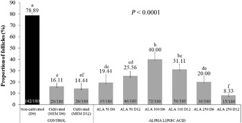
When evaluating follicular development, 6 days of culture supplemented with 100 ng/ml of alpha-lipoic acid showed the best result (40.00%; 72/180; P < 0.0001) in relation to the other treatments. The same concentration at 12 days of culture also performed well (31.11%; 56/180; P < 0.0001), although it was similar to the 50 ng/ml D12 group (25.56%; 46/180; P < 0.0001). The total proportion of all preantral ovarian follicles is shown in Fig. 3. Regarding follicular integrity, excluding the non-cultivated control (D0; primordial: 83.64%; 92/110; development: 71.43%; 50/70; P < 0.0001), supplementation with 100 ng/ml at 6 days of culture again returned the superior results, both for primordial follicles (51.28%; 20/39; P < 0.0001), and developing follicles (36.88%; 52/141; P < 0.0001) in relation to the other treatments. The proportions of intact preantral ovarian follicles are shown in Table 1.
Proportion of total preantral ovarian follicles, classified as healthy, of fragments of the ovarian cortex, of Bos taurus indicus females cultivated in vitro for 6 (D6) or 12 (D12) days in minimum essential medium (MEM) supplemented with different concentrations (50, 100 or 250 ng/ml) of alpha-lipoic acid (ALA). Different lowercase letters (a–f) indicate differences (P < 0.05) between treatments.

Percentage of intact, primordial, and developing preantral ovarian follicles, evaluated after in vitro culture of fragments of the ovarian cortex of Bos taurus indicus females, cultivated for 6 (D6) or 12 (D12) days with minimum essential medium (MEM) supplemented with different concentrations (50, 100 or 250 ng/ml) of alpha-lipoic acid (ALA)

Different lowercase letters (a–g) in the same column indicate differences (P < 0.05) between treatments.
Oxidative stress analysis
After 6 or 12 days of in vitro culture, the ovarian tissue samples showed a decrease in the antioxidant capacity evaluated by the FRAP assay [mean ± standard error of the mean (SEM)], when compared with the non-cultivated control (D0), except for treatments supplemented with 100 and 250 ng/ml of alpha-lipoic acid at 6 days of culture (44.78 ± 6.17 nmol of Trolox Eq/mg of protein; 36.91 ± 7.62 nmol of Trolox Eq/mg of protein, respectively; P ≤ 0.05). The results of the FRAP test are shown in Fig. 4. In contrast, there was no difference (P > 0.05) in the production of superoxide anions by the NBT assay between treatments with alpha-lipoic acid supplementation (Fig. 5). When evaluating the oxidative degradation of lipids using the TBARS assay, which evaluates lipid peroxidation products, we also observed that there was no difference (P > 0.05) between treatments when compared with the non-cultivated control treatment (D0; Fig. 6).
Effect of alpha-lipoic acid supplementation (MEM, 50, 100 or 250 ng/ml) on the ability of ovarian tissue to resist oxidative damage through the ability to reduce iron (FRAP) after in vitro culture of 6 or 12 days. *Differing from the non-cultivated controls (D0).

Effect of supplementation with alpha-lipoic acid (MEM, 50, 100 or 250 ng/ml) on the production of superoxide anion (NBT) after in vitro culture of 6 days (left) or 12 days (right).

Effect of alpha-lipoic acid supplementation (MEM, 50, 100 or 250 ng/ml) on lipid peroxidation (TBARS) after in vitro culture of 6 days (left) or 12 days (right).

Discussion
In the present study, it was observed that supplementation of the in vitro culture medium of bovine preantral ovarian follicles with a concentration of 100 ng/ml of alpha-lipoic acid was efficient in maintaining follicular integrity, as well as maintaining the levels of ROS stable over 12 days of in vitro culture. This is one of the few in vitro studies that used an extracellular matrix based on agarose to assess the development and integrity of preantral ovarian follicles, and to evaluate oxidative stress.
When observing follicular integrity, we noticed that supplementation with 100 ng/ml of alpha-lipoic acid proved to be effective in maintaining the integrity of the cultured follicles after 6 or 12 days of in vitro culture. Consistent with previous results, Zoheir et al. (Reference Zoheir, Harisa, Allam, Yang, Li, Liang, Abd-Rabou and Harrath2017) observed that supplementation of the culture medium with alpha-lipoic acid (250 and 500 µM) on bovine preantral ovarian follicles over 15 days also promoted an increase in development and follicular growth. Gomes et al. (Reference Gomes, Silva, González, Oliveira, Max, Lisboa, Barreiros, Santos, Sarapião, Gastal and Seneda2018) also observed positive effects on the preservation of follicular morphology and integrity with alpha-lipoic acid supplementation (50 and 100 µM) in an in vitro culture of preantral ovarian follicles of horses during 2 days of culture, corroborating our results regarding follicular integrity.
Considering that oxidative stress is a common event during in vitro culture, in the present study the production of free radicals was not observed in any of the periods and concentrations of alpha-lipoic acid tested. There was no difference between the non-cultivated control (D0), cultured control (MEM), and treatments (50, 100, and 250 ng/ml) evaluated by the NBT (superoxide anion production) and TBARS (lipid peroxidation) assays. An increase in the proportion of developing follicles, as well as a decrease in oxidative stress, was also reported in a study on preantral ovarian follicles of rats in which the culture medium was supplemented with 100 µM alpha-lipoic acid for a period of 12 days (Hatami et al., Reference Hatami, Zavareh, Salehnia, Lashkarbolouki and Karimi2014). At physiological concentrations, antioxidants protect cells against damage caused by oxidative stress. ROS are generated by the metabolism of cells and by several external factors such as high concentrations of oxygen, brightness of the environment, and manipulation of ovarian fragments in vitro, influencing the general metabolism of cells. The overproduction of free radicals led to an instability in which an excessive amount of ROS was released, leading to oxidative stress (Goto et al., Reference Goto, Noda, Narimoto, Umaoka and Mori1992; Combelles et al., Reference Combelles, Gupta and Agarwal2009; Gomes et al., Reference Gomes, Silva, González, Oliveira, Max, Lisboa, Barreiros, Santos, Sarapião, Gastal and Seneda2018), therefore we decided to supplement the culture medium with alpha-lipoic acid to reduce the oxidative stress caused by ROS.
The production of ROS in vivo is a by-product of the mitochondrial respiratory chain, balanced by the physiological presence of antioxidants in cells (Agarwal et al., Reference Agarwal, Gupta and Sharma2005, Reference Agarwal, Gupta and Sikka2006). Low concentrations of ROS can be positive for the maturation, competence, and development of oocytes (Halliwell and Gutteridge, Reference Halliwell and Gutteridge1988; Blondin et al., Reference Blondin, Coenen and Sirard1997). In this context, supplementation of the culture medium with alpha-lipoic acid appeared to improve the conditions of in vitro culture. Therefore, the results of this study will contribute to the improvement of the in vitro culture system of preantral ovarian follicles in bovine species, in addition to contributing to a better control of oxidative stress.
Given the importance of a balanced environment to promote adequate in vitro development, the objective of this study was to identify the superior concentration of alpha-lipoic acid (determined as 100 ng/ml) in the in vitro culture of preantral ovarian follicles from Bos taurus indicus females. Several benefits have already been reported with alpha-lipoic acid supplementation for other species, although in excessive amounts, alpha-lipoic acid can be harmful to follicular cells. Talebi et al. (Reference Talebi, Zavareh, Kashani, Lashgarbluki and Karimi2012) observed that 500 µM of alpha-lipoic acid caused an increase in the rate of degeneration of preantral ovarian follicles in rats. Other studies have also reported that prolonged exposure to alpha-lipoic acid resulted in increased lipid peroxidation, mitochondrial damage, and inhibition of glycogen synthesis, in addition to the interruption of the cell cycle and cell death (Moini et al., Reference Moini, Packer and Saris2002; Marsh et al., Reference Marsh, Pat, Gobe and Coombes2005; Wenzel et al., Reference Wenzel, Nickel and Daniel2005; Cakatay, Reference Cakatay2006). Therefore, adjustments in alpha-lipoic acid concentrations and culture period may be necessary to improve follicular integrity and the antioxidant capacity of the follicles in the long term. Therefore, we sought to gain a better understanding of folliculogenesis and reproductive disorders associated with animal and human infertility.
In conclusion, supplementation of the in vitro culture medium of preantral ovarian follicles with a concentration of 100 ng/ml of alpha-lipoic acid, after 6 days of culture, was effective in promoting follicular development and maintaining follicular integrity, and stability of oxidative and antioxidant parameters.
Supplementary material
To view supplementary material for this article, please visit https://doi.org/10.1017/S0967199421000502
Financial support
The authors thank the Coordination for the Improvement of Higher Education Personnel (CAPES) for supporting this research.
Conflict of interest
The authors declare none.
Ethical standards
This study was conducted according to the standards of the Ethics Committee for Animal Experimentation of the University of Londrina, following the rules established by Law 1283 for ovaries obtained from slaughterhouses.