INTRODUCTION
Acanthamoeba species are free-living amoebae (FLA) with worldwide distribution. FLA are found in soil, water and air, and can be readily isolated from these sources (Visvesvara et al. Reference Visvesvara, Moura and Schuster2007). However, these organisms are pathogenic/opportunistic amphizoic protists with potential to infect human and animal hosts (Visvesvara, Reference Visvesvara2013). Acanthamoeba spp. present two stages in their life cycles: a motile, trophic and replicating trophozoite and a resistant cyst stage. Based purely in morphological criteria, as many as 24 species have been included in the genus Acanthamoeba. Recently, species identification has relied upon sequencing of the amoeba 18S rRNA genes, and 20 different genotypes (T1–T20) of Acanthamoeba spp. have been established, which each genotype exhibits 5% or more of sequence divergence among different genotypes (Corsaro et al. Reference Corsaro, Walochnik, Köhsler and Rott2015). However, the pathogenicity of Acanthamoeba spp. could be limited to some genotypes and the majority of the human infections have been associated with the T4 genotype (Khan, Reference Khan2006).
In recent years, the incidence of infections due to Acanthamoeba spp. has shown a remarkable increase. Acanthamoeba spp. are the causative agents of a sight-threatening infection of the cornea known as Acanthamoeba keratitis (AK) in healthy patients and a fatal disease of the central nervous system (CNS) known as Granulomatous Amebic Encephalitis (GAE) in immunocompromised patients (Baig, Reference Baig2015; Lorenzo-Morales et al. Reference Lorenzo-Morales, Khan and Walochnik2015).
Early detection of the infection and treatment are critical to the outcome of the clinical course of AK and GAE infections (Szentmáry et al. Reference Szentmáry, Goebels, Matoula, Schirra and Seitz2012; Bouheraoua et al. Reference Bouheraoua, Gaujoux, Goldschmidt, Chaumeil, Laroche and Borderie2013; Lorenzo-Morales et al. Reference Lorenzo-Morales, Martín-Navarro, López-Arencibia, Arnalich-Montiel, Piñero and Valladares2013). The diagnosis of AK can often be made by in vivo confocal microscopy (IVCM). However, the direct detection of the causative agent in a corneal scrape specimen is the only reliable diagnostic method for AK. In case of GAE, magnetic resonance imaging (MRI) or computerized tomography (CT) of the brain show ring-enhancing lesions or low-density abnormalities mimicking a single or multiple space-occupying mass in the cerebral cortex (Schumacher et al. Reference Schumacher, Tien and Lane1995; Shirwadkar et al. Reference Shirwadkar, Samant, Sankhe, Deshpande, Yagi, Schuster, Sriram and Visvesvara2006). A significant delay in formulating an appropriate diagnosis before treatment is common, because of lack of suspicion and familiarity with these amoebae by clinicians and pathologists, misdiagnosed as bacterial, viral or fungal keratitis/encephalitis and the absence of rapid-specific immunodiagnostic assays (Martínez et al. Reference Martínez, Vogel, Stoppel, Charlin, Squella, Srur, Traipe, Verdaguer and Suárez1997; Khan et al. Reference Khan, Greenman, Topping, Hough, Temple and Paget2000; Clarke and Niederkorn, Reference Clarke and Niederkorn2006). Antibody detection by immunoassays may be a rapid useful diagnostic tool as an adjunct to microscopical diagnosis in detecting these parasites.
Among the species, pathogenic Acanthamoeba castellanii has been used extensively to study the molecular mechanisms of amoeboid locomotion and virulence factors involved in AK and GAE (Pollard and Ostap, Reference Pollard and Ostap1996; Leher et al. Reference Leher, Kinoshita, Alizadeh, Zaragoza, He and Niederkorn1998; Kennett et al. Reference Kennett, Hook, Franklin and Riley1999; Kong and Pollard, Reference Kong and Pollard2002; Garate et al. Reference Garate, Alizadeh, Neelam, Niederkorn and Panjwani2006).
Adherence of trophozoites to corneal epithelial cells followed by injury and invasion of tissue are thought to represent important steps in the establishment of infection. Acanthamoeba castellanii binds to mannose containing glycoproteins on the corneal epithelium through a 136-kDa mannose-binding protein on the amoebae surface (Yang et al. Reference Yang, Cao and Panjwani1997), but another surface proteins and cellular events governing adherence to the host cell are not completely elucidated (Soto-Arredondo et al. Reference Soto-Arredondo, Flores-Villavicencio, Serrano-Luna, Shibayama and Sabanero-López2014). In this study, we identified in silico an A. castellanii putative calreticulin surface protein (AcCRT) and expressed a protein domain in its recombinant form to assess its potential in the utilization in immunodiagnostic assays for detection of AK and GAE. We tested the immunodiagnostic potential of the AcCRT protein domain by ELISA using sera from infected rats with AK and GAE, and sera from patients with GAE. The results of this study, described for the first time a recombinant domain of the AcCRT that was recognized by sera from infected rats with GAE, but not by sera from patients with GAE, suggesting its immunodiagnostic potential in animal infections.
MATERIALS AND METHODS
Bacterial strains
Escherichia coli BL21 pLysE (Novagen) strain was used to express the recombinant protein domain. The E. coli KC8 was used for cloning assays.
In silico analyses of A. castellanii surface proteins
Comparative analysis between Entamoeba histolytica – the Amebozoa enteropathogen most studied – and A. castellanii amino acid sequences of surface proteins were performed using the NCBI/BLASTP tool (http://blast.ncbi.nlm.nih.gov/Blast.cgi?PROGRAM=blastp&PAGE_TYPE=BlastSearch&LINK_LOC=blasthome). The A. castellanii selected surface protein (see Results Section) was divided in amino acid portions (domains) according AmoebaDB database (http://amoebadb.org/amoeba/) and the translated coding sequences (CDS) of each exon were analyzed using the following software programs: TMHMM Server v. 2·0 (http://www.cbs.dtu.dk/services/TMHMM/) Phobius (http://phobius.sbc.su.se/), HMM-TM (http://bioinformatics.biol.uoa.gr/HMM-TM/), TopPred (http://mobyle.pasteur.fr/cgi-bin/portal.py#forms::toppred) and Predicting Antigenic Peptides (http://imed.med.ucm.es/Tools/antigenic.pl). All programs were used in default parameters. The first three programs predicted the presence of transmembrane (TM) domains, the fourth was used to predict hydropathicity and the last one antigenicity. The parameters for the domain selection were: translated CDS were considered as surface protein when they were predicted as such by at least two of the three TM predicting programs (Siqueira et al. Reference Siqueira, Thompson, Virginio, Gonchoroski, Reolon, Almeida, da Fonsêca, de Souza, Prosdocimi, Schrank, Ferreira, de Vasconcelos and Zaha2013), as well as considering the translated CDS with most hydrophilic and antigenic epitopes.
Acanthamoeba castellanii strain, cultivation and extraction of genomic DNA
Acanthamoeba castellanii trophozoites of the T4 genotype were obtained from the American Type Culture Collection (ATCC30010) (Clarke et al. Reference Clarke, Lohan, Liu, Lagkouvardos, Roy, Zafar, Bertelli, Schilde, Kianianmomeni, Bürglin, Frech, Turcotte, Kopec, Synnott, Choo, Paponov, Finkler, Heng Tan, Hutchins, Weinmeier, Rattei, Chu, Gimenez, Irimia, Rigden, Fitzpatrick, Lorenzo-Morales, Bateman, Chiu and Tang2013). Trophozoites were maintained in axenic cultures in peptone-yeast extract-glucose (PYG) medium, as previously described by Schuster (Reference Schuster2002); and samples for the genomic DNA extraction were directly taken from these cultures. Four identical and independent cultures with approximately 1 × 10⁶ trophozoites in the exponential growth phase were used for genomic DNA extraction as described by Aljanabi and Martinez (Reference Aljanabi and Martinez1997); with some modifications. Briefly, the culture was centrifuged, the cellular sediment obtained was homogenized in sterile salt buffer. SDS and proteinase K were added and the samples were incubated at room temperature overnight, after which NaCl was added to each sample. Samples were mixed in vortex at maximum speed, and tubes spun down. The supernatant was transferred to fresh tubes. An equal volume of isopropanol was added to each sample, mixed well and samples were incubated in low temperature. Samples were then centrifuged and the pellet was washed with ethanol, dried and finally resuspended in sterile dH2O and RNAse was added. The genomic DNA was quantified using Nanodrop 2000 (Thermo Scientific).
Cloning, expression and purification of the recombinant protein domain
The CDS of the selected surface protein domain was amplified by polymerase chain reaction (PCR) utilizing genomic DNA from A. castellanii. The primers were constructed using the program Vector NTI Express Software (Life Technologies) and are demonstrated in Table 1. The amplicons were cloned by in vivo homologous recombination as described by Parrish et al. (Reference Parrish, Limjindaporn, Hines, Liu, Liu and Finley2004) into pGEX-4T1 prokaryotic expression vector (GE Healthcare), linearized by EcoR1 and BamH1 restriction enzymes. The recombinant plasmids were transformed by chemical competence in E. coli strains BL21 pLysE (Novagen), BL21 RP (Stratagene), BL21 Rosetta (Novagen), BL21 Codon Plus Ril (Stratagene) e BL21 Star (Thermo Scientific). The expression of the selected recombinant surface polypeptide in fusion with glutathione S-transferase (GST) was carried out as described by Smith and Johnson (Reference Smith and Johnson1988). The bacterial cultures were growth in Circle Growth (MP Biomedicals) and induced with IPTG at final concentration of 0·1 mm shaking during 3–16 h at 37 °C. After induction, the bacterial cultures were submitted to sonication (VC601 Sonic & Material Inc. sonicator) on freeze bath during 6 cycles, 30 s with 1 min gaps in repose, after that, they were centrifuged at 20 000 g for separating soluble and insoluble fractions. The expression and solubility of the fusion protein was analysed by SDS-PAGE 12%. The fusion protein was purified by affinity chromatography using the Gluthathione-Sepharose resin (GE Healthcare) followed by thrombin (Sigma) cleavage. The purified polypeptide was quantified by Qubit™ (Thermo Scientific). The cleavage efficiency and purified polypeptide recovering were analysed by SDS-PAGE 15%.
Primer sequences used for genomic DNA amplification of A. castellanii Neff (ATCC 30010), annealing temperature and amplicon length of the recombinant protein domain CDS

Gene ID: gene code of A. castellanii selected surface protein in the Amoeba DB database. Primers: F, forward; R, reverse. Italic letters correspond to plasmid homologous sequences: pGEX-4T1. The bold letters are the complementary to target CDS.
In vivo infection models of A. castellanii
The stimulant solution (SS) for infection was prepared from cultures of A. castellanii ATCC 30010. The PYG medium containing 1 × 10⁶ trophozoites mL−1 was centrifuged at 250 g for 10 min. The pellet was washed three times with phosphate buffered saline (PBS) and resuspended in 100 µL of PBS. Adult female Wistar rats, with a mean age of 3 months, were first immunosuppressed by intraperitoneal injection with dexamethasone (5 mg kg−1) (Capilla et al. Reference Capilla, Maffei, Clemons, Sobel and Stevens2006). For the GAE infection, 10 animals were anesthetized with ketamine hydrochloride (60 mg kg−1) and xylazine hydrochloride (8 mg kg−1) and were inoculated intranasally with 100 µL of the SS. After infection, the animals were followed for 30 days. After this period, blood was collected and the rats were euthanized by barbiturate overdose (100 mg kg−1)/lidocaine (10 mg mL−1) (euthanasia solution), intraperitoneal. The parasites were re-isolated from the organ lesions and cultured in plates with non-nutrient agar 1·5% covered with inactivated E. coli (ATCC 25922).
For the AK infection model the animal corneas were scratched and challenged with A. castellanii. In this case, 10 animals were anaesthetized peritoneally as described earlier. Corneal anesthesia was obtained with topical 0·4% Oxybuprocaine hydrochloride eye drops. After routine disinfection, the corneas of right eyes were scratched three times vertically and three times horizontally with a sterile 30-gauge syringe needle and the stimulating solution, containing 1 × 10⁶ trophozoites mL−1 was applied to the scarified corneas. After infection, the animals were observed for 30 days. After this period, blood was collected and the rats were euthanized as described above. The parasites also were re-isolated from the eyes and cultured in plates with non-nutrient agar 1·5% covered with inactivated E. coli (ATCC 25922). All of the procedures were previously approved by the Ethics Committee on Animal Use of the Universidade Federal do Rio Grande do Sul with record number 27191 and followed international guidelines for the ethical use of animals in science.
Assessment of the immunodiagnostic potential of the recombinant protein domain
Serum total IgG detection were performed by indirect ELISA, as described by Virginio et al. (Reference Virginio, Hernández, Rott, Monteiro, Zandonai, Nieto, Zaha and Ferreira2003) with modifications in the dilution of sera and secondary antibodies. Briefly, microtitration plates (Maxisorp, Nunc) were coated with 0·4 µg well−1 of selected recombinant polypeptide diluted in 0·1 m carbonate/bicarbonate buffer (pH 9·6). Three AK infected rat serum samples, 8 rat serum samples infected with GAE and 4 human serum samples infected with GAE were diluted in Blotto (PBS containing 5% milk powder) at 1:1000 in case of infected rat serum samples and 1:2000 in case of infected human serum samples, and all of them tested in duplicate. Positive and negative sera and conjugate controls were included in each plate. Goat anti-rat IgG conjugated to horseradish peroxidase (Sigma) and Goat anti-human IgG (whole molecule) were used as secondary antibodies, diluted in Blotto 5% at 1:1000 and 1:30 000, respectively. OPD (Sigma)/H2O2 in citrate-phosphate buffer were used during 15 min for reaction developing. Afterwards, the reactions were interrupted for adding H2SO4 1N and absorbance were measured to 492 nm (A492) in ELISA reader (BIORAD). Acanthamoeba castellanii total protein extract was used as positive control of the ELISA in the assays with infected human serum samples for verify the serum titres as well as reject any mistake during the assays with the recombinant polypeptide. The infected human serum samples with GAE were gently granted by Govinda S. Visvesvara/Centers for Disease Control and Prevention (CDC).
Statistical analysis
Standard deviation and parametric Student's t distribution set to 95% confidence interval were performed for analysing the response levels of antibodies detected against the recombinant polypeptide between the different groups of infected sera. Differences were considered to be significant if P-values ⩽ 0·05. Statistical software packages in PRISM Version 6 (GraphPad Software In., La Jolla CA) were used.
RESULTS
In silico analyses of A. castellanii surface proteins
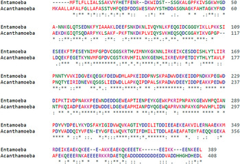
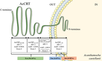
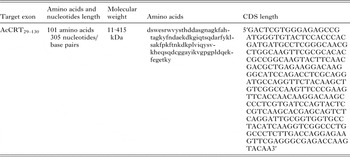
Among the different surface proteins analysed in silico for comparison between E. histolytica and A. castellanii, calreticulin from A. castellanii Neff strain (ATCC 30010) (AcCRT) with protein code XP_004349632·1 in the AmoebaDB and NCBI databases, and with 14923486 of Genbank access number exhibited 83% of query coverage and 55% of identity in the BLAST tool compared with calreticulin from E. histolytica (EhCRT) with protein code XP_655241·1 in the AmoebaDB and NCBI databases, and with 3409565 of Genbank access number (Fig. 1). The TM merged results showed that AcCRT has a small intracytoplasmic region at the beginning of the protein sequence indicating a possible signal peptide segment. There is a TM region located from the amino acid 5 until amino acid 23. The most part of the protein is extracellular. The CDS of the AcCRT protein has 3 introns and 4 exons according to the AmoebaDB database. All amino acidic sequences codified by 4 exons were analysed considering that we established working experimentally with A. castellanii genomic DNA. The exon number two, which encodes the protein portion between amino acid number 29 to amino acid number 130 (AcCRT29–130), showed antigenic, hydrophilic and extracellular amino acidic sequences (Fig. 2). The molecular characteristics of AcCRT29–130 are described in Table 2.
BLAST alignment of the calreticulin (CRT) protein coding sequences between Entamoeba histolytica and Acanthamoeba castellanii using Clustal Omega. (*) – indicates positions which have a single, fully conserved residue; (:) and (.) – indicate conservation between groups of strongly and weakly similar amino acids properties, respectively. The colours in the sequences represent physicochemical properties.

Domains representation of the calreticulin from Acanthamoeba castellanii (AcCRT) correlating with its 4 exons. The scheme depicts the predicted AcCRT with its amino end (N-terminus) inside of the amoeba and the carboxyl (C-terminus) outside. AcCRT is divided in extracellular (Ext), transmembrane (Tm) and intracellular (Int) regions, comprising 94·36, 4·65 and 0·98% of the amino acids sequences, respectively. The exon number two (circulated) encodes the domain between the amino acids (aa) 29 to 130 (AcCRT29–130). This domain is located extracellularly, and exhibits three Antigenic Determinants (AD) with an Average Antigenic Propensity (AAP) of 1·0019. The other values from the different exons also are showed. All antigenicity values were obtained in the online site http://imed.med.ucm.es/Tools/antigenic.pl.

Molecular characteristics of AcCRT29–130 exon number two

Cloning, expression and purification of the AcCRT29–130 recombinant protein domain
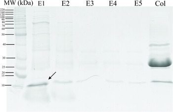
Oligonucleotides for PCR amplifications (see Table 1) were designed based on exon number two of AcCRTprotein. The amplified CDS of AcCRT29–130 was cloned into bacterial expression vector pGEX-4T1 in the E. coli strain KC8. All recombinant plasmids were sequenced using the Dyenamic ET Dye Terminator Cycle Sequencing kit for MegaBace DNA Analysis Systems (GE Healthcare). There were significant differences in the expression of recombinant plasmids (pGEX-4T1/AcCRT29–130) using the different E. coli expression strains. The BL21 pLysE (DE3) strain expressed more fusion protein compared with the other strains (data not shown). AcCRT29–130 purification was evaluated by SDS-PAGE 15% as shown in Fig. 3 in lane E1. The calculated 37 kDa molecular mass of the fusion recombinant protein AcCRT29–130-GST comprise the GST tag (26 kDa) plus purified polypeptide (11·4 kDa). The purified AcCRT29–130 polypeptide was recovered with yields of 3·64 mg L−1 of culture.
Elution pattern of AcCRT29–130 from Glutatione Sepharose resin. Purified AcCRT29–130 polypeptide band is indicated with an arrow (11·4 kDa). E1–E5: Five continuous elutions of the obtained purified polypeptide. Col: resin column sample showing part of the AcCRT29–130-GST fusion protein (37 kDa) and only GST tag protein expression (26 kDa). Lane MW, molecular weight.

Assessment of the immunodiagnostic potential of the AcCRT29–130 using human and rat sera infected with Acanthamoeba keratitis and Granulomatous Amebic Encephalitis
The AcCRT29–130 recombinant polypeptide was tested in anti-IgG indirect ELISA with sera from eight rats infected with GAE and three rats infected with AK. The non-infected rat sera were used as negative controls. The three rat serum samples infected with AK produced low levels of total IgG against AcCRT29–130 (P = 0·1502) (Fig. 4A), contrary to the significantly higher levels of total IgG from rat sera infected with GAE (P ⩽ 0.0009) (Fig. 4B) both compared with non-infected controls. Four human serum samples infected with GAE were also tested but did not produce detectable antibodies (total IgG) against AcCRT29–130 and A. castellanii total protein extract (data not shown), suggesting loss of specific antibody titres due to long-term storage or transport problems.
Total IgG responses in sera of infected rats with A. castellannii against AcCRT29–130 determined by indirect ELISA. Microtiter plates were coated with 0·4 µg well−1 of AcCRT29–130 purified recombinant polypeptide. Rat sera were collected at 30 days of infection with Acanthamoeba keratitis (AK) (A) and granulomatous amebic encephalitis (GAE) (B) and were diluted 1:1000. The data represent the mean optical density at 492 nm (OD492) (±s.d.) from three rats in the AK group and eight rats in the GAE group. The asterisks indicate significant differences of antibody responses (***P ⩽ 0·0009), compared with negative controls (non-infected rats).

DISCUSSION
The CRT, a calcium (Ca+2)-binding protein plays a variety of important roles in the regulation of key cellular functions. The identification of CRT protein homologues in endoplasmic reticulum (ER) or cellular surface of various parasites (Ferreira et al. Reference Ferreira, Molina, Valck, Rojas, Aguilar, Ramírez, Schwaeble and Ferreira2004) suggests that this protein could have many conserved roles like parasite/host interactions, phagocytosis, and modulation of host immune response (Mendlovic, Reference Mendlovic and Conconi2010). The role of CRT in host/parasite interactions has recently become an important area of research. CRT genes from a number of parasites (Trypanosoma, Leishmania, Entamoeba, Onchocerca, Schistosoma and Haemonchus) have been cloned and sequenced, revealing approximately 50% of identity with CRT human gene; (Suchitra and Joshi, Reference Suchitra and Joshi2005; González et al. Reference González, de Leon, Meza, Ocadiz-Delgado, Gariglio, Silva-Olivares, Galindo-Gómez, Shibayama, Morán, Valadez, Limón, Rojas, Hernández, Cerritos and Ximenez2011). In human trypanosomiasis, the role of TcCRT as an immune evasion mechanism is easily understood because CRT is located on the surface of the trypomastigote in blood during the acute phase of infection and accessible for binding to C1q (Ximénez et al. Reference Ximénez, González, Nieves, Silva-Olivares, Shibayama, Galindo-Gómez, Escobar-Herrera, García de León, Morán, Valadez, Rojas, Hernández, Partida and Cerritos2014). In E. histolytica, the EhCRT induces an important immunogenic response in the human host. More than 90% of patients with amoebic liver abscesses develop high levels of serum antibodies against EhCRT (González et al. Reference González, Rico, Mendoza, Ramos, García, Morán, Valadez, Melendro and Ximénez2002).
Comparative in silico analyses between CRT sequences demonstrated a mean identity of amino acid sequences (55%) between E. histolytica and A. castellanii. The in silico predictions revealed the region corresponding to the AcCRT29–130 domain is in the outside of the cell, suggesting the presence of an immunodominant epitope in this portion of the protein supported by information obtained with Predicting Antigenic program.
Recently, the presence of CRT was reported in cytoplasmic membranes of E. histolytica and Entamoeba dispar trophozoites previously activated with Jurkat cells (Vaithilingam et al. Reference Vaithilingam, Teixeira, Miller, Heron and Huston2012) or concanavalin A (Girard-Misguich et al. Reference Girard-Misguich, Sachse, Santi-Rocca and Guillén2008). It was demonstrated that trophozoites activated by human PBMC also show the presence of CRT in the surface membrane. In vivo experiments of amoebic liver abscess in hamsters showed CRT also on the surface of trophozoites. The immunohistochemical assays on trophozoites using monospecific antibodies against recombinant CRT showed that EhCRT is located in the cytoplasmic membrane (Ximénez et al. Reference Ximénez, González, Nieves, Silva-Olivares, Shibayama, Galindo-Gómez, Escobar-Herrera, García de León, Morán, Valadez, Rojas, Hernández, Partida and Cerritos2014).
Despite its localization, described primarily in ER of other parasites (González et al. Reference González, Rico, Mendoza, Ramos, García, Morán, Valadez, Melendro and Ximénez2002; Ferreira et al. Reference Ferreira, Molina, Valck, Rojas, Aguilar, Ramírez, Schwaeble and Ferreira2004), the CRT can have various isoforms and is found in many sites inside or outside the cell (Coppolino and Dedhar, Reference Coppolino and Dedhar1998; Michalak et al. Reference Michalak, Groenendyk, Szabo, Gold and Opas2009), suggesting moonlight protein functions during the pathogenesis as already reported for other parasites (Karkowska-Kuleta and Kozik, Reference Karkowska-Kuleta and Kozik2014).
Early diagnosis has always been a priority to determine the appropriate treatment and prevent fatalities in amoebic infections. In addition, no more than ever, advances in diagnostics can help prevent transmission and provide active surveillance (Ricciardi and Ndao, Reference Ricciardi and Ndao2015). Several factors have contributed to make diagnosis of E. histolytica infection difficult, including the occurrence of asymptomatic carriers and the existence of a morphologically identical, non-pathogenic amoeba: E. dispar (Laughlin and Temesvari, Reference Laughlin and Temesvari2005). Acanthamoeba castellanii infection diagnostic is not the exception. Clinical suspicion is the first and most vital step in managing A. castellanii. A detailed clinical history will usually reveal risk factors, either contact lens wear in western countries or trauma and water exposure in the developing world. Traditionally, identification of AK infection includes microscopic visualization-identification, culture and histological examination and molecular techniques as PCR (Lorenzo-Morales et al. Reference Lorenzo-Morales, Khan and Walochnik2015). In GAE the CT and MRI show single or multiple pace-occupying lesions in the brain (Visvesvara, Reference Visvesvara2013), but these assays are labour-intensive, insensitive and may not be useful in the differentiation of pathogen and non-pathogens genotypes. Despite the development of microscopic and molecular-based approaches, there is an evident need for the development of immunoassays as a rapid and early diagnostic tool.
The cloning and expression of recombinant protein domains of A. castellanii are an important alternative for the production of antigens with potential for the immunodiagnosis of AK and GAE. Here, the ability of infected rat and human sera to recognize A. castellanii recombinant AcCRT29–130 was determined by indirect ELISA. Four positive GAE infected human sera were tested against AcCRT29–130 and there was not significant total IgG antibody detection compared with the highly humoral immune response detected in rats infected with GAE. Factors like sampling age (obtained in 1989), transportation and maintenance of GAE infected human sera can alter the reactivity by loss of titres. These results are supported by ELISA where the infected human sera were tested against total protein extract from A. castellanii. There was not significant antibody detection in these sera.
Equally important, has been shown that a secretory protease from the pathogenic Acanthamoeba spp. degrades IgA, IgG and IgM. The degradation of host's defense-oriented or regulatory proteins by A. castellanii proteinase suggested that the enzyme might be an important virulence factor in the pathogenesis of GAE infections (Na et al. Reference Na, Cho, Song and Kim2002). Even though there was not significant immune response of CRT against the available GAE infected human sera samples, CRT has been recognized in other human and parasitic infections. Humans with trypanosomiasis produce antibodies against the TcCRT (Aguillón et al. Reference Aguillón, Harris, Molina, Colombo, Cortés, Hermosilla, Carreño, Orn and Ferreira1997; Marcelain et al. Reference Marcelain, Colombo, Molina, Ferreira, Lorca, Aguillón and Ferreira2000). Interestingly, parasite CRT is considered to be responsible for inducing autoimmune pathologies in infections caused by Onchocerca and Trypanosoma (Rokeach et al. Reference Rokeach, Zimmerman and Unnasch1994).
In contrast, the analysis of the antibody responses in GAE infected rats revealed that AcCRT29–130 recombinant polypeptide was strongly recognized for these sera compared with AK infected rat samples. It is known that due to the rarity of the disease and symptoms common to other pathogens causing CNS infections, the diagnosis of GAE is problematic and requires very high suspicion, one of the main reasons why it is difficult to obtain human infected serum samples. The demonstration of high levels of Acanthamoeba-specific antibodies in patient's sera may provide a useful and straightforward method to clarify suspect and get a faster diagnostic of GAE.
To the best of our knowledge, we have demonstrated for the very first time in an animal model that AcCRT29–130 recombinant polypeptide was strongly recognized by sera of the rats infected with GAE. Evaluating protein portions as antigenic determinants represent the key features responsible for crucial events such as the ability to be specifically targeted by antibodies and to induce effective immune response. Pioneer studies in protein folding (Atassi, Reference Atassi1975, Reference Atassi1978) suggest that protein surfaces contain a limited number of exclusive sites that can be immunogenic although some have a greater potential to stimulate the immune system so at present raised the question about what are the protein (or portions) traits that could define a strong epitope. Many parameters have been proposed to correlate with immunogenicity, such as hydrophilicity (Hopp and Woods, Reference Hopp and Woods1981), accessible surface area (Novotný et al. Reference Novotný, Handschumacher, Haber, Bruccoleri, Carlson, Fanning, Smith and Rose1986) and protrusion from the protein surface (Thornton et al. Reference Thornton, Edwards, Taylor and Barlow1986). In a recent study (Virginio et al. Reference Virginio, Gonchoroski, Paes, Schuck, Zaha and Ferreira2014) was showed that the expression of hydrophilic portions from recombinant proteins also helped in antigen purification by avoiding solubility problems, one of the reasons we decided express AcCRT29–130 as specific recombinant protein domain.
In conclusion, our results indicated the identification of a A.castellanii CRT, which was predicted as a surface protein, and the recombinant form of a predicted extracellular domain, AcCRT29–130, was strongly recognized in sera of rats infected with GAE. Therefore, these findings suggest a diagnostic immunoassay in animal model contributing to veterinary area in revealing new potential therapeutic targets of GAE. On the other hand, it is important to highlight that if infected human samples were obtained in an acceptable number, the presence of anti-CRT IgG antibodies could be assessed in the future, as a mean to detect recent infections. However, given the high complexity of immune response to A. castellanii and the high possibilities of detecting antibodies in healthy human patients, the assay proposed here, aimed at detecting the immunodiagnostic potential of AcCRT29–130 by ELISA may represent an important complement to diagnose these diseases. Further, research and production of new recombinant proteins/domains as antigens could open new ways for an accurate diagnosis of A. castellanii infections.
ACKNOWLEDGEMENTS
The authors thank Cancela, M., Paes, J.A. Leal, F.M. for their help in carrying out the work.
FINANCIAL SUPPORT
Funding was provided by Coordenação de Aperfeiçoamento de Pessoal de Nível Superior (Capes) and CNPq (grant 449154/2014-9, Universal 2014). Alemao Carpinteyro as a grant holder would like to thank CONACYT (Consejo Nacional de Ciencia y Tecnología) of Mexico for the support in Porto Alegre, RS-Brazil during the development of this research project. Rott, M.B.and Ferrreira, H.B. thank CNPq for fellowship, Virginio V. thank FAPERGS for fellowship.