Cardiovascular structures and airway anatomy are closely related; thus, an abnormal cardiovascular structure can cause airway compression. Abnormal blood vessels that encircle or compress the trachea and/or oesophagus are often referred to as vascular rings. However, compression can also be caused by secondary dilation of major vessels or cardiac chambers because of haemodynamic distortion of the primary thoracic cage or cardiovascular diseases, for example, congenital heart disease.Reference Lee, Cheung, Leung, Ng and Tsoi 1 , Reference Cerda, Chacon and Reichhard 2 The presenting symptoms and age at onset of cardiovascular-associated airway obstruction usually depend on the severity and location of the compression. Non-specific symptoms vary, with cough, noisy respiration, or dyspnoea often signalling the need for further diagnostic investigations. Flexible fiberoptic bronchoscopy is among the most valuable and powerful diagnostic tools and has been widely used in clinical investigation and research on airway diseases in the paediatric population for several decades.Reference Midulla, de and Barbato 3 – Reference Wood 5 Upon identification of airway stenosis or malacia on bronchoscopy, the possibility of underlying cardiovascular compression should be considered. Few comprehensive studies have been conducted on the relationship between cardiovascular structures and bronchoscopic findings.
In this study, we evaluated the clinical spectrum of cardiovascular-associated airway obstructions and correlated bronchoscopic findings with computed tomographic or magnetic resonance imaging scans. The anatomical relationship of pathogenetic cardiovascular structures with clinical bronchoscopic findings was analysed.
Patients and methods
Paediatric patients with airway-related symptoms who had undergone flexible fiberoptic bronchoscopy evaluation in the paediatric bronchoscopic laboratory of Chang Gung Memorial Hospital between 2002 and 2008 were retrospectively reviewed. Only patients having tracheobronchial obstruction associated with cardiovascular anomalies were recruited for the study. Cardiovascular diagnoses were established by echocardiography or cardiac catheterisation. Patients who had not received further computed tomographic or magnetic resonance imaging examinations were excluded. After analysing the radiographic examination, the cardiovascular aetiology of airway external compression was further classified into compression by deviated cardiovascular anatomy – anatomy associated – and compression by bulging cardiovascular structure secondary to haemodynamic changes – haemodynamics associated. Medical records were reviewed for demographic profile, underlying diseases, and presenting respiratory complaints, as well as for indications for diagnostic bronchoscopy, cardiovascular diagnoses, and bronchoscopic diagnoses.
Bronchoscopy was performed by experienced paediatric pulmonologists at either the paediatric intensive-care unit or the paediatric bronchoscopic diagnostic laboratory under conscious sedation and topical anaesthesia, consistent with recommended guidelines.Reference Midulla, de and Barbato 3 Olympus BF3C40, BFXP40, and BFN20 bronchoscopes with external diameters of 3.5, 2.8, and 2.2 millimetres, respectively, were used.Reference Wong, Lan and Lin 4 During the examinations, heart rate and oxygen saturation were continuously monitored using a pulse oximeter, and supplemental oxygen was routinely administered to avoid hypoxaemia. Airway compromise was noted in terms of static (stenosis) or dynamic (malacia) changes of tracheobronchial trees. The study protocol was approved by the Institutional Review Board of Chang Gung Memorial Hospital.
Data were expressed as proportions, and means plus or minus standard deviations. The data were compared using the Student t test or the Fisher exact test. A p-value less than 0.05 was accepted as being statistically significant. All statistical analyses were conducted using GraphPad software (GraphPad Software, Inc., San Diego California, United States of America).
Results
Patients
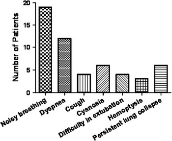
Between 2002 and 2008, a total of 1833 diagnostic fiberoptic bronchoscopies were performed, which identified 102 patients with tracheobronchial obstructions (incidence, 5.6%). After reviewing their radiographic image and clinical course, 41 patients, consisting of 22 boys (53.7%) and 19 girls (46.3%), with a median age of 17.4 months (range, 4 days to 15 years), were recruited for the study. All airway obstructions were confirmed to be due to cardiovascular compression by either computed tomographic or magnetic resonance imaging examination. In these children with cardiovascular-associated tracheobronchial obstruction, noisy breathing (19/41, 46.3%) and dyspnoea (12/41, 29.3%) were the most common indications for diagnostic bronchoscopy (Fig 1). Cyanosis (14.6%), persistent pulmonary atelectasis (14.6%), difficulty in extubation (9.8%), cough of unknown character (9.8%), and haemoptysis (7.3%) were other indications for bronchoscopy.
Indications for bronchoscopic assessment.

Cardiovascular assessment
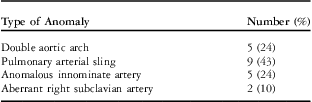
Patients were further categorised by cardiovascular diagnosis of anatomy-associated lesions (total 21 patients) or haemodynamics-associated lesions (total 20 patients). No significant difference in patient age was found between these two groups (anatomy-associated group, 11.12 plus or minus 3.0 months; haemodynamics-associated group, 24.06 plus or minus 10.48 months). Pulmonary arterial sling was the most common vascular anomaly among patients in the anatomy-associated group (9/21, 42.9%). The clinical spectrum of vascular anomalies of patients in this group has been summarised in Table 1.
Distribution of anatomy-associated lesions.

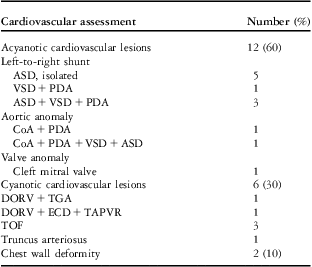
Of the patients in the haemodynamics-associated group, 12 (60%) had acyanotic cardiovascular lesions, 6 (30%) had cyanotic cardiovascular lesions, and 2 (10%) had airway compression due to vascular structures secondary to deformity of the thoracic cage. Septal defects with left-to-right shunts were the most common anomalies in the acyanotic cardiovascular group (5/12, 41.7%; Table 2). For cyanotic cardiovascular lesions, tetralogy of Fallot was the most common (3/6, 50%).
Cardiovascular assessment of haemodynamics-associated lesions.

ASD = atrial septal defect; CoA = coarctation of aorta; DORV = double outlet of right ventricle; ECD = endocardial cushion defect; PDA = patent ductus arteriosus; TAPVR = total anomalous pulmonary venous return; TGA = transposition of great arteries; TOF = tetralogy of Fallot; VSD = ventricular septal defect
Bronchoscopic and chest radiographic evaluation
On bronchoscopy, the co-existing anomalies seen were tracheal bronchus (six patients), laryngomalacia (four patients), vocal cord paralysis (one patient), pharyngomalacia (one patient), and vallecular cyst (one patient).
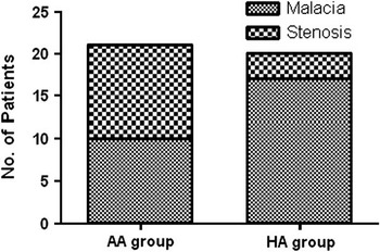
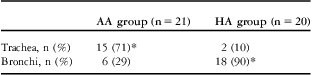
The bronchoscopic findings of airway obstruction of the anatomy-associated and haemodynamics-associated groups were compared. Malacia over the diseased airway was the most common finding in the haemodynamics-associated group (Fig 2; 17/20, p < 0.05, Fisher's exact test). However, malacia and stenosis were observed with equal frequency in the anatomy-associated group. Tracheal involvement was mostly found in all patients in the anatomy-associated group. In contrast, the bilateral main bronchi were most often involved in the haemodynamics group (Table 3).
Bronchoscopic findings in patients with cardiovascular-associated lower-airway obstruction. AA group = anatomy-associated group; HA group = hemodynamics-associated group.

Anatomical involvement distribution of cardiovascular-associated lower-airway obstruction.

AA group = anatomy-associated group; HA group = haemodynamics-associated group
*p < 0.0001 between two groups
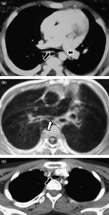
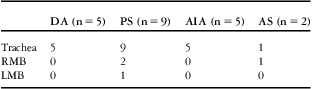
The chest radiographic images in the haemodynamics-associated group indicated that the main pulmonary artery and its distal branches were the major causes of airway compression (Fig 3a and b, Table 4). The ascending aortic arch and left atrium also contributed to large-airway obstruction. The aorta or left atrium often caused compression at the lower trachea or left main bronchus. However, the main pulmonary artery was equally involved in compression over the lower trachea and main bronchi. The right pulmonary artery and left pulmonary artery usually caused the ipsilateral compression of the main bronchus. In the anatomy-associated group, the trachea was exclusively involved in patients with double aortic arch or anomalous innominate artery (Fig 3c). In patients with a pulmonary arterial sling, lower tracheal obstruction was most common, but the involvement of both right and left main bronchi was occasionally seen (Table 5).
Tracheobronchial compression caused by haemodynamics-associated cardiovascular structures. (a) Chest computed tomographic scan of an infant with an atrial septal defect showing compression of the tracheal carina by right pulmonary arteries (long arrow) and compression of the left main bronchus by the left pulmonary artery (short arrow). (b) Chest magnetic resonance image of an infant with tetralogy of Fallot revealing compression of the left main bronchus by the prominent left pulmonary artery (arrow). (c) Chest computed tomographic scan of a child with abnormal thoracic configuration showing compression of the trachea by an innominant artery (arrow).

Cardiovascular structures involved in airway obstruction in the haemodynamics-associated group.

Ao = aorta; IA = innominate artery; LA = left atrium; LMB = left main bronchus; LPA = left pulmonary artery; MPA = main pulmonary artery; RMB = right main bronchus; RPA = right pulmonary artery
Cardiovascular structures involved in airway obstruction in the anatomy-associated group.

AIA = anomalous innominate artery; AS = aberrant right subclavian artery; DA = double aortic arch; LMB = left main bronchus; PS = pulmonary sling; RMB = right main bronchus
Discussion
Respiratory complaints such as dyspnoea and noisy respiration are commonly seen in children with cardiac or vascular abnormalities.Reference Lee, Cheung, Leung, Ng and Tsoi 1 , Reference Cerda, Chacon and Reichhard 2 , Reference Turner, Gavel and Coutts 6 – Reference Guillemaud, El-Hakim, Richards and Chauhan 8 In children with persistent noisy breathing – wheezing or stridor, recurrent respiratory distress, unexplained atelectasis, or extubation failure, the possibility of airway compression should be strongly suspected. Feeding difficulty may be found in patients with co-existing oesophageal compression, as some studies have noted.Reference Turner, Gavel and Coutts 6
The cardiovascular pathology resulting in compression of the paediatric airway can be divided into (1) congenital vascular anomalies that directly encroach and cause airway compression – for example, vascular rings; and (2) bulging cardiovascular structures, secondary to haemodynamic changes of primary anomalies, that compress the airway extrinsically. Among the anatomy-associated lesions, the double aortic arch is reported as the most commonly occurring symptomatic lesion.Reference Turner, Gavel and Coutts 6 , Reference Valletta, Pregarz, Bergamo-Andreis and Boner 7 Other causes of airway compression include anomalous innominate artery, right-sided aortic arch with aberrant subclavian artery, and left aortic arch with aberrant subclavian artery, in addition to left ductus arteriosus and pulmonary arterial sling. In this study, pulmonary arterial sling appeared most frequently in the anatomy-associated group. However, patients with a double aortic arch were not referred for bronchoscopic examination, possibly because of early echocardiographic diagnosis by a cardiologist.
Extrinsic compression of the airway due to acquired haemodynamic distortion is an important and often unrecognised cause of lower-airway obstruction. Adjacent pulmonary arteries, the aorta, and the left atrium can cause external compression as well. Consequently, cardiovascular lesions may be caused by displacement of normal structures secondary to congenital heart disease or thoracic cage anomalies.
Among patients with acyanotic cardiovascular disease with airway compression, left-to-right shunts were most commonly found. In contrast, tetralogy of Fallot was most often seen in cases of cyanotic lesions with airway obstruction. It was found that infants suffering from tetralogy of Fallot with absent pulmonary valve had aneurysmal dilatation of the pulmonary artery and its branches, which can cause compression of the tracheobronchial tree.Reference Lakier, Stanger, Heymann, Hoffman and Rudolph 9 , Reference Alsoufi, Williams and Hua 10 Partial tracheal compression due to dilated aorta was seen in two patients with double outlet of the right ventricle.
In our study, two patients with scoliosis and chest wall deformity also suffered from cardiovascular-associated airway obstruction. One child had compression of the right main bronchus by the right pulmonary artery, and another had compression of the trachea by the innominate artery. Abnormalities of chest wall structure, such as the spinal column and bony thorax, narrow the anteroposterior diameter of the mediastinum. Adjacent cardiovascular structures can then cause compression of the airway caliber.Reference Donnelly and Bisset 11 – Reference Kolilekas, Manali, Kalomenidis, Mouchtouri, Spyrou and Papiris 13
The pathology of great-vessel anomalies was conceptualised from the theoretical embryogenesis of the aortic arch and pulmonary artery.Reference Morrow and Huhta 14 Aberrant embryonic development of great vessels prompts the simultaneous aberrant development of the neighbouring airway. It may elucidate that stenosis was the major bronchoscopic finding among children in the anatomy-associated group, and tracheal involvement was most often found. However, in the haemodynamics-associated group, malacia was the predominant bronchoscopic finding. The events of airway compression caused by cardiovascular pathology may occur in the postnatal stage when airway embryogenesis has already been completed.
The location of airway compression depends on the site of the cardiovascular pathology. In anatomy-associated anomalies, different pathologic lesions are associated with unique bronchoscopic and radiographic findings.Reference Furuya, Vargas and Ramirez-Figueroa 15 Involvement of the upper trachea is always found in children with a double aortic arch. In pulmonary arterial sling lesions, the course of the left pulmonary artery usually results in compression of the lower trachea until the carina and right main bronchus, sometimes simultaneously with the left main bronchus.
The site of pulmonary artery branching is closely related to the involved airway segment. In our study, the main pulmonary artery, which was located just below the level of the carina, could cause compression of the tracheal carina and its main branches. The left pulmonary artery caused involvement of only the ipsilateral main bronchus. However, because of the anatomy of the mildly left-deviated main pulmonary artery,Reference Berlinger, Long, Foker and Lucas 16 the right pulmonary artery could cause compression of the right main bronchus and sometimes of the left main bronchus. Considering the anatomy of the aorta, aortic impression is just over the left lateral aspect of the lower trachea until the left main bronchus. Our study revealed that airway pathology originating from compression of the aorta was usually located in the lower trachea and left main bronchus. In this study, airway compression caused by the left atrium was usually located in the tracheal carina or left main bronchus.
In conclusion, stenosis or malacia of the tracheobronchial tree on bronchoscopy warrants further investigation for underlying cardiovascular anomalies. Malacia of the involved airway is the predominant bronchoscopic finding of secondary haemodynamics-associated anomalies. Primary anatomy-associated lesions are usually associated with tracheal airway obstruction. In contrast, secondary haemodynamics-associated lesions are associated with obstructions of the airway below the carina.