Introduction
Morphokinetic information provided by time-lapse microscopy (TLM) has been increasingly utilized in embryo selection over the last decade, despite insufficient evidence of a significant effect on live-birth rates (Pribenszky et al., Reference Pribenszky, Nilselid and Montag2017; Armstrong et al., Reference Armstrong, Bhide, Jordan, Pacey, Marjoribanks and Farquhar2019; Del Gallego et al., Reference Del Gallego, Remohi and Meseguer2019). A considerable advance in embryo selection efficacy is foreseen for the next years with the combination of morphological, metabolic and morphokinetic data by integrative systems using artificial intelligence (Curchoe and Bormann, Reference Curchoe and Bormann2019). Understanding the strengths, constraints and relative accuracy of different markers of embryonic developmental competence is of great relevance for the improvement of current methods and development of new strategies for embryo selection in reproductive medicine. In addition, more accurate approaches for embryo quality assessment could highly benefit basic studies on the mechanisms regulating embryo developmental competence by providing more solid parameters to measure it.
During the last decade, TLM technology has permitted a more detailed and dynamic assessment of in vitro embryo development, avoiding the risk of exposing embryos to uncontrolled atmospheric conditions for repeated microscopic examinations (Lemmen et al., Reference Lemmen, Agerholm and Ziebe2008). Several studies have assessed the relationship between TLM-derived morphokinetic parameters and IVF/ICSI outcomes. Faster fertilization kinetics was associated with better embryo development (Coticchio et al., Reference Coticchio, Mignini Renzini, Novara, Lain, De Ponti, Turchi, Fadini and Dal Canto2018), while faster cleavage kinetics, in addition to improved embryo development (Dal Canto et al., Reference Dal Canto, Coticchio, Mignini Renzini, De Ponti, Novara, Brambillasca, Comi and Fadini2012; Kirkegaard et al., Reference Kirkegaard, Kesmodel, Hindkjaer and Ingerslev2013; Motato et al., Reference Motato, de los Santos, Escriba, Ruiz, Remohi and Meseguer2016), was also associated with higher implantation (Meseguer et al., Reference Meseguer, Herrero, Tejera, Hilligsoe, Ramsing and Remohi2011; Chamayou et al., Reference Chamayou, Patrizio, Storaci, Tomaselli, Alecci, Ragolia, Crescenzo and Guglielmino2013; Basile et al., Reference Basile, Nogales Mdel, Bronet, Florensa, Riqueiros, Rodrigo, Garcia-Velasco and Meseguer2014) and pregnancy rates (Lemmen et al., Reference Lemmen, Agerholm and Ziebe2008; Meseguer et al., Reference Meseguer, Rubio, Cruz, Basile, Marcos and Requena2012; Rubio et al., Reference Rubio, Galan, Larreategui, Ayerdi, Bellver, Herrero and Meseguer2014). More recently, faster blastulation kinetics was demonstrated to be associated with higher live-birth rates (Fishel et al., Reference Fishel, Campbell, Montgomery, Smith, Nice, Duffy, Jenner, Berrisford, Kellam, Smith, Foad and Beccles2018).
Although several studies have suggested that TLM-based embryo selection may improve IVF outcomes (Pribenszky et al., Reference Pribenszky, Nilselid and Montag2017; Shebl et al., Reference Shebl, Haslinger, Kresic, Enengl, Reiter, Oppelt and Ebner2021), it has been argued that, as its accuracy varies between different settings and studies, its clinical relevance is yet to be confirmed (Armstrong et al., Reference Armstrong, Bhide, Jordan, Pacey, Marjoribanks and Farquhar2019; Del Gallego et al., Reference Del Gallego, Remohi and Meseguer2019). In fact, we have recently provided evidence that patient profile may affect the predictive performance of time-lapse information in ICSI cycles with embryo transfers performed on day 2/3 ET; fertilization and cleavage morphokinetic parameters were reached significantly earlier in embryos providing a live birth compared with those not providing a live birth in patients younger that 37 years, but not in patients aged 37 years or more (Dal Canto et al., Reference Dal Canto, Bartolacci, Turchi, Pignataro, Lain, De Ponti, Brigante, Mignini Renzini and Buratini2021). Nevertheless, a recent robust study assessing 1810 embryo transfers suggested that blastulation kinetics as assessed by time of blastulation start and duration constituted a superior predictor of live birth than classical morphology (Fishel et al., Reference Fishel, Campbell, Foad, Davies, Best, Davis, Smith, Duffy, Wheat, Montgomery, Wachter and Beccles2020). This is in contrast with a previous smaller study including 235 patients, in which the utilization of blastulation and cleavage morphokinetics information combined did not improve implantation or clinical pregnancy rates in relation to classical morphology (Goodman et al., Reference Goodman, Goldberg, Falcone, Austin and Desai2016).
Previous publications are also discrepant with regard to the potential prognostic value of TLM-derived pre-blastulation morphokinetic data. Notably, higher live-birth rates were observed with embryo selection based on cleavage morphokinetics compared with classical morphology in the first study published on this topic (Siristatidis et al., Reference Siristatidis, Komitopoulou, Makris, Sialakouma, Botzaki, Mastorakos, Salamalekis, Bettocchi and Palmer2015). In a subsequent study, earlier tPNf, t2 and t4 were associated with higher live-birth occurrence in a univariate analysis, although only t2 was considered useful to predict live birth in a model combining morphokinetic with morphological information (fragmentation grade) (Ahlstrom et al., Reference Ahlstrom, Park, Bergh, Selleskog and Lundin2016). In contrast, in a more recent study, no morphokinetic parameters encompassing fertilization and cleavage stages were associated with live birth in embryos previously selected by classical morphology (Barberet et al., Reference Barberet, Bruno, Valot, Antunes-Nunes, Jonval, Chammas, Choux, Ginod, Sagot, Soudry-Faure and Fauque2019). These discrepancies render the live-birth prognostic potential of fertilization and cleavage morphokinetic data still unclear.
In the face of the aforementioned gaps in the literature and aiming to shed light on the potential clinical value of TLM-derived morphokinetic information, we tested the hypothesis that early morphokinetic parameters encompassing fertilization and cleavage are more predictive of live birth than classical morphology. With this aim, we confronted the prognostic accuracy of morphological and morphokinetic data obtained from 1801 transferred embryos with a known live-birth outcome, utilizing a novel strategy including receiver operating characteristic (ROC) curves derived from morphological and morphokinetic scores, as well as a multivariate analysis assessing the relationship between different combinations of morphological and morphokinetic quality degrees with live-birth occurrence.
Materials and methods
Patients and design
This is a retrospective cohort study conducted at the Biogenesi Reproductive Medicine Centre, Monza, Italy, from 2015 to 2019. Eligible patients were those undergoing their first homologous ICSI cycle achieving a fresh embryo transfer (ET) after embryo culture in a time-lapse system. Here, 1801 embryos were produced and transferred fresh 3 days after fertilization in 1102 ET, 403 of which were in single transfers (SET) and 699 in double (DET) transfers. Only embryos providing a known live-birth outcome coupled with morphokinetic information were included in the study. Therefore, embryos transferred in DET resulting in a single live birth were excluded from the analysis, while embryos transferred in SET and in DET and resulting in single and twin live births, respectively, were included in the analysis as LB embryos (embryos achieving a live birth with trackable morphokinetics; n = 168). Conversely, embryos transferred in SET or DET resulting in a negative implantation diagnosis were included in the analysis as NLB embryos (embryos NOT achieving a live birth; n = 1633). It is important to point out, therefore, that LB embryos are not simply those resulting in a live birth, but those resulting in a live birth and allowing the identification of their morphokinetic information retrospectively. In our centre, as DETs are predominantly performed, a relatively small percentage of the embryos achieving a live birth has their morphokinetic information trackable. Embryos resulting in pregnancy losses between implantation and birth were excluded from the study.
To test the hypothesis that morphokinetic quality is more predictive of live birth than morphological quality, ROC curves based on morphokinetic or morphological quality scores were generated and the respective areas under the curve (AUC) were compared. Moreover, the association between live-birth achievement and 12 possible combinations of four morphokinetic quality degrees (A–D) with three morphological quality degrees (A–C) was assessed using multivariate analysis. All procedures and protocols were approved by the locally competent ethical committee (ASST, Monza).
Ovarian stimulation, ICSI and embryo culture
Ovarian stimulation and pituitary downregulation were induced with rFSH (Puregon, Merck Sharp & Dohme, Rome, Italy or Gonal-F, Merck, Rome, Italy) and GnRH antagonist (Ganirelix, Merck Sharp & Dohme, Rome, Italy), respectively. Hormone doses were decided and adjusted considering patients’ characteristics and treatment response. Oocyte maturation was triggered with 6500 IU hCG (Ovitrelle Merck, Rome, Italy) 36 h prior to oocyte pickup, when at least three follicles ≥ 17–18 mm were first detected using ultrasound monitoring. ICSI and embryo culture were carried out according to conventional methodology as previously described (Bartolacci et al., Reference Bartolacci, Buratini, Moutier, Guglielmo, Novara, Brambillasca, Renzini and Dal Canto2019). Briefly, after oocyte collection, cumulus–oocyte complexes were transferred into fertilization medium (Sequential Fert, Origio, Måløv, Denmark). Cumulus cells were removed by brief exposure to cumulase (80 U/ml; ICSI Cumulase, Origio, Måløv, Denmark), followed by mechanical action with the use of denuding plastic pipettes (Vitromed), at approximately 2–3 h after collection. ICSI was performed with oocytes held with the first polar body at the 12 o’clock position, and insertion of the injecting needle at the 3 o’clock position. Microinjected oocytes were transferred to Embryoslide™ Culture slides (Vitrolife, Göteborg, Sweden), into 30-μl microdrops of cleavage medium (Origio, Måløv, Denmark), and covered with paraffin oil. Embryos were cultured in an integrated embryo culture TLM system (EmbryoScope™ Time-lapse System; Vitrolife, Göteborg, Sweden), with image acquisition programmed for every 10 min at seven different focal planes for each embryo. All embryos were cultured in the same TLM device at identical culture conditions.
Embryo assessment
Conventional morphological embryo evaluation was performed during embryo culture without removing embryos from the TLM incubator. Fertilized oocytes were confirmed by the presence of two pronuclei at 16–18 h and cleavage was assessed 25 to 27 h post-ICSI. On day 3, embryos were classified according to blastomere number and size, degree of fragmentation and presence of multinucleation (ALPHA Scientists In Reproductive Medicine, 2011) in three morphological quality groups: A = top, B = intermediate and C = poor, which were then used to assess the association between live-birth achievement and 12 possible combinations of different morphological and morphokinetic quality degrees, as detailed below in ‘Statistical analysis’. Top quality embryos, defined as those with six to eight blastomeres, less than 10% fragmentation and no multinucleation, were primarily selected for transfer. Intermediate embryos also had six to eight blastomeres, but with 11–25% fragmentation, and poor quality embryos presented six to eight uneven blastomeres with fragmentation greater than 25%. Morphological scores relative to live-birth potential were generated using a training dataset, and were subsequently assigned to embryos of an external dataset for data validation, as detailed below in ‘Statistical analysis’.
Evaluation of time-lapse images
The EmbryoViewer image analysis software (Vitrolife, Göteborg, Sweden), was used to annotate the time of developmental events. Cleavage time was considered as the moment at which cell division was completed and the originating cells were completely segregated and invested by their respective cytoplasmic membranes, following the guidelines proposed by Ciray et al. (Reference Ciray, Campbell, Agerholm, Aguilar, Chamayou, Esbert, Sayed and Time Lapse User2014). Annotation of morphokinetic information was performed daily by different operators, according to the laboratory work shifts. All operators were trained by a senior embryologist and previously evaluated in their abilities to perform the analysis independently and accurately. Internal quality evaluation checks are routinely performed by a senior embryologist to ascertain annotation accuracy and homogeneity within the team. Annotations were performed daily and were all blinded in relation to clinical outcomes. The definitions of the timepoints assessed in the study were as follows: tPNf, time of pronuclei breakdown; t2, time at which the embryo presented two separate and distinct cells; t3, time at which a three-blastomeres embryo was achieved; t4, time at which a four-blastomeres embryo was achieved; t5, time at which a five-blastomeres embryo was achieved; t8, time at which an eight-blastomere embryo was achieved. Additionally, PN fading was used as an alternative start time to calculate time to t2 cell stage, i.e., t2-tPNf.
Embryo transfer
Embryo selection was exclusively based on morphological assessment as described above, and ET strategy was decided considering maternal age, couple history and embryo quality. Selected embryos were transferred fresh on the third day after fertilization in SET or DET according with the American Society for Reproductive Medicine guidelines (Practice Committee of American Society for Reproductive Medicine, 2013) with abdominal ultrasound guidance. Remaining embryos were further cultured or immediately frozen for eventual later transfers, but were not included in the study. Embryo implantation was diagnosed 12 days after ET with a βhCG test.
Statistical analysis
All continuous variables are presented as means ± standard deviation (SD) or medians and interquartile range (IQR). Patient variables and morphokinetic parameters of LB and NLB embryos were compared using the Mann–Whitney non-parametric test. Live-birth prediction models for morphokinetic and morphological scores were developed by splitting the data into training and validation datasets: 723 observations were utilized for model development (training dataset) and 1078 observations for model evaluation (validation dataset). The validation dataset was external and did not overlap with the training dataset. Live-birth predictive performances of embryo morphokinetics and morphology were assessed through ROC curves, generated by plotting true positive rates (sensitivity) against false-positive rates (1 − specificity) across all possible threshold values, comparing predicted confidence scores with actual live-birth outcome. The AUC was used to determine the discriminating performance of the model, varying from 0.5 (random prediction) to 1 (perfect discrimination) (Hosmer and Lemeshow, Reference Hosmer and Lemeshow2000).
A generalized linear model (GLM) was used to generate morphological scores based on live-birth predictive values of categorical variables, such as blastomere number and the degree of fragmentation. The logistic model revealed that blastomere number increased live-birth logit by 0.487 (SE = 0.086, P = 0.001), while fragmentation degree decreased it by 0.044 (SE = 0.018, P = 0.03). Morphological scores were obtained with the inverse function logit p = (a), p = exp (a)/[1+exp (a)], and ranged from of 1.2 to 25.3. These scores, obtained from each combination of blastomere number and fragmentation degree, were then used to generate ROC curves, from which AUCs were calculated to determine live-birth prediction performances of training (model development) and validation/external data, separately.
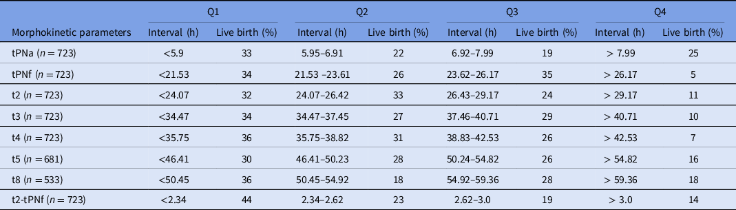
The same training dataset, previously used to generate the morphological scores, was used to create the morphokinetic scores. To obtain morphokinetic scores, time values were converted from continuous to categorical variables by dividing morphokinetic intervals in quartiles as previously described (Meseguer et al., Reference Meseguer, Herrero, Tejera, Hilligsoe, Ramsing and Remohi2011). Quartiles 1–4 represent time intervals at which the 25% fastest, 25% second fastest, 25% third fastest and 25% slowest embryos achieved the morphokinetic parameter, respectively. The percentage of embryos reaching a live birth was calculated for each quartile (Table 3), and for each morphokinetic parameter an optimal range was defined as that spanning the two consecutive quartiles with the highest accumulated live birth rate. A logistic regression analysis was used to select morphokinetic parameters expressed as binary variables (inside or outside the optimal range) according to their association with live birth. The selected parameters were t4 [OR = 2.10 (95% CI 1.10–4.23)] and t2-tPNf [OR = 1.86 (95% CI 1.18–2.94)], which were then used to create four morphokinetic scores of live-birth potential: embryos presenting t4 and t2-tPNf within the aforementioned optimal range were classified as A (top morphokinetics), while those presenting t4 within and t2-tPNf out of the optimal range were classified as B, those presenting t4 out and t2-tPNf within the optimal range were classified as C and those presenting both parameters out of the optimal range were classified as D (poor morphokinetics). Each individual embryo was classified according to these scores for the generation of the morphokinetics-based ROC curve in training and validation/external datasets, separately. In addition, the morphological and morphokinetic scores were also applied to generate ROC curves based on combined morphology/morphokinetics information, again utilizing both datasets (training and validation) separately.
The comparative predictive power of morphological and morphokinetic data was further investigated by comparing live-birth occurrence associated with the different combinations of three morphological quality groups and four morphokinetic quality groups described above (12 combined groups: AA, AB, AC, AD, BA, BB, BC, BD, CA, CB, CC, CD). A multivariate analysis was then performed using the GLM, to account for dependencies within patients due to transfer of different numbers of embryos. Maternal age, maternal body mass index (BMI), number of oocytes recovered, number of embryos transferred, and cause of infertility were included as variables potentially affecting live-birth occurrence in the GLM model, to adjust estimations of differences among the 12 groups. Association with live birth is expressed as odds ratio (OR) with 95% confidence interval (95% CI) in relation to the reference group (AA; top morphology/top morphokinetics).
Statistical power analysis was performed using the MedCalc Software (v.19.5.3) and revealed that the study has a power equal to 80% assuming a type I error of 0.05. Data analysis was carried out with the Statistical Package for Social Sciences (SPSS) v.21.0 (SPSS Inc., USA), and differences were considered statistically significant when P-values were < 0.05.
Results
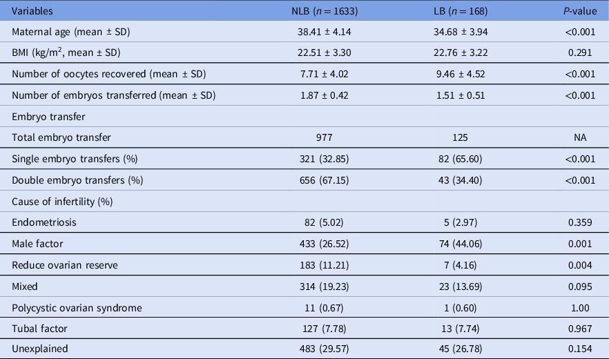
Patient and cycle characteristics for LB and NLB embryos are shown in Table 1. Maternal age and number of embryos transferred were lower, while number of oocytes recovered was higher in patients providing LB embryos compared with patients providing NLB embryos. No difference was observed with regard to maternal BMI between groups. Patients providing LB embryos had a higher percentage of single transfers and a higher incidence of infertility due to male factor, while the incidence of reduce ovarian reserve was higher in patient providing NLB embryos.
Patient characteristics according to ICSI outcome (LB: patients that produced embryos reaching a live birth; NLB: patients producing embryos that were transferred but did not implant)

NA, not applicable.
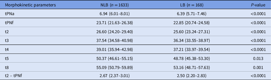
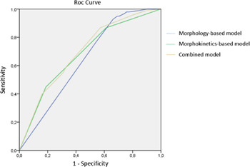
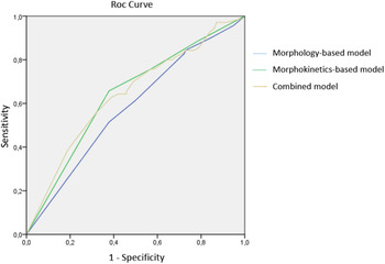
All morphokinetic parameters assessed were reached earlier in LB compared with NLB embryos (Table 2). Highest live-birth rates were observed in the first two consecutive temporal quartiles for all morphokinetic parameters, with the exception of tPNf, for which slightly higher live-birth rates were observed in the two central quartiles (Table 3). The morphokinetic parameters more strongly associated with live birth were t2-tPNf [OR = 1.86 (95% CI 1.18–2.94)] and t4 [OR = 2.10 (95% CI 1.10–4.23)], which were then utilized to create morphokinetic scores used to generate ROC curves predicting live-birth chance with an AUC = 0.69 (95% CI 0.63–0.75) with the training dataset for model development (Figure 1) and with an AUC = 0.64 (95% CI 0.58–0.70) with external validating data (Figure 2). The ROC curves based on morphological scores presented inferior live-birth predictive performances in comparison with those provided by morphokinetic scores [AUC = 0.64 (95% CI 0.59–0.69) with training data (Figure 1); AUC = 0.58 (95% CI 0.51–0.65) with external validating data (Figure 2)]. Finally, the ROC curve based on scores combining morphokinetic and morphological information achieved an AUC = 0.70 (95% CI 0.65–0.75) during model development with training data (Figure 1), and an AUC = 0.65 (95% CI 0.59–0.71) with external validating data (Figure 2), therefore holding a predictive live birth power slightly superior than that of the ROC curve exclusively based on morphokinetic scores.
Fertilization and cleavage morphokinetic parameters of embryos grouped according to ICSI outcome (LB: embryos reaching a live birth; NLB: embryos that were transferred but did not implant)

Data presented in hours [median and interquartile range (IQR)].
Distribution of live-birth rates in intervals corresponding to the four quartiles (Q1, Q2, Q3, Q4) for each of the morphokinetic parameters assessed on training data set

Live-birth prediction according to ROC curves derived from models based on morphokinetic [AUC = 0.69 (95% CI 0.63–0.75), P < 0.001], conventional morphology [AUC = 0.64 (95% CI 0.59–0.69), P < 0.01], and both information combined [AUC = 0.70 (95% CI 0.65–0.75), P < 0.001] utilizing a training dataset.

Live-birth prediction according to ROC curves derived from models based on morphokinetics [AUC = 0.64 (95% CI 0.58–0.70), P < 0.001], conventional morphology [AUC = 0.58 (95% CI 0.51–0.65), P < 0.01], and both information combined [AUC = 0.65 (95% CI 0.59–0.71), P < 0.001] utilizing a validation/external dataset not used for model development.

The multivariate analysis of groups of embryos with different combinations of morphokinetic quality (A–D; corresponding to the morphokinetic scores described above) and morphological quality (A–C; according to the Istanbul consensus; ALPHA Scientists In Reproductive Medicine, 2011) revealed that embryos with poor morphokinetics, regardless their morphological score (AD, BD and CD), presented significantly lower live-birth chance than embryos with top morphology and top morphokinetics (AA/reference group; adjusted P < 0.001, Table 4). Of 1164 embryos with top morphology, 444 (38.1%) also presented top morphokinetics, while 234 (20.1%) presented poor morphokinetics. Conversely, of 587 embryos with top morphokinetics, 444 (75.6%) also presented top morphology, while only 34 (5.8%) presented poor morphology.
Live-birth chance [OR (95%IC)] of embryos grouped according to their morphokinetic (A–D) and morphological (A–C) evaluation in relation to the reference group (AA; bold) providing top morphokinetics, top morphology and highest live-birth chance

*P < 0.05, **P < 0.01, ***P < 0.001 versus reference group (AA). OR, odds ratio.
Discussion
Despite the increasing application of TLM-derived morphokinetic information in embryo selection, its ability to improve ICSI/IVF outcomes remains questionable in the face of discrepant studies and varying accuracy reported by different clinics (Armstrong et al., Reference Armstrong, Bhide, Jordan, Pacey, Marjoribanks and Farquhar2019; Del Gallego et al., Reference Del Gallego, Remohi and Meseguer2019). The data presented here represent novel and solid indication that early morphokinetic parameters are more predictive of live birth following ET on day 3 than conventional morphology. Therefore, the present findings may serve as important references for the improvement of embryo selection methods and therefore ICSI/IVF success.
Earlier morphokinetic parameters from tPNa to t8 were associated with competence to provide a live birth in the present study. This is in line with numerous previous studies, either preceding or utilizing TLM technology, indicating that faster cleavage dynamics reflects higher embryo morphological quality (Shoukir et al., Reference Shoukir, Campana, Farley and Sakkas1997; Sakkas et al., Reference Sakkas, Shoukir, Chardonnens, Bianchi and Campana1998; Dal Canto et al., Reference Dal Canto, Coticchio, Mignini Renzini, De Ponti, Novara, Brambillasca, Comi and Fadini2012; Kirkegaard et al., Reference Kirkegaard, Kesmodel, Hindkjaer and Ingerslev2013; Motato et al., Reference Motato, de los Santos, Escriba, Ruiz, Remohi and Meseguer2016), as well as competence to reach implantation (Van Royen et al., Reference Van Royen, Mangelschots, De Neubourg, Laureys, Ryckaert and Gerris2001; Meseguer et al., Reference Meseguer, Herrero, Tejera, Hilligsoe, Ramsing and Remohi2011; Chamayou et al., Reference Chamayou, Patrizio, Storaci, Tomaselli, Alecci, Ragolia, Crescenzo and Guglielmino2013; Basile et al., Reference Basile, Nogales Mdel, Bronet, Florensa, Riqueiros, Rodrigo, Garcia-Velasco and Meseguer2014), pregnancy (Van Montfoort et al., Reference Van Montfoort, Dumoulin, Kester and Evers2004; Lemmen et al., Reference Lemmen, Agerholm and Ziebe2008; Meseguer et al., Reference Meseguer, Rubio, Cruz, Basile, Marcos and Requena2012; Rubio et al., Reference Rubio, Galan, Larreategui, Ayerdi, Bellver, Herrero and Meseguer2014) and live birth (Lundin et al., Reference Lundin, Bergh and Hardarson2001; Ahlstrom et al., Reference Ahlstrom, Park, Bergh, Selleskog and Lundin2016). More importantly, the ROC curves and respective AUC generated in this study indicate that morphokinetic parameters encompassing fertilization and cleavage hold superior live-birth predictive power than conventional morphology. This indication was reinforced by a multivariate analysis, in which a poor morphokinetic evaluation, regardless of embryo morphological quality, determined lower live-birth rates.
The present findings are in agreement with a previous study in which embryo selection based on cleavage parameters from t2 to t8 provided a live-birth rate higher than that resulting from embryo selection according to conventional morphology (Siristatidis et al., Reference Siristatidis, Komitopoulou, Makris, Sialakouma, Botzaki, Mastorakos, Salamalekis, Bettocchi and Palmer2015). In addition, the present data are also in line with another previous study in which earlier tPNf, t2 and t4 were associated with higher live-birth rate, although only t2 was found to be predictive of live birth (Ahlstrom et al., Reference Ahlstrom, Park, Bergh, Selleskog and Lundin2016). Conversely, our results disagree with a recent study, in which neither fertilization nor cleavage parameters were associated with live birth following single transfers of embryos morphologically selected (Barberet et al., Reference Barberet, Bruno, Valot, Antunes-Nunes, Jonval, Chammas, Choux, Ginod, Sagot, Soudry-Faure and Fauque2019). This discrepancy might be related to the different number of embryos assessed [232 in Barberet et al. (Reference Barberet, Bruno, Valot, Antunes-Nunes, Jonval, Chammas, Choux, Ginod, Sagot, Soudry-Faure and Fauque2019) versus 1801 embryos in the present study], different embryo/patient population profiles and/or variation in stringency levels of morphological selection between studies.
A slightly higher live-birth predictive performance was observed when morphokinetic and morphological scores were combined in the same model in the present study. The relatively limited contribution of morphological information is consistent with the distribution and live-birth rates of embryos classified according to morphokinetic and morphological scores simultaneously (Table 4). The present data suggest that embryo selection based on morphokinetics automatically promotes selection for morphology. Conversely, selection according to morphology does not appear to select for morphokinetics as efficaciously. Notably, in the present study, only 38.1% of the embryos presenting top morphology also presented top morphokinetics, whereas 75.6% of the embryos presenting top morphokinetics also presented top morphology. Interestingly, conversely, while only 5.8% of the embryos presenting top morphokinetics presented poor morphology, 20.1% of the embryos presenting top morphology presented poor morphokinetics, which was associated with a drastically reduced live-birth rate. This observation holds relevant clinical value as it points to a high chance of an incorrect choice (approximately one out of five embryos could be mis-selected), when embryo selection is exclusively based on conventional morphology. Furthermore, this is in agreement with our finding that embryos with top morphokinetics but poor morphology, although not common, hold greater chances to achieve a live birth than embryos with top morphology and poor morphokinetics.
A higher degree of subjectivity in the assessment of morphological compared with morphokinetic parameters may have accounted for the superior predictive performance of time-lapse data in the present study. This interpretation is supported by previous studies that assessed interoperator annotation homogeneity of morphokinetic and morphological parameters. While high interoperator homogeneity has been consistently reported for morphokinetic parameters (Sundvall et al., Reference Sundvall, Ingerslev, Breth Knudsen and Kirkegaard2013; Martinez et al., Reference Martinez, Santalo, Rodriguez and Vassena2018), for morphological parameters, except for a first report suggesting good reproducibility (Arce et al., Reference Arce, Ziebe, Lundin, Janssens, Helmgaard and Sorensen2006), the following studies converged to indicate a relevant degree of interoperator annotation variability, and are likely to reflect the high intrinsic subjectivity of the morphological analysis (Baxter Bendus et al., Reference Baxter Bendus, Mayer, Shipley and Catherino2006; Paternot et al., Reference Paternot, Wetzels, Thonon, Vansteenbrugge, Willemen, Devroe, Debrock, D'Hooghe and Spiessens2011; Storr et al., Reference Storr, Venetis, Cooke, Kilani and Ledger2017).
We acknowledge that our study is limited by its retrospective nature and by the utilization of data generated in a single IVF centre. Therefore, the present results might be influenced by local patient/embryo profiles and embryo culture methodology. Nevertheless, the number of transferred embryos assessed and the utilization of live birth as the primary endpoint are major strengths of this study and support its clinical relevance. Our findings may also be useful for clinics not utilizing TLM technology by providing references for the incorporation of morphokinetic information obtained by regular microscopy in embryo assessment. More specifically, the data reported here may serve as valuable parameters to choose the best possible time points for embryo examination to obtain relevant morphokinetic information while assessing embryo morphology.
In conclusion our study provides novel evidence that TLM-derived morphokinetic information encompassing fertilization and cleavage holds higher accuracy in determining competence to achieve a live birth than conventional morphology. In addition, the present data alert the relatively high risk of unsuccessful post-ICSI embryo selection when the strategy is exclusively based on classical morphology. Therefore, our study provides valuable parameters to improve embryo selection in ICSI/IVF practice, as well as embryo quality assessment in studies on mechanisms regulating embryo developmental competence.
Acknowledgements
The authors thank the Biogenesi Reproductive Center embryologists team for technical support during data generation and Stefano Castellano for assistance in manuscript formatting.
Financial support
This research did not receive any specific grant from any funding agency in the public, commercial or not-for-profit sector.
Conflict of interest
The authors declare no conflict of interest.
Ethical approval
All procedures performed in studies involving human participants were in accordance with the ethical standards of the institutional and/or national research committee and with the 1964 Helsinki declaration and its later amendments or comparable ethical standards.