

Introduction
The consumption of polyphenols derived from vegetables, fruits and cereals has been associated with numerous beneficial disease prevention effects. In some cases, however, the solubility and bioaccessibility of the polyphenols may be a challenge to the delivery of these components in food matrices. In this study, the possible use of high pressure as a strategy to increase the encapsulation of hydrophobic molecules within protein particles was assessed, using casein micelles and curcumin as model systems. Curcumin is a bis- α, β-unsaturated β-diketone (Anand et al. Reference Anand, Kunnumakkara, Newman and Aggarwal2007) present in the spice turmeric (Sahu et al. Reference Sahu, Kasoju and Bora2008), and has shown a number of anti-proliferative, anti-microbial, anti-inflammatory, and antioxidant properties (Sharma et al. Reference Sharma, Gescher and Steward2005; Maheshwari et al. Reference Maheshwari, Singh, Gaddipati and Srimal2006).
The major challenge for curcumin application in food and cosmetics is its poor solubility in aqueous solutions, as this limits its clinical efficacy and cellular uptake (Anand et al. Reference Anand, Kunnumakkara, Newman and Aggarwal2007; Yu & Huang, Reference Yu and Huang2011). Several methods have been designed to increase the solubility and bioavailability of curcumin, including complexation with proteins, maltodextrins or lipophilic compounds (see, for example: Reddy et al. Reference Reddy, Sudharshan, Rao and Lokesh1999; Manju & Sreenivasan, Reference Manju and Sreenivasan2011; Wu et al. Reference Wu, Xu, Huang and Wen2011). Most of these studies analyse the formation of protein-ligand complexes assuming equilibrium between the free ligand (present in solution at low concentrations) and protein-bound ligand (typically being the vast majority of these species in the system).
Casein micelles have been suggested as applicable carriers for the delivery of curcumin in cell culture models (Sahu et al. Reference Sahu, Kasoju and Bora2008). Casein micelles are stabilised by a polyelectrolyte layer of κ-casein which imparts steric stabilisation (Holt & Horne, Reference Holt and Horne1996), and have a porous structure wherein molecules may be able to diffuse, and many clefts which greatly increase the surface area available for interactions with small molecules (Dalgleish, Reference Dalgleish2011).
It has been previously shown that casein proteins are avid binders of polyphenols (Aguie-Beghin et al. Reference Aguie-Beghin, Sausse, Meudec, Cheynier and Douillard2008; Yuksel et al. Reference Yuksel, Avci and Erdem2010) and could be an ideal carrier for delivery of molecules such as small tannins, curcumin, and vitamins (Livney & Dalgleish, Reference Livney and Dalgleish2007; Sahu et al. Reference Sahu, Kasoju and Bora2008; Shukla et al. Reference Shukla, Narayanan and Zanchi2009). The binding properties of curcumin to casein micelles have been characterized by fluorescence spectroscopy (Sahu et al. Reference Sahu, Kasoju and Bora2008; Rahimi Yazdi & Corredig, Reference Rahimi Yazdi and Corredig2012), but further studies are required to assess the effect of processing on the ability of casein micelles to associate with hydrophobic molecules. The present study stems from the idea that high hydrostatic pressure, by causing rearrangements to the proteins and to the supramolecular structure of the micelles, may improve the binding of curcumin to the casein micelles.
Considerable information is available on the effects of static high pressure treatment on milk proteins (Gaucheron et al. Reference Gaucheron, Famelart, Mariette, Raulot, Michel and Le Graet1997; Gebhardt et al. Reference Gebhardt, Doster, Friedrich and Kulozik2006; Huppertz et al. Reference Huppertz, Fox, de Kruif and Kelly2006a). High pressure causes denaturation of the two main serum proteins, α-lactalbumin and β-lactoglobulin, the extent of modification being affected by treatment time, temperature and pH (Needs et al. Reference Needs, Capellas, Bland, Manoj, Macdougal and Paul2000; Huppertz et al. Reference Huppertz, Fox, de Kruif and Kelly2006a). High pressure processing also affects the supramolecular structure of the casein micelles, due to the flexible nature of the structure of the casein proteins. The micelles can be disrupted through solubilization of the colloidal calcium phosphate and the decrease of protein–protein interactions (Gebhardt et al. Reference Gebhardt, Doster, Friedrich and Kulozik2006; Huppertz et al. Reference Huppertz, Kelly and de Kruif2006b). A significant decrease in the size of casein micelles has been shown to occur at pressures between 100–600 MPa and temperatures between 10–40 °C, along with the formation of small micellar fragments during the process (Gaucheron et al. Reference Gaucheron, Famelart, Mariette, Raulot, Michel and Le Graet1997; Anema et al. Reference Anema, Lowe and Stockmann2005a; Gebhardt et al. Reference Gebhardt, Doster, Friedrich and Kulozik2006). These structural changes have been exploited also to incorporate lipid-soluble vitamins into casein micelles (Menéndez-Aguirre et al. Reference Menéndez-Aguirre, Stuetz, Grune, Kessler, Weiss and Hinrichs2011).
The objective of this research was to study the effect of high pressure on the affinity of casein micelles for curcumin, this molecule is regarded as the epitome of lipid-soluble bioactives. Increasing affinity would result in an increase of the capacity of the micelles as nano-delivery systems for this class of bioactives. To determine the details of the binding between curcumin and milk proteins, fluorescence spectroscopy was employed, using both direct steady-state fluorescence, and tryptophan quenching.
Materials and methods
Materials
Chemicals were from Sigma (St Louis, MO, USA) unless otherwise indicated. Ultrapure water was prepared with a filtration system (Millipore, Billerica, MA, USA). Curcumin (⩾94% (curcuminoid content), ⩾80% curcumin) was purchased from Sigma-Aldrich (Sigma-Aldrich Canada Ltd., Oakville, Ontario, Canada). Stock solutions of curcumin (1 mm, 100 and 10 μm) were prepared in ethyl alcohol (96%), and the actual curcumin concentration was checked at 420 nm by spectrophotometry.
Sample preparation
Fresh pasteurized skim milk was purchased from Centrale del Latte di Milano (Granarolo, Bologna, Italy). This milk contained 3·1% protein. Sodium azide (0·02% w/v) was immediately added to prevent bacterial growth. Milk permeate was prepared using an Amicon ultrafiltration stirred cell unit (Model 8200, 200 ml, Millipore), equipped with a Ultra cell YM-10 ultrafiltration membrane (10 kDa nominal cut-off and a nominal area of 0·0064 m2) under nitrogen at 4 °C.
In fluorescence experiments, curcumin was added to milk at concentrations ranging from 0·5 to 50 μm. The high concentrations representing saturating ligand concentrations (Rahimi Yazdi & Corredig, 2011). In some experiments, curcumin was added to milk at a concentration of 100 μm prior to high pressure treatment. To minimize the effect of ethanol (O'Connell et al. Reference O'Connell, Saracino, Huppertz, Uniake, de Kruif, Kelly and Fox2006), the maximum concentration of ethanol was kept below 5%.
High hydrostatic pressure treatment
Patented FoodSaver® Bags (Model# T010-00034-098, FoodSaver, Jarden Corporation, Rye, NY, USA) were filled with skimmed milk in the presence/absence of 100 μm curcumin, vacuum packaged, and sealed in a sealing system (Model CGM Matic, GANDUS Company, Milan, Italy). Hyperbaric treatment was carried out on the sealed bags at SSICA in a QFP 35L-600, (Avure Technologies, Franklin, TN, USA). The system has a 35 litres useful volume and can reach a pressure of 600 MPa. The compression times to reach the maximum pressure starting with an empty chamber are around 2 min, while decompression is practically instantaneous. The system permits the execution of treatments in adiabatic conditions under controlled temperatures of between 4 to 90 °C, for the studies using three different pressures: 200, 400, 600 MPa. The treatment time was 20 min and the temperature was 20 °C.
SDS-PAGE
Sodium dodecyl sulphate polyacrylamide gel electrophoresis (SDS-PAGE) was performed on 12% acrylamide gels, run in a Bio-Rad (Hercules, CA, USA) Mini protean III apparatus, at 16 mA for each gel. About 0·01 mg protein were loaded in each sample lane, and molecular weight markers were from GE Healthcare (Montreal, Canada).
Fluorescence spectroscopy
Fluorescence measurements were carried out in a Perkin Elmer LS50B spectrometer (Perkin Elmer, Waltham, MA, USA). Control skim milk and pressurized milk were diluted (1 : 20) in milk permeate, to maintain the chemical environment as close as possible to that of the original milk.
Direct fluorescence measurements were performed by adding curcumin (0·1 to 20 μm, final concentration) to milk, as done in previous work (Rahimi Yazdi & Corredig Reference Rahimi Yazdi and Corredig2012). To minimize the addition of ethanol during titration, different stock solutions were used (10 μm, 100 μm, and 1 mm), so that volume change in the cuvette never exceeded 2%. Binding parameters were calculated according to the Benesi-Hildebrand equation (Benesi & Hildebrand, Reference Benesi and Hildebrand1949). To determine whether the addition of curcumin to milk samples before high pressure would increase binding with the milk proteins, the fluorescence intensity of milk samples treated at high pressure in the presence of 100 μm curcumin was measured, and the fluorescence intensity was compared with the values of milk samples with curcumin added after high pressure treatment. The release of protein-bound curcumin upon storage was determined by measuring fluorescence intensity in appropriate samples with curcumin, immediately after the treatment and after one week of storage at 4 °C in the dark.
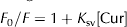
To further characterise the binding affinity of curcumin to milk proteins, fluorescence quenching was also employed, as it is a powerful technique fluorescence of proteins by curcumin was measured at increasing curcumin concentrations (0–50 μm) in the presence of variously treated milk samples diluted in milk permeate (1:20). In these measurements, samples were excited at 280 nm and emission spectra were collected from 300 to 450 nm. Quenching of tryptophan were made at 25 °C and repeated three times. In the quenching studies, controls were separately considered for each measurement. A linear dependence of quenching on the quencher concentration may be described using the Stern–Volmer equation (Lakowicz, Reference Lakowicz2006),
 $$F_0 /F = {\rm 1} + K_{{\rm sv}} [{\rm Cur}]$$
where F 0 is the fluorescence intensity of individual control samples, F is the fluorescence intensity after addition of the quencher, [Cur] is the concentration of curcumin, and K sv is the Stern–Volmer quenching constant (K sv−1 corresponds to the concentration of curcumin at which 50% of the peak intensity is reduced).
For high pressure-treated samples, the quenching dependence on the concentration of curcumin was no longer linear, but showed an exponential trend, usually suggest the simultaneous occurrence of static and dynamic quenching, that may be described by a modified form of the Stern–Volmer equation,
 $$F_0 = F \times {\rm e}^{(K_{\rm app}{\rm [Cur]})} $$
where K app is the apparent static quenching constant.
Statistical analysis
All assays were performed in triplicate. Statistical differences were evaluated using analysis of variance (ANOVA), and mean values were compared by using a Tukey test. All statistics were performed by using S-PLUS software (Insightful Corp., TIBCO Software Inc., Palo Alto, CA, USA).
Results and discussion
Direct fluorescence
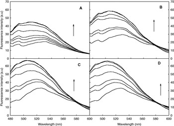
Changes in the fluorescence emission spectra of skim milk control and high pressure treated milk after addition of curcumin, at concentration gradually increasing from 0·1 to 20 μm, are shown in Fig. 1. It has to be noted that addition of curcumin in the absence of milk proteins results in negligible fluorescence increase or in curcumin precipitation at the highest curcumin concentrations. The increase in curcumin fluorescence intensity upon curcumin addition was higher in pressure treated milk, and the position of the emission maximum in these samples showed a marked blue shift. This shift has been previously reported and it has been attributed to an increased hydrophobicity of the chemical environment of curcumin (Sahu et al. Reference Sahu, Kasoju and Bora2008; Rahimi Yazdi & Corredig, 2011). Along the same lines, it has also to be noted that the addition of curcumin in the absence of physical treatments did not alter the average apparent diameter of casein micelles, that remained at 160 nm as measured by dynamic light scattering (not shown).
Emission spectra for untreated skim milk (A); and for skim milk treated by high pressure at 200 MPa (B); 400 MPa (C); 600 MPa (D). The samples were diluted in permeate (1:20) and curcumin was added (1·0, 1·5, 3·0, 5·0, 8·0 and 10·0 μm). Arrow indicates increasing concentrations of curcumin.

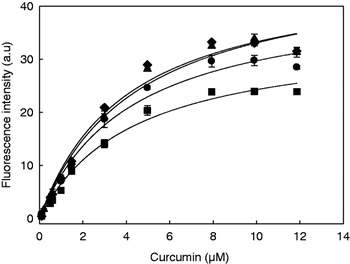
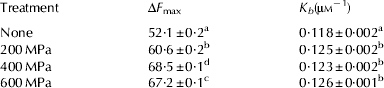
Figure 2 analyses the fluorescence increase in the various samples as a function of added curcumin. By assuming equilibrium between limited amounts of free curcumin and a curcumin–protein complex, these changes may be quantitatively analysed by using the Benesi & Hildebrand (Reference Benesi and Hildebrand1949). It is then possible to calculate the fluorescence value at saturating ligand (F max) and the apparent binding constant K b. As summarized in Table 1, the lowest F max was found in skim milk control, and this parameter was significantly higher in all pressure-treated milk samples. However, the data in Fig. 2 and Table 1 indicate that rearrangements occurring above a given pressure threshold (400 MPa) and the reported reduction in the size of the casein micelles under these conditions (Gebhardt et al. Reference Gebhardt, Doster, Friedrich and Kulozik2006) did not improve binding of curcumin to protein hydrophobic sites. The values of K b increased in the pressure-treated samples, but the observed increase was independent of the treatment intensity.
Changes in fluorescence intensity (measured at λem.520 nm) as a function of concentration of curcumin for untreated skim milk (■), and for skim milk treated at 200 MPa (●), 400 MPa (⧫), and 600 MPa (▴). Values are the average of 3 replicate experiments, bars represent standard deviation. Lines are drawn to guide the eye.

Binding parameters for curcumin calculated from direct fluorescence experiments. Apparent binding constant (K b) and maximum increase in fluorescence intensity (ΔF max) were derived from titration experiments and calculated according to Benesi & Hildebrand (1949). Results are the average of data obtained on three independent milk batches. Within a column, different superscript letter indicate statistical difference at P < 0·05
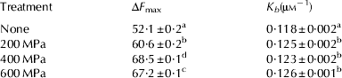
It is worth noting that the increase in the binding capacity of milk proteins due to high pressure treatment was much higher (∼30% increase in F max) than that reported for heat-treated skim milk (<2%, according to Rahimi Yazdi & Corredig, 2011). In addition, in heat treated milk the K b increased markedly (Rahimi Yazdi & Corredig, 2011), whereas only a ∼4% increase in K b was evident in high pressure treated milk (Table 1).
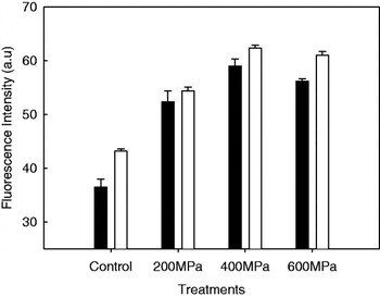
To evaluate whether curcumin could be efficiently associated by some ‘transient’ protein species formed during pressure treatment, milk samples were pressure treated in the presence of 100 μm curcumin, and the fluorescence of these samples was compared with that measured on samples where 100 μm curcumin was added to milk after the pressure treatment. Appropriate controls were also set up, being represented by milk treated at high pressure in the absence of curcumin, and by untreated milk containing 100 μm curcumin. Results are shown in Fig. 3, and confirm the higher binding capacity of the pressure-treated milk. There was a modest increase of binding when curcumin was incorporated during high pressure processing, suggesting that the (transient) rearrangements occurring during high pressure treatment may favour curcumin incorporation. Once again, pressures above 200 MPa seemed to be more effective, most probably because of the modest extent of rearrangements in the casein micelles at this pressure (Huppertz et al. Reference Huppertz, Fox and Kelly2004). Given the fact that we used skim milk in this study for its practical relevance, we did not consider here other parameters that reportedly affect casein micelle behaviour at high pressure (Merel-Rausch et al. Reference Merel-Rausch, Kulozik and Hinrichs2007).
Values of curcumin fluorescence intensity for milk samples. The concentration of curcumin was 100 μm in all cases. Curcumin was added before (empty bars) or after (solid bars) pressure treatment. Controls: pressure treated milk with no curcumin (solid bar) and untreated milk in the presence of 100 μm curcumin (empty bar). Samples were diluted 1:20 in permeate immediately before analysis.

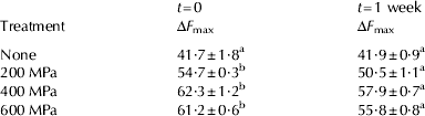
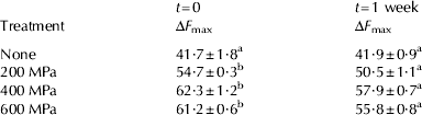
The stability of the emission spectra of milk samples treated by high pressure in the presence of 100 μm curcumin was determined by comparing the spectroscopic results obtained immediately after the treatment with those after 1 week storage at 4 °C in the dark. The maximum fluorescence intensity in untreated milk was unchanged after 1 week of storage (Table 2), whereas it decreased (about 8%) in all pressure treated samples almost regardless of treatment intensity.
Storage dependent changes in fluorescence intensity. Where indicated, milk samples were treated at high pressure in the presence of 0·1 mm curcumin, and curcumin fluorescence was measured immediately or after one week of storage. Results are the average of three independent milk batches. Within a row, different superscript letter indicate statistical difference at P < 0·05
The change in maximum fluorescence during storage may be attributed to rearrangements occurring to casein micelles after high pressure treatment, as already reported (Huppertz et al. Reference Huppertz, Kelly and de Kruif2006b). SDS–PAGE electrophoresis gave no indication of proteolysis during storage of the HP treated samples as a possible consequence of microbial contamination (data not shown). Therefore, slow structural changes in the binding properties of the involved proteins were the cause of the changes observed here.
Quenching of tryptophan fluorescence
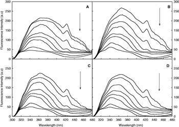
Binding of ligands to proteins is often accompanied by changes in the fluorescence properties of intrinsic protein fluorophores, most often resulting in geometry-sensitive quenching. Progressive curcumin addition led to a gradual decrease in the intensity of the tryptophan fluorescence. This may be appreciated from the spectra in Fig. 4, that also indicate how ‘buried’ tryptophan residues (i.e., those emitting at the shortest wavelengths) were the ones least sensitive to addition of the quencher, regardless of the presence and intensity of the pressure treatment.
Tryptophan fluorescence spectra for untreated skim milk (A); and for skim milk treated by high pressure at 200 MPa (B); 400 MPa (C); 600 MPa (D). Samples were diluted in permeate (1:20) and curcumin was added at 2·5, 5·0, 10·0, 15·0, 20·0 and 45 μm concentration. Arrow indicates increasing concentrations of curcumin.

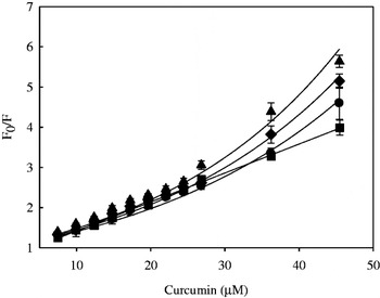
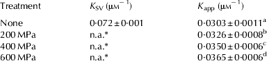
A further analysis of the quenching effects in the various samples is presented in Fig. 5, showing the variation of F 0/F for the various samples as a function of added curcumin. In the case of skim milk there was a linear relation between F 0/F and curcumin concentration. An upward curvature was instead evident in pressurized milk, suggesting the presence of multiple binding sites – characterized by a different quenching mechanism – after pressure treatment (Lakowicz, Reference Lakowicz2006). A similar effect was observed in heat-treated milk (Rahimi Yazdi & Corredig, 2011). An apparent binding constant for curcumin may be calculated from the quenching data by using the appropriate form of the Stern–Volmer equation (Lakowicz, Reference Lakowicz2006). Although these data (Table 3) cannot be directly compared with those obtained through direct titration (see Table 1), they indicate that the formation of multiple binding sites with a characteristic quenching behaviour is a pressure-dependent event.
Stern–Volmer plot for skim milk (■), 200 MPa (●), 400 MPa (⧫), and 600 MPa (▴) re-diluted in permeate. Values are the average of 3 replicate experiments, bars represent standard deviation. Lines are drawn to guide the eye.

Stern–Volmer constants (K SV and K app) derived from quenching experiments. Results are the average of three independent milk batches. Within a column, different superscript letter indicate statistical difference at P < 0·05
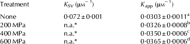
n.a.* not applicable: quenching dependence is not linear (see Fig. 5)
According to Huppertz et al. (Reference Huppertz, Kelly and de Kruif2006b), high pressure dissociates casein micelles by initiating the solubilisation of the colloidal calcium phosphate, with simultaneous release of αS1-, αS2-, β- and κ-caseins. The size reduction of the micelles may be the reason for the increased maximum fluorescence intensity measured in milk samples treated with high pressure and of the change in the quenching pattern discussed above, as smaller micelles would have an increased number of sites available for interaction with curcumin. In particular, between 100 and 300 MPa the size distribution pattern changes from a monomodal to bimodal (Knudsen & Skibsted, Reference Knudsen and Skibsted2010). In general, dissociation of casein micelles and their size reduction increases the surface hydrophobicity of the particles, as demonstrated by increased binding of the fluorescent hydrophobic probe ANS (Gaucheron et al. Reference Gaucheron, Famelart, Mariette, Raulot, Michel and Le Graet1997).
Other than these macroscopic effects, quenching data indicated that the persistent structural modification induced by high-pressure treatment result in a rearrangement of residues, such as tryptophan, that may be considered as internal markers of the hydrophobic regions of the proteins. The treatment-dependent prevalence of dynamic quenching in pressure-treated milk as opposed to that of static quenching in untreated milk suggests that tryptophan and curcumin do not actually form a ground-state complex in the treated proteins, but curcumin must in this case be adjacent to the tryptophan at the moment of excitation.
In the frame of quenching studies it has to be noted that the quenching constants reported here for untreated skim milk were statistically identical to those measured on casein micelles isolated by ultracentrifugation (not shown). This preliminary observation suggests that whey proteins only play a marginal role in curcumin binding, at least in the absence of physical modification of the structure of milk proteins.
Conclusions
High Hydrostatic Pressure increases the binding of curcumin to skim milk proteins by increasing the protein hydrophobicity. The structural modifications responsible for increased curcumin binding by pressure treated milk are different (and to some extent more effective) than those reported for heat treatment (Rahimi Yazdi & Corredig, Reference Rahimi Yazdi and Corredig2012). Binding could be further improved by pressure treatment of milk in the presence of curcumin, although in this case rearrangements occurred during storage after high pressure treatment that resulted in significant release of the bound curcumin.
Tryptophan quenching data indicate that high pressure treatment facilitates the access of curcumin to suitable binding regions in the involved proteins. The role played by pressure-induced dissociation of caseins remain to be ascertained, as is the role of whey proteins, that are however much less abundant than casein in skim milk. Whey proteins may contribute additional binding sites in their partially denatured form (Anema et al. Reference Anema, Stockmann and Lowe2005b) or when present as transiently modified species during the treatment (Lozinsky et al. Reference Lozinsky, Iametti, Barbiroli, Likhtenshtein, Kálai, Hideg and Bonomi2006), but they may also hinder the access of the ligand to casein micelles when treatments modify either or both these protein systems.
The results reported here and in previous work on skim milk highlight the possible practical relevance of our results when it comes to increasing the bioactive ‘payload’ in milk-based systems for the delivery of nutrients, pharmaceuticals, and nutraceuticals. Also, the approaches reported here seem to be flexible and robust enough to be useful in further mechanistic investigation on these systems.
The authors would like to thank the Natural Sciences and Engineering Council of Canada as well as the Ontario Graduate Scholarship Fund for partly funding this work.