Implications
Soy isoflavones are constituents of soybeans used widely in human food and livestock nutrition. However, the effects of soy isoflavones on metabolism or endocrine function in skeletal muscle or adipose tissue have received very little attention. The results presented here may be useful not only for application to animal nutrition but also for understanding the effects of soy foods on the health of human beings, especially the effects of soy-based formulas on the development of human infants.
Introduction
Soy components contain protein, lipids, fiber and phytochemicals including isoflavones (genistein and daidzein), which are also phytoestrogens (Mesiano et al., Reference Mesiano, Katz, Lee and Jaffe1999). The first report of the association between soybeans and disease control (glycemic) was in the year of 1910 (Friedenwald and Ruhrah, Reference Friedenwald and Ruhrah1910). Recently, there has been an increasing interest in the potential role of soy isoflavones in the treatment of chronic diseases. Studies in humans and animals have shown that the consumption of soybean or soybean-derived products may help to protect against cardiovascular diseases, certain types of cancer, obesity and diabetes (Bhathena and Velasquez, Reference Bhathena and Velasquez2002). The ability of isoflavones to prevent these diseases depends on their absorption, metabolism, distribution to target tissues and excretion of waste compounds, that is, their bioavailability. However, the underlying mechanism of the effects of isoflavones on metabolism or endocrine function in the skeletal muscle or adipose tissue has not been unraveled.
The similarities in morphology and physiology between human and pig organs make pig an appropriate animal model for clinical studies. The Chinese Guangxi minipig is a well-known indigenous breed that has been used for basic research (Zhang et al., Reference Zhang, Yu, Li, Li, Zeng, Cheng and Bu2004; Wang et al., Reference Wang, Gu, Tang, Geng, Fan, Li, Chu, Shi, Huang, Zhang and Yin2009) due to its small size and biological characteristics. Thus, the minipig model may be one of the best choices for research on soybean isoflavones.
In this study, we sought to determine whether soy isoflavones affect changes in body or muscle and adipose tissue weights, as well as insulin-like growth factor I (IGF-I) and peroxisome proliferator-activated receptor-γ (PPARγ) expression profile in muscle or adipose tissue in Chinese Guangxi minipigs.
Material and methods
Animals and diets
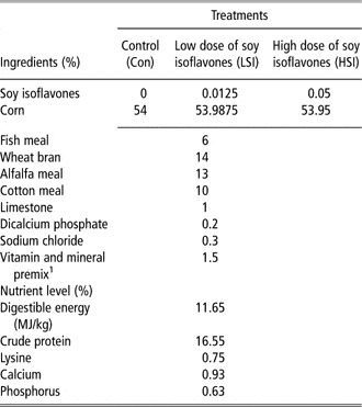
This study was conducted according to the guidelines of the Animal Care and Use Committee of the Institute of Subtropical Agriculture, the Chinese Academy of Science (Yin et al., Reference Yin, Yao, Liu, Gong, Ruan, Deng, Tan, Liu and Wu2010). A total of 72 male Chinese Guangxi minipigs weaned at 21 days (2.50 ± 0.54 kg) were assigned into three groups in a randomized complete-block design based on BW and litter, with six replicates per group and four piglets per replicate (Yao et al., 2008). The piglets in the three groups were fed the following diets: basal diet (control, Con), 125 mg/kg soy isoflavones (as soy isoflavone extracts; Wang Zhong Hua Biotechnology, Hunan, China; low dose of soy isoflavones, LSI) and 500 mg/kg soy isoflavones (high dose of soy isoflavones, HSI); the soy isoflavone levels of HSI were equal to the diet in which the protein was supplied by soybean meal. Only the protein component of the experimental diets was modified; all other nutrients met the nutrient requirements for minipigs (Gao and Liu, Reference Gao and Liu2009; Table 1). All piglets had ad libitum access to diet and water and consumed the diets for 2 months. BW and feed intake data were recorded weekly after an overnight fast (Tan et al., Reference Tan, Yin, Liu, Li, Xu, Kong, Huang, Tang, Shinzato, Smith and Wu2009).
Composition of the experimental diets

1Premix provided per kilogram diet: retinol acetate, 3600 mg; cholecalciferol, 62.5 mg; dl-α-tocopheryl acetate, 30 mg; menadione, 3 mg; vitamin B-12, 12 mg; riboflavin, 4.0 mg; niacin, 40 mg; pantothenic acid, 15 mg; choline chloride, 11 g; folic acid, 700 mg; thiamin, 1.5 mg; pyridoxine, 3.0 mg; biotin, 100 mg. Mn, 20 mg (MnO); Fe, 83 mg (FeSO4·H2O); Cu, 20 mg (CuSO4·5H2O); I, 0.48 mg (ethylene diamine dihydroiodide); Se, 0.36 mg (Na2SeO3); Zn, 100 mg (ZnO).
Sample collection
One pig from each replicate was chosen, and blood from the overnight fasting minipigs was collected by vein puncture at 0800 h, heparinized and centrifuged at 1000 × g for 15 min. Serum aliquots were then stored at −20°C until analysis. Then, the minipigs were electrically stunned (250 V, 0.5 A, for 5 s), exsanguinated and eviscerated in a slaughterhouse (Deng et al., Reference Deng, Yoa, Chu, LI, Huang, Yin, Liu, Zhang and Wu2009). Immediately, the left side of each carcass and perirenal fat were weighed. Then, skeletal muscle and fat were dissected from the left half of the carcass and also weighed separately. The weights were used to calculate total percentages of these components in the carcass (Li et al., Reference Li, Yin, Liu, Hou, Hou, Yang and Yang2007).
Samples of longissimus lumborum muscle, subcutaneous adipose tissue (3rd lumbar vertebral level) and perirenal fat were rapidly excised from the right side of the carcass. Visible intermuscular adipose tissue between muscles was carefully removed. Samples were immediately frozen in liquid nitrogen, and then stored at −80°C until further analysis (Tan et al., Reference Tan, Yin, Liu, Tang, Xu, Konga, Li, Yao, Gu and Smith2011).
Blood parameters
The serum concentrations of IGF-I, interleukin 6 (IL-6) and tumor necrosis factor-α (TNF-α) were determined using a radioimmunoassay method with kits for porcine assay and were commercially available from Chemclin Biotech Co., Ltd (Beijing, China; Kong et al., Reference Kong, Wu, Liao, Hou, Liu, Yin, Li, Huang, Zhang, Deng, Xie, Deng, Xiong, Ruan, Kang, Yang, Yin and Fan2007). All samples were measured in six replicates.
Real-time PCR
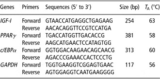
Total RNA was extracted from longissimus lumborum muscle, subcutaneous adipose tissue and perirenal fat using TRIZOL Reagent (Invitrogen, Carlsbad, CA, USA). Primers for selected genes (Table 2) were designed using Oligo 6.0 software. Real-time quantitative PCR analyses were performed with 5 ng of reverse-transcribed RNA and both sense and anti-sense primers in a final volume of 10 μl using SYBR Green I as a PCR core reagent. A total of 35 cycles of amplification were conducted using an ABI 7900 (ABI Biotechnology, Foster City, CA, USA). Standard curves were generated using 10-fold dilutions of purified amplicons obtained by real-time PCR. A melting curve analysis was performed to ensure that only the expected products had been generated. The amplification of glyceraldehyde 3-phosphate dehydrogenase (GAPDH) was used for each sample to normalize the expression of the selected genes.
Primers used for real-time PCR

TA = annealing temperature.
Western blotting
Approximately 100 mg of muscle and adipose tissues were powdered in liquid nitrogen and treated with 500 to 1000 μl RIPA lysis buffer (50 mM Tris–HCl (pH = 7.4), 150 mM NaCl, 1% NP-40, 0.1% SDS) including 1% complete protease inhibitor (Roche, San Francisco, CA, USA). The tissue samples were homogenized and centrifuged at 13 000 × g for 15 min and the supernatant was collected and stored under −80°C for further analysis. Protein levels of the extracts were measured using a BCA protein assay kit (Pierce, Rockford, IL, USA). Aliquots of 30 μg of total protein with SDS loading buffer (Takara, Dalian, China), approximately 30 μl in total, were subjected to electrophoresis (Bio-Rad, Hercules, CA, USA) and separated on a 12% SDS–PAGE gel; they were then transferred using a Trans-Blot Electrophoretic Transfer Cell (Bio-Rad) to polyvinylidene difluoride membranes (Millipore, Bedford, MA, USA) under the condition of 30 mA at 4°C overnight. After blocking with 5% non-fat milk in Tris-buffered saline (TBS; 20 mM Tris, 500 mM NaCl, pH = 7.6), the membranes were then washed in TBS plus 0.1% Tween-20 (TBST) and incubated with rabbit anti-PPARγ polyclonal antibody (No. 2435; Cell Signal Technology, Danvers, MA, USA) at a 1 : 1000 dilution for 2 h at room temperature. The membranes were then rinsed in TBST three times and incubated with peroxidase-conjugated goat anti-rabbit IgG (Santa Cruz Biotechnology, Santa Cruz, CA, USA) for 1 h at a dilution of 1 : 5000. Mouse anti-GAPDH monoclonal antibody (sc-66163; Santa Cruz Biotechnology) diluted at 1 : 1000 was used to normalize the amount of proteins. An AlphaImager 2200 (Alpha Innotech Corporation, Leandro, CA, USA) software was used to determine the density of the protein bands.
Statistical analyses
Data were analyzed with one-way ANOVA (SAS version 8.2). Differences among treatments were evaluated by Duncan's Multiple Range test and were considered significant at P < 0.05.
Results
Growth performance and carcass characteristics
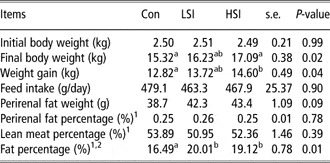
Minipigs fed with the HSI diet gained more BW than either the LSI and Con groups (P < 0.05; Table 3), and the soybean isoflavone treats did not affect animals’ feed intake (P > 0.05). According to the average feed intake data, we gained the amount of isoflavone intake per day per pig, of 57.9 and 234.0 mg for LSI and HSI groups, respectively. The weight change was accompanied by a significant increase in fat percentage (P = 0.01) in response to the increase in soy isoflavones, but not in lean meat percentage (P > 0.05). In addition, there tended to be an increase in the weight of perirenal fat in the minipigs in the LSI and HSI groups compared with the group fed the Con diet (P = 0.09). However, no significance was noted for perirenal fat percentage of the three groups.
Growth performance and carcass characteristics of Chinese Guangxi minipigs fed Con, LSI or HSI diets

Con = control; LSI = low dose of soy isoflavones (125 mg/kg); HSI = high dose of soy isoflavones (500 mg/kg).
a,b,cValues with different letters are significantly different (P < 0.05).
1The ratio of perirenal fat, lean meat and fat weight to half carcass weight.
2Fat (by manual dissection of the carcass) including subcutaneous fat, abdominal fat and intermuscular fat.
Serum contents of hormone and cytokines
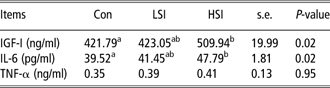
Minipigs that were fed the HSI diet had enhanced serum concentrations of IGF-I and IL-6 (P < 0.05; Table 4). In addition, no significant difference in the serum level of TNF-α was observed among the three groups (P > 0.05).
IGF-I, IL-6 and TNF-α levels in serum (at 0800 h) of Chinese Guangxi minipigs fed control, low- or high-dose isoflavone diets
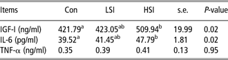
IGF-I = insulin-like growth factor I; IL-6 = interleukin-6; TNF-α = tumor necrosis factor-α; Con = control; LSI = low dose of soy isoflavones (125 mg/kg); HSI = high dose of soy isoflavones (500 mg/kg).
a,bValues with different letters are significantly different (P < 0.05).
Regulation of gene expressions in muscle and adipose tissue
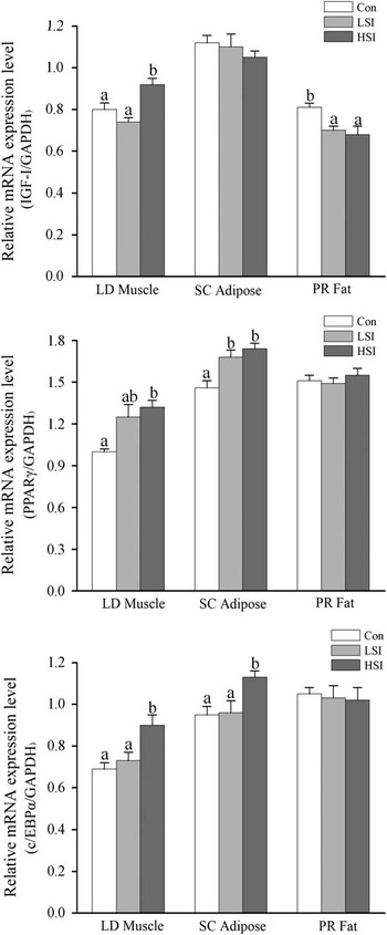
As shown in Figure 1, the minipigs that consumed the HSI diet had higher levels of IGF-I mRNA in longissimus lumborum muscle (P < 0.05), but not in subcutaneous adipose tissue. On the other hand, both the LSI and HSI diets significantly decreased the amounts of IGF-I mRNA in perirenal fat (P < 0.05). Furthermore, minipigs fed with the LSI and HSI diets showed increased PPARγ mRNA levels in subcutaneous adipose tissue (P < 0.05). Nevertheless, only the HSI diet enhanced PPARγ mRNA levels in longissimus lumborum muscle (P < 0.05) and CCAAT enhancer-binding protein α (c/EBPα) mRNA levels in both muscle and subcutaneous adipose tissue (P < 0.05). No significant difference was observed in the amounts of PPARγ and c/EBPα mRNA in perirenal fat of minipigs fed with diets consisting of low and high doses of soy isoflavones.
Effects of soy isoflavones on mRNA expression levels of insulin-like factor I, peroxisome proliferator-activated receptor-γ and CCAAT enhancer-binding protein-α in muscle and adipose tissue of Chinese Guangxi minipigs fed with a standard diet or with diets supplemented with soy isoflavones. (LM = longissimus muscle; SCAT = subcutaneous adipose tissue; PR fat = perirenal fat; Con = control; LSI = low dose of soy isoflavones (125 mg/kg); HSI = high dose of soy isoflavones (500 mg/kg); values with different letters on bars are significantly different (P < 0.05), mean ± s.e. (n = 6 for each group).)

Protein levels of PPARγ in muscle and adipose tissue
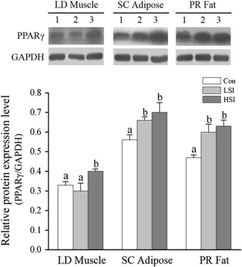
As shown in Figure 2, the PPARγ protein expression level was significantly greater in longissimus lumborum muscle of minipigs that consumed the HSI diet (P < 0.05). Moreover, both the HSI and LSI diets increased PPARγ protein expression levels in perirenal fat of minipigs (P < 0.05).
Effects of soy isoflavones on protein expression levels of peroxisome proliferator-activated receptor-γ in muscle and adipose tissue of Chinese Guangxi minipigs fed with a standard diet or with diets supplemented with soy isoflavones. A representative Western blot is shown. (LM = longissimus muscle; SCAT = subcutaneous adipose tissue; PR fat = perirenal fat; 1 = Control (Con); 2 = low dose of soy isoflavones (LSI; 125 mg/kg); 3 = high dose of soy isoflavones (HSI; 500 mg/kg); values with different letters on bars are significantly different (P < 0.05), mean ± s.e. (n = 6 for each group).)

Discussion
This study demonstrated that, compared with the Con or LSI diet (57.9 mg/day), the higher level of soy isoflavones in the HSI diet (234.0 mg/day) improved the growth performance of Chinese Guangxi minipig, which also showed changes in the IGF-I system. The data regarding weight gain were also consistent with those in obese Zucker rats (Mezei et al., Reference Mezei, Banz, Steger, Peluso, Winters and Shay2003), which showed that high levels (0.116%) of soy isoflavones increased weight gain in female rats, but did not influence visceral fat weight. On the other hand, soy isoflavones stimulate the growth of weanling pigs and increase lean accretion while decreasing ham fat deposition in barrows (Payne et al., Reference Payne, Bidner, Southern and and Geaghan2001). Therefore, the results of in vivo studies conflict, and the underlying mechanisms need to be investigated further. IGF-I show high-sequence homology to insulin and are known to exert biological effects in an endocrine, a paracrine and an autocrine manner. A recent in vitro study reported that, while low concentrations (< 10 μM) of genistein and daidzein had no effects on IGF-I mRNA levels in porcine skeletal muscle cells, high concentrations (100 μM) decreased the mRNA levels of the gene (Kalbe et al., Reference Kalbe, Mau and Rehfeldt2008). Porcine muscle satellite cells are directly exposed to 100 μM (∼26 mg/l) isoflavones in the culture medium, and this concentration causes muscle cell death and DNA damage (Mau et al., Reference Mau, Viergutz and Rehfeldt2006). However, the concentration of 10 μM increases the values. These data suggest the dose-dependent effects of isoflavones. In our in vivo research, high concentrations of soy isoflavones enhanced the amount of IGF-I mRNA in muscle tissue without affecting the proportion of lean meat, but decreased the gene levels in perirenal fat.
Soy isoflavones have been considered to exert estrogenic activity, and estrogens have been reported to enhance the immune response (Ridker et al., Reference Ridker, Hennekens, Rifai, Buring and Manson1999). Hence, the dietary intake of isoflavones may influence the host's immune system. Post-menopausal women showed significantly greater serum concentrations of IL-6 after consuming a high-isoflavone (73 mg/day) soy diet compared with a Con group, whereas no significant effects were observed in TNF-α; these effects were different in male subjects (Jenkins et al., Reference Jenkins, Kendall, Connelly, Jackson, Parker, Faulkner and Vidgen2002). The experiments in male minipigs showed similar results that the HSI diet stimulated the serum level of IL-6, but not TNF-α. Interestingly, the soy isoflavones genistein and daidzein have been shown to bind to PPARγ, as well as PPARα and δ (Mezei et al., Reference Mezei, Banz, Steger, Peluso, Winters and Shay2003; Dang and Löwik, Reference Dang and Löwik2004). PPARγ activation regulates the expression of several key genes involved in lipid metabolism and endocrine function in adipocytes (Arner, Reference Arner2003). One of the most important transcription factors is c/EBPα (Gregoire et al., Reference Gregoire, Smas and Sul1998). Studies provide strong evidence for the role of natural PPARγ ligands (e.g. fatty acids) in anti-inflammation (Allred et al., Reference Allred, Talbert, Southard, Wang and Kilgore2008). Therefore, PPARγ is a target of the pharmaceutical industry and has attracted interest in the food industry.
This study indicated that the addition of soy isoflavones dose-dependently increased the PPARγ mRNA and protein expression in skeletal muscle and adipose tissue, and a similar trend was noted for the c/EBPα gene. A previous study demonstrated that the soy isoflavone daidzein and its metabolite equol regulate adipocyte differentiation through PPARγ activation (Cho et al., Reference Cho, Lee, Banz, Moustaid-Moussa, Shay and Kim2010). In murine macrophage-like RAW 264.7 cells expressing a peroxisome proliferator response element-containing reporter and either PPARα or PPARγ plasmid, unconjugated genistein and daidzein enhanced the gene expression of both PPARα and PPARγ (Mezei et al., Reference Mezei, Banz, Steger, Peluso, Winters and Shay2003). These studies provide an in vitro molecular basis for our results. Another report showed that treatment with low levels of soy isoflavones (75 mg/day) increased PPARα and PPARγ expressions in skeletal muscle compared with the Con, whereas treatment with high levels of soy isoflavones (150 mg/day) had no effect (Wagner et al., Reference Wagner, Zhang, Shadoan, Kavanagh, Chen, Tresnasari, Kaplan and Adams2008). This suggests that low doses of soy isoflavones can have fully different effect than high doses. Hence, this is very important for the soy isoflavone study.
Conclusions
In summary, this study showed that a diet containing soy isoflavones regulated BW and fat percentage of male Chinese Guangxi minipigs, which also showed the changes in the IGF-I system and PPARγ. However, the results of this study are case specific and are not easily extrapolative to other circumstances and species, including humans. Further studies will be needed to clarify the causative relationship and to determine whether soy isoflavones increase the serum levels of IL-6 to modulate the immune system.
Acknowledgments
This study was supported in part by the National Natural Science Foundation of China (30972118, 31001015), the Hunan Provincial Natural Science Foundation (08JJ3080, 31001015, 31001016 and 31072042), the Major Scientific Projects Foundation of Hunan Province (2009FJ1005) and the State Commission of Science Technology of Guangdong (2006B200330005).