





Introduction
Small ruminants such as sheep and goats play an essential role in sustaining rural livelihoods across Africa, Asia, and the Middle East, providing meat, milk, hides, and manure that contribute to food security and income generation (Wodajo et al., Reference Wodajo, Gemeda, Kinati, Mulem, Van Eerdewijk and Wieland2020). In many low-income settings, they are often referred to as the ‘poor man’s bank’, serving as a readily available source of cash in times of need (de Vries, Reference De Vries2008). Because ownership is frequently dominated by women and smallholder farmers, improving the health and productivity of small ruminants has a direct socio-economic impact on household resilience, gender empowerment, and poverty alleviation (Ogolla et al., Reference Ogolla, Chemuliti, Ngutu, Kimani, Anyona, Nyamongo and Bukachi2022).
Among the diseases that constrain small-ruminant production, peste des petits ruminants (PPR) remains one of the most devastating. PPR is a transboundary, highly contagious viral disease caused by peste des petits ruminants virus (PPRV), a member of the genus Morbillivirus within the family Paramyxoviridae, closely related to Rinderpest virus (RPV), Measles virus (MeV), and Canine distemper virus (CDV; Tounkara et al., Reference Tounkara, Kwiatek, Niang, Sidibe, Sery, Dakouo, Salami, Lo, Ba, Diop, El Mamy, El Arbi, Barry, Isselmou, Habiboullah, Lella, Doumbia, Gueya, Savadogo, Ouattara, Minougou, Libeau and Bataille2019). The disease, first identified in Côte d’Ivoire in 1942, is now endemic across much of Africa, the Middle East, and South Asia, with increasing detection in new regions, including parts of North and Central Asia (Legnardi et al., Reference Legnardi, Raizman, Beltran-alcrudo, Cinardi, Robinson, Falzon, Kapnang Djomgang, Okori, Parida, Njeumi and Benfield2022). While sheep and goats are the principal hosts, recent studies have shown infection in camels and several wild ruminants, raising concerns about cross-species transmission and potential wildlife reservoirs (Rahman et al., Reference Rahman, Dhama, Ali, Hussain, Oneeb, Chaudhary, Wensman and Shabbir2020).
Clinically, PPR is characterized by high fever, mucosal erosions, diarrhoea, and severe respiratory distress, with morbidity approaching 100% and mortality up to 90% in naïve flocks (Mansour et al., Reference Mansour, Hussain, Al-husseiny, Abid and Kshash2024). In endemic areas, repeated outbreaks lead to reduced fertility, poor weight gain, and chronic productivity losses. The global economic impact of PPR – through mortality, production losses, and control measures – has been estimated at between USD 1.4 and 2.1 billion annually (Chukwudi et al., Reference Chukwudi, Weka, Inusa, Luka, Munir and Abubakar2025; Jones et al., Reference Jones, Rich, Mariner, Anderson, Jeggo, Thevasagayam, Cai, Peters and Roeder2016).
In response to this threat, the Food and Agriculture Organization of the United Nations (FAO) and the World Organisation for Animal Health (WOAH, formerly known as the Office International des Epizooties (OIE)) launched in 2015 the PPR Global Eradication Programme (PPR-GEP), targeting eradication of the disease by 2030. The initiative builds on the historical success of rinderpest eradication and emphasizes mass vaccination, surveillance, and strengthening of veterinary services. Currently available live-attenuated vaccines, including the Nigeria 75/1 and Sungri 96 strains, provide solid and long-lasting immunity against all four PPRV lineages (Hodgson et al., Reference Hodgson, Moffat, Hill, Flannery, Graham, Baron and Darpel2018). However, their effectiveness is hindered by two major limitations: thermolability, which necessitates continuous cold-chain maintenance, and the absence of a DIVA (Differentiating Infected from Vaccinated Animals) capability, which complicates post-vaccination surveillance (Balamurugan et al., Reference Balamurugan, Ojha, Kumar, Asha, Ashraf, Dsouza, Pal, Bokade, Harshitha, Deshpande, Swathi, Suresh, Govindaraj, Hasnadka, ChandraSekar, Hemadri, Guha, Felix, Parida and Gulati2024; Jia et al., Reference Jia, Wang, Liu, Meng and Fan2020).
To achieve sustainable eradication, there is an urgent need for next-generation vaccines that combine thermostability, safety, and DIVA compatibility. Current research efforts include the development of recombinant viral-vectored vaccines using adenovirus or capripoxvirus backbones expressing the PPRV F and H proteins, subunit and virus-like-particle (VLP) formulations with enhanced stability, and reverse-genetics-engineered marker vaccines suitable for serological differentiation (Darpel et al., Reference Darpel, Corla, Stedman, Bellamy, Flannery, Rajko-nenow, Powers, Wilson, Charleston, Baron and Batten2024; Rojas et al., Reference Rojas, Sevilla and Martín2021). Parallel advances in formulation science – such as lyophilized and stabilizer-enhanced preparations – have shown promise in maintaining potency at ambient temperature for weeks, potentially transforming field vaccination logistics (Diallo et al., Reference Diallo, Motsoane, Gelaw, Baziki, Boukary, Melesse, Chitsungo, Gebresillassie, Tessema, Olugasa, Ishola, Nwankpa and Bodjo2024).
In this review, we summarize recent progress in the development of innovative PPRV vaccine strategies and highlight ongoing efforts to improve thermostability and DIVA design. These advances are expected to underpin sustainable disease control and support the global goal of eradicating PPR by 2030, thereby securing the livelihoods of millions who depend on small-ruminant production for survival.
Biology and lineage diversity of PPRV
PPRV is a pleomorphic, enveloped, negative-sense single-stranded RNA virus classified within the genus Morbillivirus (family Paramyxoviridae, order Mononegavirales) (Amarasinghe et al., Reference Amarasinghe, Ayllón, Bào, Basler, Bavari, Blasdell, Briese, Brown, Bukreyev, Balkema-Buschmann, Buchholz, Chabi-Jesus, Chandran, Chiapponi, Crozier, de Swart, Dietzgen, Dolnik, Drexler, Dürrwald, Dundon, Duprex, Dye, Easton, Fooks, Formenty, Fouchier, Freitas-Astúa, Griffiths, Hewson, Horie, Hyndman, Jiāng, Kitajima, Kobinger, Kondō, Kurath, Kuzmin, Lamb, Lavazza, Lee, Lelli, Leroy, Lǐ, Maes, Marzano, Moreno, Mühlberger, Netesov, Nowotny, Nylund, Økland, Palacios, Pályi, Pawęska, Payne, Prosperi, Ramos-González, Rima, Rota, Rubbenstroth, Shī, Simmonds, Smither, Sozzi, Spann, Stenglein, Stone, Takada, Tesh, Tomonaga, Tordo, Towner, van den Hoogen, Vasilakis, Wahl, Walker, Wang, Whitfield, Williams, Zerbini, Zhāng, Zhang and Kuhn2019). It shares extensive structural and genomic homology with other morbilliviruses such as MeV, RPV, and CDV, reflecting conserved replication strategies and gene organization (Noton and Fearns, Reference Noton and Fearns2015).
Morphology and structural organization
The PPRV virion ranges from 150–700 nm in diameter and comprises a helical ribonucleoprotein (RNP) complex encapsidated by a lipid envelope derived from the host plasma membrane. The RNP contains the genomic RNA tightly associated with the nucleoprotein (N), phosphoprotein (P), and large RNA-dependent RNA polymerase (L), which together form the polymerase complex responsible for transcription and replication (Muniraju et al., Reference Muniraju, Munir, Parthiban, Banyard, Bao, Wang, Ayebazibwe, Ayelet, El Harrak, Mahapatra, Libeau, Batten and Parida2014). Two surface glycoproteins – hemagglutinin (H) and fusion (F) – project from the viral envelope and mediate host-cell attachment and membrane fusion, respectively (Parida et al., Reference Parida, Muniraju, Mahapatra, Muthuchelvan, Buczkowski and Banyard2015; Zou et al., Reference Zou, Niu, Cheng, Wu, Li, Luo and Huang2025). These glycoproteins are the principal targets of neutralizing antibodies and strongly influence host specificity and virulence (Rojas et al., Reference Rojas, Sevilla and Martín2021).
Beneath the envelope lies the matrix (M) protein, which connects the RNP to the viral membrane and coordinates virion assembly and budding. Structural modeling suggests that M interacts directly with both H and F to synchronize viral egress and maintain virion integrity (Prajapati et al., Reference Prajapati, Alfred, Dou, Yin, Prajapati, Li and Zhang2019). Cryo-electron microscopy of morbilliviruses reveals the RNP arranged in a herringbone-like helical pattern with a pitch of approximately 6.5 nm per turn, a feature conserved across the genus (Noton and Fearns, Reference Noton and Fearns2015; Yunus and Shaila, Reference Yunus and Shaila2012).
Genome organization and molecular features
The PPRV genome (∼15,948 nt) is a non-segmented, negative-sense RNA that obeys the ‘rule of six’, meaning efficient replication occurs only when the total genome length is divisible by six (Bailey et al., Reference Bailey, Chard, Dash, Barrett and Banyard2007). Genes are arranged in the conserved morbillivirus order 3′–N–P/C/V–M–F–H–L–5′, encoding six structural and two non-structural proteins (C and V), the latter produced via co-transcriptional RNA editing of the P gene (Muniraju et al., Reference Muniraju, Munir, Parthiban, Banyard, Bao, Wang, Ayebazibwe, Ayelet, El Harrak, Mahapatra, Libeau, Batten and Parida2014; Parida et al., Reference Parida, Muniraju, Mahapatra, Muthuchelvan, Buczkowski and Banyard2015). Conserved gene-start and gene-end motifs regulate transcriptional reinitiation, while short intergenic sequences act as transcriptional spacers (Bailey et al., Reference Bailey, Chard, Dash, Barrett and Banyard2007). The 3′- and 5′-untranslated regions contain promoter elements required for replication of both genomic and antigenomic strands (Jamin and Yabukarski, Reference Jamin and Yabukarski2017).
Comparative analyses show PPRV has one of the largest genomes among morbilliviruses, second only to feline morbillivirus (Rajko-nenow et al., Reference Rajko-Nenow, Tabitha, Cunliffe, Flannery, Ropiak, Avaliani, Donduashvili, Michael and Carrie2017). Evolutionary studies indicate that PPRV diverged from RPV roughly 1,000 years ago, maintaining overall genomic stability but undergoing adaptive evolution in the H and F genes, which are under episodic positive selection (Courcelle et al., Reference Courcelle, Salami, Tounkara, Lo, Ba, Diop, Niang, Sidibe, Sery, Dakouo, Kaba, Sidime, Keyra, Diallo, El Mamy, El Arbi, Barry, Isselmou, Habiboullah, Doumbia, Gueya, Awuni, Odoom, Ababio, TawiahYingar, Coste, Guendouz, Kwiatek, Libeau and Bataille2024; Ramesh et al., Reference Ramesh, Suresh, Mambully, Rani, Ojha, Kumar and Balamurugan2024). The F protein is synthesized as an inactive precursor (F₀) that must be cleaved into F₁ and F₂ subunits for membrane fusion and infectivity (Zou et al., Reference Zou, Niu, Cheng, Wu, Li, Luo and Huang2025).
Genetic diversity and lineage evolution
Molecular epidemiology divides PPRV into four genetically distinct lineages (I–IV) based primarily on partial or complete nucleotide sequences of the N, F, and H genes (Banyard et al., Reference Banyard, Parida, Batten, Oura, Kwiatek and Libeau2010; Dhar et al., Reference Dhar, Sreenivasa, Barrett, Corteyn, Singh and Bandyopadhyay2002; Libeau et al., Reference Libeau, Diallo and Parida2014). Historically, lineages I and II circulated in West and Central Africa (Banyard et al., Reference Banyard, Parida, Batten, Oura, Kwiatek and Libeau2010; Dou et al., Reference Dou, Liang, Prajapati, Zhang, Yanmin and Zhang2020), lineage III in East Africa and the Middle East, and lineage IV across South and East Asia (Ali et al., Reference Ali, Ahmed, Osman, Eldin and Osman2023; Legnardi et al., Reference Legnardi, Raizman, Beltran-alcrudo, Cinardi, Robinson, Falzon, Kapnang Djomgang, Okori, Parida, Njeumi and Benfield2022). Over the last decade, lineage IV has expanded westward into Africa – including Nigeria, Ethiopia, Sudan, and North Africa – indicating ongoing viral adaptation and transboundary spread (Bello et al., Reference Bello, Kazeem, Oladele, Fatihu, Tambuwal and Jibril2018a; Mantip et al., Reference Mantip, Sigismeau, Shamaki, Woma, Kwiatek, Libeau, Farougou and Bataille2022).
Among the commonly used genetic markers, the N gene offers the highest phylogenetic resolution and is now the preferred target for lineage classification and outbreak tracing (Kumar et al., Reference Kumar, Maherchandani, Kashyap, Singh, Sharma, Chaubey and Hinh2014). The H gene, though more variable, provides key insights into antigenic evolution, receptor usage, and immune escape (Dhar et al., Reference Dhar, Sreenivasa, Barrett, Corteyn, Singh and Bandyopadhyay2002; Dou et al., Reference Dou, Liang, Prajapati, Zhang, Yanmin and Zhang2020). The global predominance of lineage IV – associated with high transmissibility and intercontinental dissemination – makes it central to current vaccine-matching and eradication strategies (Anand et al., Reference Anand, Mahadevappa, Ojha, Pal, Priya, Dsouza, Bokade, Kumar, Hemadri, Gulati and Balamurugan2025; Woma et al., Reference Woma, Adombi, Yu, Qasim, Sabi, Maurice, Olaiya, Loitsch, Bailey, Shamaki, Dundon and Quan2016).
Immunity to PPRV
Innate immune response
The innate immune system provides the first line of defense against invading pathogens through germline-encoded pattern recognition receptors (PRRs) that detect conserved microbial components known as pathogen-associated molecular patterns (PAMPs) (Takeuchi and Akira, Reference Takeuchi and Akira2010). Upon recognition of PAMPs, PRRs initiate signaling cascades that result in the production of pro-inflammatory cytokines, chemokines, and type I interferons, which establish an antiviral state and orchestrate subsequent adaptive immune responses (Kawai and Akira, Reference Kawai and Akira2010, Reference Kawai and Akira2011). In mammals, PRRs are categorized into several families, including Toll-like receptors (TLRs), RIG-I-like receptors, NOD-like receptors, C-type lectin receptors, and cytosolic DNA sensors such as cyclic GMP–AMP synthase (Diebold et al., Reference Diebold, Kaisho, Hemmi, Akira and Reis e Sousa2004). Among these, the TLR family is the most extensively characterized. Humans express at least 10 TLRs (TLR1–TLR10), while mice possess 12 (TLR1–TLR9, TLR11–TLR13) (Sameer and Nissar, Reference Sameer and Nissar2021). TLRs are broadly divided into cell-surface receptors (e.g., TLR1, TLR2, TLR4, TLR5, TLR6, TLR11) that recognize extracellular or membrane-associated ligands, and intracellular receptors (e.g., TLR3, TLR7–TLR9, TLR12, and TLR13) located within endosomal or lysosomal compartments. Several TLRs, particularly TLR3, TLR7–TLR9, are key players in antiviral sensing and interferon induction (El-Zayat et al., Reference El-Zayat, Sibaii and Mannaa2019).
PPRV is both lymphotropic and epitheliotropic, replicating initially in lymphoid tissues and subsequently in epithelial cells of the respiratory and gastrointestinal tracts. Infection triggers early induction of inducible nitric oxide synthase, interferon-gamma (IFN-γ), and tumor necrosis factor-alpha (TNF-α) within affected tissues, reflecting the activation of infiltrating immune and inflammatory cells that amplify antiviral signaling (Atmaca and Kul, Reference Atmaca and Kul2012; Prajapati et al., Reference Prajapati, Alfred, Dou, Yin, Prajapati, Li and Zhang2019; Ul-Rahman et al., Reference Ul-Rahman, Abubakar, Razaq, Raza, Munir, Munir and Abubakar2025). PPRV infection also upregulates multiple cytokines, including IFN-β, IFN-γ, interleukin (IL)-1β, IL-4, IL-6, IL-8, IL-10, and IL-12, consistent with engagement of both Th1- and Th2-type immune pathways. Histopathologically, strong IFN-γ staining appears in epithelial cells of the oral mucosa, respiratory tract, tongue, monocytes, and syncytial cells, indicating broad activation of antiviral signaling across mucosal and lymphoid compartments (Atmaca and Kul, Reference Atmaca and Kul2012). However, PPRV has evolved multiple immune evasion strategies. Although IFN-β can be induced transiently, its induction is inherently weak and actively antagonized by the viral V and C proteins, which suppress MDA-5- and RIG-I-mediated signaling (Chang et al., Reference Chang, Guo, Liu, Zhang, Feng, Ma and Shang2019; Wang et al., Reference Wang, Wang, Duan, Chen, Hu, Wang, Jia, Chu, Liu, Wang, Zhang, Xiao, Wang, Dang and Yang2022). In addition, the V and C proteins interfere with NF-κB p65 nuclear translocation under TNF-α stimulation, dampening inflammatory gene activation (Linjie et al., Reference Linjie, Xiaoling, Xiaoxia, Xin, Ali and Jialin2021). Structural proteins also contribute: the N protein can bind MyD88 and promote proinflammatory cytokine induction (Li et al., Reference Li, Yang, Ma, Wu, Qin, Cao, Zhou, Jin, He, Zheng, Liu, Li and Shang2022). Moreover, PPRV engages extracellular vesicles as a stealth dissemination route, aiding immune evasion and spread (Chen et al., Reference Chen, Wang, Yang, Fang, Zhao, Zeng, Lv, Zhang, Zhang, Xue, Chen, Wang and Qi2022). Intriguingly, PPRV-induced ferroptosis further amplifies proinflammatory cytokine expression even while suppressing type I IFN responses (Hou et al., Reference Hou, Cheng, Zhijun, Lei, Chen, Mingzhuo, Liu, Chen, Wang, Xue and Qi2025). At the transcriptomic level, susceptible small ruminants mount strong induction of downstream interferon-stimulated genes (ISGs), such as ISG15, MX1, and RSAD2, even when upstream IFN signals are dampened (Tang et al., Reference Tang, Tang, Du, Jia, Zhu, Chuanfeng, Meng and Liu2022; Tirumurugaan et al., Reference Tirumurugaan, Pawar, Raj, Thangavelu, Hammond and Parida2020). Collectively, these findings illustrate a finely balanced interplay: PPRV triggers innate antiviral and inflammatory pathways, but simultaneously deploys multiple countermeasures to blunt their effectiveness, thereby enabling viral replication and pathogenesis.
Humoral immunity to PPRV
The surface of PPRV is dominated by a mosaic of envelope glycoproteins that elicit strong antibody-mediated immune responses in the host (Kumar et al., Reference Kumar, Maherchandani, Kashyap, Singh, Sharma, Chaubey and Hinh2014). The most important of these are the H and F proteins (Diallo et al., Reference Diallo, Minet, Le Goff, Berhe, Albina, Libeau and Barrett2007). PPR is a highly contagious animal disease caused by a virus in the genus Morbillivirus, family Paramyxoviridae. This infection is responsible for high morbidity and mortality in sheep and goats and in some small wild ruminant species. The huge number of small ruminants, which are reared in the endemic areas, makes PPR a serious disease threatening the livelihood of poor farmers. Taking advantage of the close relationship between rinderpest and PPR viruses, the attenuated rinderpest vaccine was used in the control of PPR. It is now replaced by the homologous attenuated PPR vaccine. Unfortunately, animals that have received this vaccine cannot be distinguished serologically from infected animals. With the advent of DNA recombinant technology, efforts are being made to develop effective PPR marker vaccines to enable such differentiation, which would allow countries to implement both vaccination and disease surveillance programmes at the same time (Taheri Asl et al., Reference Taheri Asl, Moghbeli, Kargar, Lotfi and Kafilzadeh2024). Neutralizing antibodies can block viral entry by preventing attachment or fusion with host cell membranes (Apsana et al., Reference Apsana, Balamurugan, Veeregowda, Abraham, Raju, Rathnamma, Byregowda, Rahman and Shaila2016) and can also recruit Fc-dependent effector mechanisms such as complement activation and antibody-dependent cell-mediated cytotoxicity (ADCC) to clear infected cells (Rojas et al., Reference Rojas, Rodríguez-martín, Avia, Martín and Sevilla2019).
The H protein of PPRV is structurally composed of an N-terminal cytoplasmic tail, a transmembrane domain, a stalk, and a C-terminal globular head that contains the receptor-binding site (Rojas et al., Reference Rojas, Sevilla and Martín2021). Unlike the hemagglutinin-neuraminidase protein of other paramyxoviruses, the H protein of morbilliviruses may lack neuraminidase activity (Hashiguchi et al., Reference Hashiguchi, Kajikawa, Maita, Takeda, Kuroki, Sasaki, Kohda, Yanagi and Maenaka2007). Conserved T-cell epitopes have been identified within the PPRV H glycoprotein that are recognized by both CD4⁺ helper and CD8⁺ cytotoxic T cells in cattle, indicating that this protein elicits broad cellular immune responses (Mitra-Kaushik et al., Reference Mitra-Kaushik, Nayak and Shaila2001). Although less extensively characterized, the F protein also contains neutralizing epitopes and contributes to humoral protection through the generation of fusion-inhibiting antibodies (Gaafar et al., Reference Gaafar, Ali, Abd-elrahman and Almofti2019).
Following natural infection or vaccination, animals develop strong PPRV-specific immunoglobulin G (IgG) responses. These antibodies not only neutralize homologous strains but also exhibit cross-neutralizing activity against multiple viral lineages (Hodgson et al., Reference Hodgson, Moffat, Hill, Flannery, Graham, Baron and Darpel2018), a property critical to the FAO-WOAH Global Eradication Programme. However, recent challenge studies indicate that while high neutralizing antibody titers are strongly protective, they may not always prevent subclinical infection or transient viral RNA detection, suggesting a cooperative role for cellular and innate mechanisms in complete protection (Schmitz et al., Reference Schmitz, Eblé, van Gennip, Maris-veldhuis, De Vries, van Keulen, De Swart and van Rijn2023).
In addition to vaccine-induced immunity, passive transfer of maternal antibodies from infected or vaccinated dams provides temporary protection to neonates. Colostrum-derived antibodies can persist above protective thresholds for variable durations depending on antibody concentration and species. Early studies reported persistence for 3–4.5 months in kids and lambs (Awa et al., Reference Awa, Ngagnou, Tefiang, Yaya and Njoya2002), while more recent findings indicate a broader range: antibodies may persist up to 18 weeks in lambs born to vaccinated ewes but often decline substantially by 14 weeks (Amanova et al., Reference Amanova, Turyskeldy, Kondybaeva, Sametova, Usembai, Kerimbayev and Bulatov2023). In some regions, earlier decay – within 8–10 weeks – has been observed (Markus et al., Reference Markus, Adamu, Kazeem, Olaolu and Woma2019), emphasizing the need for local evaluation of maternal antibody kinetics before scheduling vaccination. Consequently, most control programmes recommend primary vaccination of kids and lambs at 3–4 months of age to avoid interference from maternal immunity while ensuring timely protection.
Recent studies confirm that neutralizing antibodies elicited against the PPRV H and F glycoproteins exhibit extensive cross-reactivity across viral lineages, supporting the feasibility of global eradication (Hodgson et al., Reference Hodgson, Moffat, Hill, Flannery, Graham, Baron and Darpel2018). Fine epitope mapping and conservation analyses further indicate that major linear B-cell epitopes within the H protein remain structurally conserved among circulating isolates (Yu et al., Reference Yu, Zhu, Gao, Zhang, Dong, Chen, Yu, Xie, Jiang and Li2017). Serological analyses reveal that IgM antibodies emerge within the first week of infection, followed by long-lived IgG – predominantly IgG1 – by 3–4 weeks post-exposure (Kinimi et al., Reference Kinimi, Odongo, Muyldermans, Kock and Misinzo2020). These humoral markers underpin diagnostic assays such as virus neutralization tests and competitive enzyme-linked immunosorbent assay (ELISA), which are essential for surveillance programmes; however, assay performance declines in atypical and wildlife hosts (Tully et al., Reference Tully, Batten, Ashby, Mahapatra, Parekh, Parida, Njeumi, Willett, Bataille, Libeau, Kwiatek, Caron, Berguido, Lamien, Cattoli, Misinzo, Keyyu, Mdetele, Gakuya, Bodjo, Taha, Elbashier, Khalafalla, Osman and Kock2023). Altogether, these findings highlight the central role of humoral immunity in both protection and epidemiological monitoring of PPRV.
Cell-mediated immune response to PPRV
PPRV infection involves strong lymphoid tropism, causing transient lymphopenia through viral cytolysis and immune-cell redistribution (Gaur et al., Reference Gaur, Jain, Chaudhary and Kaul2024). Recovery from this lymphodepletion coincides with activation of antigen-specific CD4⁺ and CD8⁺ T cells, which are crucial for viral clearance and the establishment of long-term protection (Milovanović et al., Reference Milovanović, Dietze, Wernery and Hoffmann2023).
Following infection or vaccination, CD4⁺ helper T cells secrete ILs such as IL-2 and IFN-γ that support both B-cell maturation and cytotoxic T-cell activation, driving a Th1-skewed response (Gaur et al., Reference Gaur, Jain, Chaudhary and Kaul2024). Increased IFN-γ and TNF-α expression has been reported in PPRV-infected goats, consistent with activation of cell-mediated pathways (Atmaca and Kul, Reference Atmaca and Kul2012). CD8⁺ T cells recognize PPRV peptides presented on MHC class I and destroy infected epithelial and lymphoid cells via perforin–granzyme effector mechanisms. In goats vaccinated with live-attenuated PPRV, IFN-γ-producing CD8⁺ cells are detected within two weeks post-vaccination, supporting their role in protection (Milovanović et al., Reference Milovanović, Dietze, Wernery and Hoffmann2023). In vitro, infected PBMCs show expansion of both CD4⁺ and CD8⁺ subsets together with up-regulation of pro-inflammatory cytokines IL-1β, IL-6, and TNF-α (Li et al., Reference Li, Yang, Ma, Wu, Qin, Cao, Zhou, Jin, He, Zheng, Liu, Li and Shang2022).
Early innate cytotoxic responses also contribute to antiviral defense. Qi et al. (Reference Qi, Li, Li, Wang, Zhang and Wang2019) demonstrated that PPRV infection suppresses peripheral NK-cell cytotoxicity in goats through a miR-1–TWEAK signaling axis, leading to reduced expression of perforin and IFN-γ (Qi et al., Reference Qi, Li, Li, Wang, Zhang and Wang2019). This suggests that PPRV actively modulates NK cells to evade early immune clearance. Conversely, in the presence of anti-PPRV antibodies, NK-cell–enriched fractions mediate ADCC against PPRV-infected cells, targeting the viral H and F glycoproteins on infected surfaces (Rojas et al., Reference Rojas, Rodríguez-martín, Avia, Martín and Sevilla2019). Together, these findings imply that NK cells play dual roles—direct antiviral effectors early in infection and mediators of ADCC once antibodies appear.
Beyond humoral immunity, evidence from morbillivirus infections indicates that CMI is indispensable for complete viral clearance. Morbillivirus-specific T-cell and NK-cell responses limit replication and remove infected cells, paralleling observations in measles, where cellular immunity governs recovery and long-term virus control (Griffin, Reference Griffin2021; Morris et al., Reference Morris, Yates, De Swart, De Vries, Mina, Nelson, Lin, Kouyos, Griffin and Grenfell2018). Accordingly, vaccines that elicit robust IFN-γ-secreting T cells and functional NK responses are more likely to confer durable protection and accelerate eradication efforts.
Immunoprophylaxis of PPRV
Heterologous vaccines
Morbilliviruses exhibit a remarkable degree of antigenic and structural conservation, particularly between PPRV and RPV, which share high sequence homology in the hemagglutinin (H) and fusion (F) glycoproteins responsible for viral entry and neutralization (Baron et al., Reference Baron, Diop, Njeumi, Willett and Bailey2017; de Vries et al., Reference De Vries, Duprex and De Swart2015). This antigenic similarity underlies the historical success of heterologous immunization approaches, where vaccines originally developed against RPV were found to confer cross-protection against PPRV.
The tissue-culture rinderpest vaccine (TCRV)—an attenuated RPV strain—was widely deployed from the late 1980s through the 1990s across West Africa, the Middle East, and South Asia. Experimental and field studies demonstrated that the TCRV induced cross-neutralizing antibodies capable of inhibiting PPRV replication in vitro and protecting sheep and goats against virulent challenge (Diallo et al., Reference Diallo, Taylor, Lefèvre and Provost1989; Kumar et al. Reference Kumar, Barua, Riyesh and Tripathi2017). Animals vaccinated with TCRV mounted strong and long-lasting humoral and cell-mediated immune responses comparable to those elicited by homologous PPRV vaccines, with protective immunity lasting up to one year in some cases (Liu et al., Reference Liu, Wu, Liu, Li and Wang2014).
A key advantage of the heterologous RPV vaccine was its excellent safety profile: it could be administered to pregnant ewes and does without adverse effects, and offspring of vaccinated dams received passively transferred maternal antibodies that protected them for approximately three months (Awa et al., Reference Awa, Ngagnou, Tefiang, Yaya and Njoya2002). These practical benefits, combined with the wide availability of TCRV during the early period of PPR emergence, made it the vaccine of choice for large-scale control campaigns across endemic regions.
At the molecular level, the cross-protection afforded by TCRV has been attributed to conserved epitopes within the F and H glycoproteins. Recombinant vaccinia viruses co-expressing RPV F and H proteins conferred protection against PPRV challenge in goats, confirming that these glycoproteins alone were sufficient to mediate cross-neutralizing immunity (Giavedoni et al., Reference Giavedoni, Jones, Mebus and Yilma1991). The phenomenon is bidirectional: cattle vaccinated with wild-type PPRV were protected against rinderpest, further illustrating the high degree of antigenic overlap between the two viruses (Holzer et al., Reference Holzer, Hodgson, Logan, Willett and Baron2016).
Despite its effectiveness, the use of rinderpest-based vaccines was progressively discontinued following the launch of the Global Rinderpest Eradication Programme (GREP). Continued field use posed diagnostic challenges, as serological cross-reactivity risked confounding rinderpest surveillance and certification efforts (Morens et al., Reference Morens, Holmes, Davis and Taubenberger2011). As a result, TCRV was withdrawn from routine use in small ruminants, marking the end of the heterologous vaccination era. Nevertheless, the experience demonstrated the feasibility of PPR control through vaccination and provided critical immunological and logistical insights that informed subsequent eradication strategies.
Homologous vaccines
The cessation of rinderpest-based vaccination after the launch of the GREP prompted the development of homologous vaccines derived directly from PPRV. The first such vaccine originated from a Nigerian isolate obtained from a goat that died during a PPR outbreak in 1975. Through extensive attenuation by serial passage of the virulent virus in Vero cells, the strain—designated PPRV Nigeria 75/1—completely lost its pathogenicity while retaining strong immunogenicity (Diallo et al., Reference Diallo, Taylor, Lefèvre and Provost1989).
Field trials involving nearly 98 000 sheep and goats demonstrated high seroconversion rates and almost 100% protection against challenge with multiple virulent PPRV isolates representing different lineages (Diallo et al., Reference Diallo, Minet, Le Goff, Berhe, Albina, Libeau and Barrett2007). Immunity develops rapidly, with neutralizing antibodies detectable within seven days post-vaccination and protection persisting for at least three years (Santhosh et al., Reference Santhosh, Gomes, Hegde, Rathnamma, Veeregowda, Byregowda, Renukaprasad, Bhanuprakash, Prabhudas, Hegde and Isloor2013). The vaccine has shown no adverse effects, including in pregnant animals, and confers passive protection to offspring via maternal antibodies. Owing to its stability, safety, and broad efficacy, the Nigeria 75/1 strain remains the prototype and globally recommended PPR vaccine, particularly across sub-Saharan Africa and the Middle East (Kamel and El-Sayed, Reference Kamel and El-Sayed2019).
Following its success, several additional lineage-specific attenuated vaccines were developed from local field isolates in South Asia. Among these, Sungri-96, Coimbatore (CBE)-97, and Arasur-87 are the best characterized (Sen et al., Reference Sen, Saravanan, Balamurugan, Rajak, Sudhakar, Bhanuprakash and Singh2010). The Sungri-96 vaccine was derived from a virulent PPRV isolate collected from goats in the Sungri region of Himachal Pradesh, India, in 1994. The wild-type virus was passaged 10 times in marmoset lymphoblastoid cells, followed by 59 passages in Vero cells with defective interferon signaling pathways, producing a stable, non-reverting attenuated strain (Sen et al., Reference Sen, Saravanan, Balamurugan, Rajak, Sudhakar, Bhanuprakash and Singh2010). The vaccine induces robust humoral and cellular immunity, with ≥90% of vaccinated animals developing protective neutralizing antibodies under field conditions (Hodgson et al., Reference Hodgson, Moffat, Hill, Flannery, Graham, Baron and Darpel2018).
Similarly, the Coimbatore-97 and Arasur-87 vaccines – derived from goat and sheep isolates, respectively – have shown excellent safety and immunogenicity profiles, producing protection levels comparable to the Nigeria 75/1 vaccine (Saravanan et al., Reference Saravanan, Sen, Balamurugan, Rajak, Bhanuprakash, Palaniswami, Nachimuthu, Thangavelu, Dhinakarraj, Hegde and Singh2010). The attenuated viruses are genetically stable, showing no reversion to virulence even after five serial passages in susceptible hosts, and provide solid cross-lineage protection (Rojas et al., Reference Rojas, Sevilla and Martín2021). Collectively, these homologous vaccines have become the cornerstone of the FAO-WOAH GEP-PPR, underpinning mass-vaccination campaigns across Africa and Asia and enabling measurable progress towards the goal of PPR eradication by 2030.
Reverse-genetics-based marker vaccines
Reverse genetics has revolutionized the ability to manipulate RNA virus genomes, providing a platform for both functional genomics and vaccine development (Pfaller et al., Reference Pfaller, Cattaneo and Schnell2015). This technology enables the generation of infectious viruses entirely from cloned cDNA, allowing precise insertion, deletion, or modification of genes to enhance immunogenicity or introduce marker traits. Initially, reverse genetics was more straightforward in positive-sense RNA viruses because their genomic RNA can directly serve as mRNA (Zhou et al., Reference Zhou, Liu, Yang, Dong, Xie, Ou, Chen, Yang, Ye, Ivanov, Liu, Zou, Li and Guo2025). In contrast, negative-sense RNA viruses, including members of the genus Morbillivirus, require an RNP complex composed of genomic RNA tightly associated with nucleoprotein (N), phosphoprotein (P), and polymerase (L) for infectivity (Pekosz et al., Reference Pekosz, He and Lamb1999). Consequently, the successful rescue of such viruses required co-expression of these helper proteins alongside the full-length antigenomic cDNA clone (Figure 1).
Generation of recombinant live attenuated PPRV vaccine using reverse genetics. Helper plasmids (expression constructs of NP, P, and L) and the full-length PPRV antigenomic construct (with a genetic tag) are co-transfected into Vero cells. After 4–5 days of incubation, newly rescued PPRV is harvested and propagated in fresh Vero cells to obtain a higher concentration of the engineered virus.

Figure 1 Long description
The process of generating a recombinant live attenuated PPRV vaccine using reverse genetics is illustrated. At the top left, the PPR virus is depicted as a circular structure. To the right, the PPRV full-length cDNA plasmid is shown as a circular blue structure, alongside helper plasmids labeled N, P and L, representing replicative machinery. An arrow labeled Co-transfection points from these plasmids to a dish containing vero cells, indicating the co-transfection process. Below, a syringe labeled Live attenuated PPRV Vaccine is connected to a dish labeled Fresh vero cells, showing the propagation step. Arrows lead from the fresh vero cells to a group of circular structures labeled Newly rescued genetically modified PPRV and then to another dish labeled Vero cells, indicating viral assembly and release. The steps involved in the creation and propagation of the engineered virus are visually represented.
The first major success came when rabies virus was rescued from cloned cDNA in 1994 using a T7-RNA polymerase system (Conzelmann and Schnell, Reference Conzelmann and Schnell1994), leading to the rapid adaptation of reverse genetics to other Mononegavirales. However, early attempts to rescue PPRV using the same approach repeatedly failed, largely because of inefficient transcription initiation at the genomic termini (Beaty et al., Reference Beaty, Park, Won, Hong, Lyons, Vigant, Freiberg, tenOever, Duprex and Lee2017). The breakthrough came in 2012, when Hu et al. established a functional reverse-genetics system for the Nigeria 75/1 vaccine strain. By using a cytomegalovirus promoter to drive Pol II-mediated transcription and flanking the antigenomic construct with hammerhead and hepatitis-delta ribozymes to generate precise termini, they successfully rescued infectious recombinant PPRV (rPPRV) expressing green fluorescent protein (GFP) between the P and M genes (Hu et al., Reference Hu, Chen, Huang, Baron and Zhigao2012). The recombinant virus replicated efficiently and induced immunity comparable to the parental live-attenuated vaccine, while the anti-GFP antibody response provided a DIVA marker (Table 1).
Comparative overview of current and next-generation vaccine platforms for peste des petits ruminants virus (PPRV)

Table 1 Long description
Columns list platform, example constructs or strains, antigens expressed, host species, main immunological findings, DIVA potential, and key references. Heterologous live vaccine uses tissue culture rinderpest virus with the full rinderpest genome in goats and sheep, reporting cross protective immunity and safety for pregnant animals, with DIVA potential marked no. Homologous live attenuated vaccine lists Nigeria 75 slash 1, Sungri 96, Coimbatore 97, and Arasur 87, expressing the full attenuated PPRV genome in goats and sheep, reporting strong lifelong protection, high seroconversion, and variable thermostability, with DIVA potential marked no. Reverse genetics based vaccines list rPPRV GFP, rPPRV VP1, Delta C77 H mutant, and DIVA exchange constructs, expressing H and F with marker insertions or deletions in experimental goats and sheep, reporting live like immunity, DIVA differentiation, and strong humoral and cellular responses, with DIVA potential marked yes with inserted markers. Recombinant viral vectored vaccines list Ad5 H slash F, NDV HKur, BoHV 4 H, and GTPV H slash F, expressing H and F in mice, goats, and sheep, reporting durable immunity and neutralizing antibodies, thermostability, and potential dual protection for GTPV and PPRV, with DIVA potential marked partial or yes. Virus like particle vaccines list insect cell derived constructs combining M with H, M with F, or M with H, F, and N, expressing structural proteins M, H, F, and N in mice and goats, reporting robust neutralizing and interferon gamma responses, with DIVA potential marked yes with absence of N gene. Recombinant DNA vaccines list pSCA1 H, pSCA1 F, and an anti idiotype plasmid, expressing H, F, or anti idiotype mimotope regions in mice and sheep, reporting PPRV specific antibodies and Th1 and Th2 cytokines, with DIVA potential marked possible using plasmid based serology. Recombinant subunit and peptide vaccines list recombinant H, recombinant F, and peptide epitopes from H and F domains, expressing purified recombinant H and F domains in mice and goats, reporting moderate antibody titers with adjuvants required and noting safety and scalability, with DIVA potential marked yes using recombinant antigen based ELISA.
DIVA, Differentiating Infected from Vaccinated Animals; F, fusion glycoprotein; H, hemagglutinin glycoprotein; M, matrix protein; N, nucleocapsid protein; VLP, virus-like particle; rPPRV, recombinant peste des petits ruminants virus; NDV, Newcastle disease virus; MVA, modified vaccinia Ankara; BoHV-4, bovine herpesvirus 4; Ad5, adenovirus serotype 5; ELISA, enzyme-linked immunosorbent assay; IFN-γ, interferon gamma.
Following this, PPRV was engineered to express foreign antigens such as the FMDV VP1 gene (rPPRV/VP1), generating bivalent vaccine candidates that simultaneously induced strong neutralizing antibody responses against both PPRV and foot-and-mouth disease virus in goats (Yin et al., Reference Yin, Chen, Hu, Wen, Wang, Ge, Yin, Zhi, Xia and Bu2014). Similarly, Kumar et al. (Reference Kumar, Barua, Riyesh and Tripathi2017) reviewed recombinant rPPRV constructs expressing heterologous antigens (e.g., eGFP, luciferase) for live tracing and as multivalent vaccine vectors. Muniraju et al. (Reference Muniraju, Mahapatra, Buczkowski, Batten, Banyard and Parida2015) created a negatively marked rPPRV by deleting the binding epitope of monoclonal antibody C77 on the hemagglutinin (H) protein, though this approach offered limited DIVA value because the loss of C77 recognition could not be easily translated into field diagnostics.
A major advancement came with the development of antigen-swapping DIVA vaccines. Selvaraj et al. (Reference Selvaraj, Mahapatra and Parida2021) employed reverse genetics to construct two chimeric live-attenuated PPRV vaccines – Sungri/96 DIVA and Nigeria/75/1 DIVA – by exchanging the C-terminal variable region of the N protein with that of dolphin morbillivirus. These chimeric viruses replicated to high titers, remained fully attenuated, and conferred complete clinical and virological protection in goats following challenge. Crucially, vaccinated animals could be differentiated from wild-type infected ones using a novel ELISA detecting antibodies against the DMV N-tail, absent in natural PPRV infection (Selvaraj et al., Reference Selvaraj, Mahapatra and Parida2021). This innovative ‘antigenic swap’ approach provides a robust marker vaccine strategy without the need for foreign gene insertion, maintaining genome stability and regulatory simplicity. Another innovative extension was reported by Liu et al. (Reference Liu, Li, Liu, Sun, Liu, Wu and Wang2019), who established a reverse-genetics system for small ruminant morbillivirus (SRMV; Nigeria 75/1 strain) expressing the EG95 antigen from Echinococcus granulosus. The rescued rSRMV-EG95 expressed the parasitic antigen stably in vitro, highlighting the potential for multivalent recombinant vaccines targeting both PPRV and zoonotic parasitic infections such as hydatidosis.
Collectively, these studies demonstrate that reverse-genetics-based PPRV vaccines combine the immunogenicity of live-attenuated strains with molecular traceability, DIVA compatibility, and the potential for thermostable and multivalent formulations – attributes essential for achieving the FAO-WOAH GEP-PPR goals by 2030.
VLP-based vaccines
VLPs are self-assembled, genome-free nanostructures that conformationally mimic the architecture of authentic viruses (Kushnir et al., Reference Kushnir, Streatfield and Yusibov2012). They are produced by co-transfection of cell-based expression systems – most commonly the baculovirus–insect cell system (BEVS) – with recombinant plasmids encoding key structural proteins. Although incapable of replication, VLPs display the native antigenic topology of the virus surface, allowing them to elicit strong immune responses comparable to those induced by inactivated viruses (Mejía-Méndez et al., Reference Mejía-Méndez, Vazquez-duhalt, Hernández, Sánchez-arreola and Bach2022). VLP platforms have been successfully applied to several Paramyxoviridae members, including Newcastle disease virus (NDV; Bello et al., Reference Bello, Kazeem, Oladele, Fatihu, Tambuwal and Jibril2018), and are now regarded as a leading next-generation approach for PPRV vaccination.
During the last decade, multiple studies have described the generation of PPRV-derived VLPs. In the pioneering work of Li et al. (Reference Li, Jin, Sui, Zhao, Yang, Wang, Junping and Gang2014), co-expression of the M protein with either the F or H glycoprotein in BEVS yielded VLPs morphologically indistinguishable from native virions. Subcutaneous immunization of mice and goats with these particles induced high titers of virus-neutralizing antibodies and strong lymphocyte proliferation responses. Notably, unadjuvanted H-VLPs (10 µg dose) elicited potent humoral immunity, attributed to the repetitive, dense array of conformational H epitopes displayed on the particle surface (Li et al., Reference Li, Jin, Sui, Zhao, Yang, Wang, Junping and Gang2014). Because commercial diagnostic ELISAs target the internal N protein, these VLP vaccines could, in principle, support DIVA surveillance.
More recently, Yan et al. (Reference Yan, Banadyga, Zhao, Zhao, Schiffman, Huang, Li, Wang, Gao, Feng, Wang, Wang, Xia, Wang, Yang and Qiu2019) successfully generated complex M + H + F + N VLPs using the baculovirus–insect cell expression system. These VLPs displayed authentic PPRV morphology and incorporated all major structural proteins necessary for antigenic mimicry. Immunization of goats and mice with purified VLPs led to the development of high titers of virus-neutralizing antibodies and a marked increase in PPRV-specific IgG, demonstrating that these particles could effectively stimulate both humoral and cellular branches of immunity. The vaccinated goats also exhibited significantly elevated IFN-γ responses, indicative of Th1-type activation, and lymphocyte proliferation assays confirmed strong T-cell recall activity. Remarkably, the magnitude of neutralizing antibody titers and cytokine induction were comparable to those elicited by the conventional Nigeria 75/1 live-attenuated vaccine, demonstrating the potential of this non-replicating platform as a safe and effective alternative for PPR control.
Building on this work, Yan et al. (Reference Yan, Li, Li, Schiffman, Huang, Zhang, Li, Jin, Wang, Zhang, Gao, Feng, Zhao, Wang and Xia2020) performed a direct comparative evaluation between VLPs derived from the virulent lineage IV strain (Tibet/30) and those from the attenuated lineage II vaccine strain (Nigeria 75/1). Both formulations induced robust immune responses in goats and sheep, including high neutralizing antibody titers and enhanced secretion of IFN-γ and IL-4, reflecting balanced Th1/Th2 activation. Notably, the Tibet/30-derived VLPs demonstrated slightly stronger antigenicity and higher neutralizing potency, which the authors attributed to subtle differences in surface glycoprotein conformation and epitope presentation between the two lineages (Yan et al., Reference Yan, Li, Li, Schiffman, Huang, Zhang, Li, Jin, Wang, Zhang, Gao, Feng, Zhao, Wang and Xia2020). These results collectively establish VLPs as a highly immunogenic, lineage-independent platform capable of eliciting balanced and durable protective immunity in small ruminants.
Overall, these findings confirm that PPRV VLPs can safely induce protective humoral and cellular responses while avoiding the risks associated with live vaccines. Their scalability, thermostability potential, and compatibility with existing DIVA diagnostics make them an attractive platform for sustainable PPR control and eradication initiatives.
Recombinant viral-vectored vaccines
Recombinant viral vectors have become a central platform in modern vaccinology owing to their capacity for high transgene expression, broad safety margins, and ability to elicit balanced humoral and cellular immunity (Travieso et al., Reference Travieso, Li, Mahesh, Mello and Blasi2022). Among these, adenoviral, poxviral, herpesviral, and NDV backbones have been successfully engineered to express PPRV antigens and evaluated in small ruminants (Table 1). Their non-integrating nature, tolerance for large foreign genes, and capacity to trigger both innate and adaptive responses make them particularly well suited for field deployment (Vrba et al., Reference Vrba, Kirk, Brisse, Liang and Hinh2020; Wang et al., Reference Wang, Liang, Wang, Li, Feng, Zhao, Wang, Yan, Yang and Xia2023).
Replication-defective human adenovirus type 5 (Ad5) expressing the PPRV F and H glycoproteins was among the first vectored PPR vaccines to demonstrate full protection in goats. A single Ad-H immunization, with or without co-delivery of Ad constructs encoding ovine granulocyte–macrophage colony-stimulating factor and IL-2, induced potent virus-specific and neutralizing antibodies, activated CD8⁺ T-cell responses, and completely protected animals against virulent challenge four months post-vaccination (Herbert et al., Reference Herbert, Baron, Batten, Baron and Taylor2014). Equivalent protection was later achieved in goats (Wang et al., 2013) and sheep (Rojas et al., Reference Rojas, Moreno, Valcárcel, Peña, Sevilla and Martín2014). Extending this work, Darpel et al. (Reference Darpel, Corla, Stedman, Bellamy, Flannery, Rajko-nenow, Powers, Wilson, Charleston, Baron and Batten2024) demonstrated that Ad5-H–immunized goats maintained strong antibody and memory T-cell responses for at least 15 months, with a booster at 12 months restoring titers to peak levels. These findings confirmed that adenoviral vectors can provide durable, long-term immunity in the natural host. Similarly, a replication-competent canine adenovirus type 2 (CAV-2) expressing the PPRV H gene induced long-lasting neutralizing antibodies in goats, supporting the use of heterologous adenoviral backbones for ruminant immunization (Qin et al., Reference Qin, Huang, Ruan, Hou, Yang, Wang, Huang, Wang, Feng, Gao and Xia2012).
Other viral vectors have also been explored for DIVA-compatible PPR vaccination. Bovine herpesvirus 4, which establishes a latent non-integrative infection, was engineered to express a codon-optimized PPRV H gene and shown to induce both cytotoxic T-lymphocyte activity and seroneutralizing antibodies in sheep (Rodríguez-Martín et al., Reference Rodríguez-Martín, Manuel Rojas, Macchi, Franceschi, Russo, Sevilla, Donofrío and Martín2021). Poxvirus-based vectors have proven equally promising: while recombinant fowlpox viruses expressing PPRV F or H produced weak immune responses, modified vaccinia Ankara expressing the same antigens generated strong IFN-γ and antibody responses and afforded complete clinical protection in goats following two immunizations (Rojas et al., Reference Rojas, Sevilla and Martín2021).
More recently, attention has turned to NDV as a thermostable, dual-use vaccine vector (Bello et al., Reference Bello, Yusoff, Ideris, Hair-bejo, Jibril, Peeters and Omar2020). The recombinant NDV Clone 30 expressing PPRV H replicated efficiently in both avian and caprine cells and maintained infectivity after heat exposure (Murr et al., Reference Murr, Hoffmann, Grund, Römer-Oberdörfer and Mettenleiter2020). Two subcutaneous doses induced high neutralizing antibody titers and completely protected goats from viremia, clinical signs, and viral shedding after challenge. In a follow-up study, Fakri et al. (Reference Fakri, Bamouh, Elmejdoub, Elkarhat, Tadlaoui, Chen, Bu and Elharrak2021) showed that recombinant NDV constructs expressing PPRV glycoproteins induced long-term immunity and sustained antibody titers, further confirming their potential as thermostable, DIVA-compatible vaccines for low-resource environments.
Beyond adenoviral and NDV platforms, poxvirus vectors have also shown strong potential for multivalent vaccine development against PPRV. Chen et al. (Reference Chen, Hu, Qu, Hu, Zhang, Zhi, Huang and Bu2010) constructed a recombinant goatpox virus (GTPV) co-expressing the PPRV H and F genes, which induced high and durable neutralizing antibody titers in both goats and sheep. Immunized animals remained protected for up to 12 months post-vaccination without exhibiting adverse reactions or virus shedding. The GTPV-PPRV recombinant therefore represents a promising dual-protective vaccine capable of simultaneously preventing goatpox and PPR, with excellent field stability and safety in endemic regions.
Taken together, these recombinant vector systems – adenoviral, herpesviral, poxviral, and NDV – consistently demonstrate high immunogenicity, durable protection, and strong safety profiles. Their ability to elicit both antibody and T-cell immunity, combined with scalable production and adaptability to DIVA diagnostics, positions them as a critical component of next-generation PPR vaccines. When integrated with live-attenuated and VLP approaches, these vectored platforms are likely to accelerate progress towards the FAO-WOAH goal of global PPR eradication.
Recombinant DNA vaccines
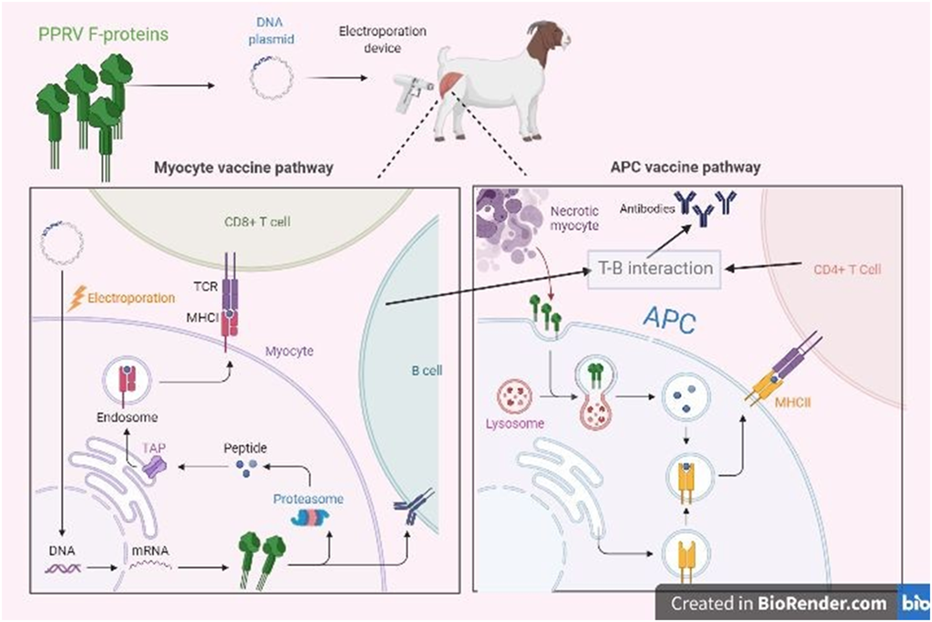
Recombinant DNA vaccines have emerged as promising, safe, and adaptable tools for immunization against PPRV. These vaccines consist of plasmid DNA encoding viral antigens under strong eukaryotic promoters, which are expressed by host cells after delivery and presented via both MHC class I and II pathways (Figure 2), thereby eliciting balanced humoral and cellular immune responses (Bello et al., Reference Bello, Alsaadi, Naeem, Almahboub, Bosaeed and Aljedani2025; Kutzler and Weiner, Reference Kutzler and Weiner2008).
Mechanism of action of PPRV-based DNA vaccine. The gene encoding PPRV F protein is cloned into a mammalian expression plasmid and administered via the intramuscular route. The protein is intracellularly generated in the myocyte, processed via the major histocompatibility complex class I pathway, and activates a CD8+ cell immune response. PPRV F protein from the necrotic myocyte may also be engulfed by the antigen-presenting cells, processed via the MHC class II pathway, and activated antibody-mediated immune response.

Figure 2 Long description
The mechanism of action of a PPRV-based DNA vaccine is divided into two pathways: the myocyte vaccine pathway and the APC vaccine pathway. On the left, the myocyte vaccine pathway begins with PPRV F-proteins and a DNA plasmid, which is introduced into a goat using an electroporation device. The pathway continues with the electroporation process leading to the entry of DNA into the myocyte. Inside the myocyte, the DNA is transcribed into mRNA, which is then translated into proteins. These proteins are processed by the proteasome into peptides. The peptides are transported by TAP into the endoplasmic reticulum, where they bind to MHC class I molecules. The MHC class I-peptide complex is presented on the surface of the myocyte, activating CD8 plus T cells. On the right, the APC vaccine pathway involves the necrotic myocyte releasing PPRV F proteins, which are engulfed by antigen-presenting cells (APCs). Inside the APC, the proteins are processed in the lysosome and presented on MHC class II molecules. This presentation leads to T-B cell interaction, activating CD4 plus T cells and antibody production. The diagram highlights the interaction between T cells and B cells, emphasizing the immune response activation through both pathways.
DNA vaccines offer several distinct advantages: they are non-replicating, non-integrating, and inherently biosafe. They can be produced rapidly at low cost, remain stable under moderate storage conditions, and permit repeated immunization without antivector immunity (Eusébio et al., Reference Eusébio, Neves, Costa, Biswas, Alves, Cui and Sousa2021). These attributes make DNA vaccination particularly attractive for use in endemic and resource-limited regions where cold-chain maintenance and large-scale livestock vaccination campaigns present logistical challenges.
Experimental DNA vaccines against PPRV have demonstrated encouraging immunogenicity in small animal models. Wang et al. (Reference Wang, Liu, Shi, Li, Li, Chen, Jin, Xu and Li2013b) first constructed a Semliki Forest virus replicon-based DNA vector expressing the PPRV hemagglutinin (H) gene (pSCA1-H), which elicited high titers of PPRV-specific and neutralizing antibodies as well as lymphocyte proliferation in BALB/c mice. A subsequent study using pSCA1-F, encoding the fusion (F) protein, confirmed similar humoral and cellular activation, with significant induction of IL-2, IL-10, IFN-γ, and TNF-α, reflecting a mixed Th1/Th2 immune profile (Wang et al., Reference Wang, Yue, Jin, Liu, Pan, Wang, Guo, Li and Li2015). In addition to direct expression of viral glycoproteins, innovative DNA immunogens have also been explored. Apsana et al. (Reference Apsana, Isloor and Shaila2015) demonstrated that immunization of sheep with a plasmid encoding the variable region of an anti-idiotypic antibody – designed to mimic PPRV epitopes – successfully induced both virus-specific humoral and cell-mediated immune responses. This study provided proof that DNA-encoded mimotopes can trigger specific immunity in the natural host without expressing the native viral antigen, broadening the conceptual scope of PPRV DNA vaccine design.
Although PPRV DNA vaccines remain in early development, their safety, thermostability, and adaptability make them valuable candidates for future refinement. Optimization of antigen design, delivery systems (e.g., electroporation, nanoparticle vectors), and validation in the target host species are critical next steps towards a practical DIVA-compatible platform.
Conclusion and way forward
The sustainable control and eventual eradication of PPR will yield enormous socio-economic benefits, particularly in low- and middle-income countries where small ruminants represent a primary source of livelihood, nutrition, and financial security. Achieving this global goal hinges on the availability of safe, thermostable, and DIVA-compatible vaccines capable of inducing long-lasting protective immunity across diverse viral lineages and production environments.
Significant progress has been achieved in the development of next-generation PPRV vaccines, spanning reverse-genetics–derived live attenuated constructs, viral-vectored platforms, VLP formulations, and recombinant plasmid DNA vaccines. Among these, reverse-genetics–based vaccines – which retain the replication competence and immunogenicity of traditional live vaccines while incorporating DIVA markers – currently hold the greatest potential for near-term application in eradication programmes. Viral-vectored vaccines, particularly those based on adenovirus, NDV, and goatpox virus backbones, have also demonstrated durable immunity and safety, providing promising alternatives for dual or multivalent protection. Non-replicating systems such as VLPs and DNA vaccines offer unmatched biosafety and design flexibility, and their immunogenicity can be further enhanced through rational epitope engineering, codon optimization, and the incorporation of novel adjuvants or delivery systems.
Given the scientific momentum and coordinated global initiatives under the FAO-WOAH (OIE) GEP-PPR, there is strong reason for optimism that PPR could become the second animal disease to be eradicated worldwide after rinderpest. Continued investment in modern vaccinology, genomic surveillance, and international cooperation will be critical to realize this milestone within the coming decade.



 Open access

