Introduction
The genus Micarea Fr. is a cosmopolitan group of crustose lichens, which has recently attracted increased interest among researchers around the world. According to various sources (Kirk et al. Reference Kirk, Cannon, Minter and Stalpers2008; Lücking et al. Reference Lücking, Hodkinson and Leavitt2017; Wijayawardene et al. Reference Wijayawardene, Hyde, Dai, Sánchez-García, Goto, Saxena, Erdoğdu, Selçuk, Rajeshkumar and Aptroot2022), the diversity of the Micarea is estimated at c. 100 species. Obviously, this number is an underestimate, since 69 Micarea species have been described in the last 10 years alone (Aptroot & Cáceres Reference Aptroot and Cáceres2014, Reference Aptroot and Cáceres2024; Brand et al. Reference Brand, van den Boom and Sérusiaux2014; Córdova-Chávez et al. Reference Córdova-Chávez, Aptroot, Castillo-Camposa, Cáceres and Pérez-Pérez2014; van den Boom & Ertz Reference van den Boom and Ertz2014; Brackel Reference Brackel2016; Guzow-Krzemińska et al. Reference Guzow-Krzemińska, Czarnota, Łubek and Kukwa2016, Reference Guzow-Krzemińska, Sérusiaux, van den Boom, Brand, Launis, Łubek and Kukwa2019; McCarthy & Elix Reference McCarthy and Elix2016a, Reference McCarthy and Elixb, Reference McCarthy and Elix2020a, Reference McCarthy and Elixb; Etayo Reference Etayo2017; van den Boom et al. Reference van den Boom, Brand, Coppins and Sérusiaux2017a, Reference van den Boom, Sipman, Divakar and Ertzb, Reference van den Boom, Guzow-Krzemińska and Kukwa2020, Reference van den Boom, Etayo and de Silanes2023; Elix & McCarthy Reference Elix and McCarthy2018; Kantvilas Reference Kantvilas2018; Hyde et al. Reference Hyde, Tennakoon, Jeewon, Bhat, Maharachchikumbura, Rossi, Leonardi, Lee, Mun and Houbraken2019; Kantvilas & Coppins Reference Kantvilas and Coppins2019; Launis et al. Reference Launis, Malíček, Svensson, Tsurykau, Sérusiaux and Myllys2019a, Reference Launis, Pykälä, van den Boom, Sérusiaux and Myllysb; Launis & Myllys Reference Launis and Myllys2019; Coppins et al. Reference Coppins, Kashiwadani, Moon, Spribille and Thor2021; Kantelinen et al. Reference Kantelinen, Hyvärinen, Kirika and Myllys2021, Reference Kantelinen, Svensson, Malíček, Vondrák, Thor, Palice, Svoboda and Myllys2024; van den Boom Reference van den Boom2021; Vondrák et al. Reference Vondrák, Svoboda, Malíček, Palice, Kocourková, Knudsen, Mayrhofer, Thüs, Schultz and Košnar2022; Schumm & Aptroot Reference Schumm and Aptroot2024). According to our estimation, the genus Micarea has more than 160 species.
Today the Micarea prasina group is one of the most studied in the genus. Based on molecular data, it includes 32 species, of which 26 have been described over the past 10 years (Guzow-Krzemińska et al. Reference Guzow-Krzemińska, Czarnota, Łubek and Kukwa2016, Reference Guzow-Krzemińska, Sérusiaux, van den Boom, Brand, Launis, Łubek and Kukwa2019; van den Boom et al. Reference van den Boom, Brand, Coppins and Sérusiaux2017a, Reference van den Boom, Guzow-Krzemińska and Kukwa2020; Launis et al. Reference Launis, Malíček, Svensson, Tsurykau, Sérusiaux and Myllys2019a, Reference Launis, Pykälä, van den Boom, Sérusiaux and Myllysb; Launis & Myllys Reference Launis and Myllys2019; Kantelinen et al. Reference Kantelinen, Hyvärinen, Kirika and Myllys2021). Micarea corallothallina Cáceres et al., M. hyalinoxanthonica Brand et al., M. kartana Kantvilas & Coppins and M. melanoprasina Brand et al. (Cáceres et al. Reference Cáceres, Mota, de Jesus and Aptroot2013; Brand et al. Reference Brand, van den Boom and Sérusiaux2014; Kantvilas Reference Kantvilas2018) probably belong to the M. prasina group as well, but due to the lack of molecular data their phylogenetic relationships are unclear.
The Micarea prasina group is characterized by effuse thalli composed of goniocysts and a ‘micareoid’ photobiont (a coccoid green alga with cells 4–7.5 μm diam.). Another important characteristic is the presence of the Sedifolia-grey pigment often produced in the epihymenium, pycnidial walls and dark-coloured parts of the thallus. The species of the M. prasina group are also characterized by immarginate apothecia of various colours, a hyaline hypothecium, branched paraphyses, and Micarea-type asci, with a K/I+ blue amyloid tholus and a more lightly staining axial body often with a darkly stained lining (Coppins Reference Coppins1983; Hafellner Reference Hafellner1984; Czarnota Reference Czarnota2007; Ekman et al. Reference Ekman, Andersen and Wedin2008; Czarnota & Guzow-Krzemińska Reference Czarnota and Guzow-Krzemińska2010; Guzow-Krzemińska et al. Reference Guzow-Krzemińska, Czarnota, Łubek and Kukwa2016, Reference Guzow-Krzemińska, Sérusiaux, van den Boom, Brand, Launis, Łubek and Kukwa2019; Launis et al. Reference Launis, Malíček, Svensson, Tsurykau, Sérusiaux and Myllys2019a, Reference Launis, Pykälä, van den Boom, Sérusiaux and Myllysb). According to molecular studies (e.g. Guzow-Krzemińska et al. Reference Guzow-Krzemińska, Czarnota, Łubek and Kukwa2016, Reference Guzow-Krzemińska, Sérusiaux, van den Boom, Brand, Launis, Łubek and Kukwa2019; Launis et al. Reference Launis, Malíček, Svensson, Tsurykau, Sérusiaux and Myllys2019a, Reference Launis, Pykälä, van den Boom, Sérusiaux and Myllysb; Kantelinen et al. Reference Kantelinen, Hyvärinen, Kirika and Myllys2021), the M. prasina group is monophyletic and divided into two main lineages, namely the M. micrococca clade and the M. prasina clade, with M. tomentosa Czarnota & Coppins and M. pusilla Launis et al. basal to these two clades. Launis et al. (Reference Launis, Pykälä, van den Boom, Sérusiaux and Myllys2019b) introduced a new character for species separation, the presence (Pol+) or absence (Pol−) of crystalline granules visible in polarized light in thallus and apothecia sections in the M. prasina group. This, combined with morphological and chemical characteristics, made possible a reliable separation of species in this group (Guzow-Krzemińska et al. Reference Guzow-Krzemińska, Sérusiaux, van den Boom, Brand, Launis, Łubek and Kukwa2019; Launis et al. Reference Launis, Malíček, Svensson, Tsurykau, Sérusiaux and Myllys2019a, Reference Launis, Pykälä, van den Boom, Sérusiaux and Myllysb; Kantelinen et al. Reference Kantelinen, Hyvärinen, Kirika and Myllys2021). For other taxa of the genus Micarea, the significance of this character remains poorly studied (Konoreva et al. Reference Konoreva, Chesnokov, Kuznetsova and Stepanchikova2019, Reference Konoreva, Chesnokov, Stepanchikova, Spribille, Björk and Williston2021a, Reference Konoreva, Chesnokov and Tagirdzhanovab).
In Russia, until recently most of the species with a goniocystose thallus and pale apothecia were referred to Micarea prasina Fr. s. lat. and required revision. Currently, 16 species of the M. prasina group are reliably known in Russia (Stepanchikova et al. Reference Stepanchikova, Andreev, Himelbrant, Motiejūnaitė, Schiefelbein, Konoreva and Ahti2017, Reference Stepanchikova, Himelbrant, Kuznetsova, Motiejūnaitė, Chesnokov, Konoreva and Gagarina2020, Reference Stepanchikova, Himelbrant, Chesnokov, Konoreva and Timofeeva2022; Urbanavichene & Urbanavichus Reference Urbanavichene and Urbanavichus2017, Reference Urbanavichene and Urbanavichus2021; Urbanavichus & Urbanavichene Reference Urbanavichus and Urbanavichene2017; Konoreva et al. Reference Konoreva, Chesnokov, Kuznetsova and Stepanchikova2019, Reference Konoreva, Chesnokov, Korolev and Himelbrant2020, Reference Konoreva, Chesnokov and Tagirdzhanova2021b; Launis et al. Reference Launis, Malíček, Svensson, Tsurykau, Sérusiaux and Myllys2019a; Tarasova et al. Reference Tarasova, Konoreva, Zhurbenko, Pystina, Chesnokov, Androsova, Sonina, Semenova and Valekzhanin2020; Urbanavichus et al. Reference Urbanavichus, Vondrák, Urbanavichene, Palice and Malíček2020; Davydov et al. Reference Davydov, Yakovchenko, Konoreva, Chesnokov, Ezhkin, Galanina and Paukov2021). To study the diversity of the genus Micarea in southern parts of the Russian Far East, extensive material was collected that was morphologically similar to M. isidioprasina van den Boom et al. Further study of the morphology, anatomy, secondary metabolites and molecular data led to the conclusion that the specimens belong to a new species, described here as M. svetlanae Konoreva & Chesnokov.
Materials and Methods
Field and herbarium studies
This study is based on the fieldwork of Liudmila Konoreva and Sergey Chesnokov in the Russian Far East in 2017–2020. The specimens collected and presented in this paper are deposited in the herbaria of the Komarov Botanical Institute of the Russian Academy of Sciences (LE), the Botanical Garden-Institute of the Far Eastern Branch of the Russian Academy of Sciences (VBGI), the Polar-Alpine Botanical Garden-Institute (separate department of the Kola Science Centre of the Russian Academy of Sciences) (KPABG) and the V. F. Kuprevich Institute of Experimental Botany of the National Academy of Science (MSK). A total of 74 specimens were studied, 18 of which were sterile. More detailed information about the locations of the samples studied is presented in the Supplementary Material (available online). The material was examined using standard microscopic techniques and spot tests with 10% potassium hydroxide (K), calcium hypochlorite (C) and paraphenylenediamine (PD) (Smith et al. Reference Smith, Aptroot, Coppins, Fletcher, Gilbert, James and Wolseley2009). Crystalline granules were investigated using a Zeiss Axio Scope.A1 compound microscope with polarizing filters. High performance thin-layer chromatography (HPTLC) was performed at the Laboratory of Lichenology and Bryology of the Komarov Botanical Institute, according to standard procedures using solvent systems A and C (Orange et al. Reference Orange, James and White2001). The names of the pigments observed in Micarea species follow Meyer & Printzen (Reference Meyer and Printzen2000). Photographs of the species were taken with a Motic SMZ-171-LED stereoscopic microscope with an attached MotiCam S6 camera and an Axio Scope.A1 with Axiocam 506 colour camera. The distribution maps were prepared using the GIS Axioma 5.1 program.
Nomenclature of vascular plants corresponds to the book ‘Flora of the Kuril Islands’ (Barkalov Reference Barkalov2009).
DNA extraction, amplification and sequencing
DNA was extracted directly from pieces of thalli or apothecia using the modified CTAB method (Guzow-Krzemińska & Węgrzyn Reference Guzow-Krzemińska and Węgrzyn2000) and used for PCR amplification of mtSSU rDNA. The primers mrSSU1 and mrSSU3R (Zoller et al. Reference Zoller, Scheidegger and Sperisen1999) were used as PCR and sequencing primers. PCR amplification was performed as follows: initial denaturation at 95 °C for 10 min and six cycles at 95 °C for 1 min, 62 °C for 1 min and 72 °C for 105 s, followed by 40 cycles at 95 °C for 1 min, 56 °C for 1 min and 72 °C for 1 min, and a final extension step at 72 °C for 10 min (Czarnota & Guzow-Krzemińska Reference Czarnota and Guzow-Krzemińska2010). Amplicons were sequenced by Eurogen (Moscow, Russia; https://evrogen.ru/). Newly generated sequences were deposited in NCBI (GenBank) (Table 1).
Micarea specimens used in this study with voucher information and GenBank Accession numbers. Sequences newly generated for this study are given in bold.

Sequence alignment and phylogenetic analysis
The mtSSU alignment was compiled with all the species of the Micarea prasina group and several closely related species were used as an outgroup following Guzow-Krzemińska et al. (Reference Guzow-Krzemińska, Sérusiaux, van den Boom, Brand, Launis, Łubek and Kukwa2019). The dataset was aligned online using MAFFT v. 7 (Katoh & Standley Reference Katoh and Standley2013; available at http://mafft.cbrc.jp/alignment/server/), with the L-INS-i method (Katoh et al. Reference Katoh, Kuma, Toh and Miyata2005) selected automatically by the program. To exclude ambiguously aligned positions, alignment was subsequently analyzed by the automated1 algorithm as implemented in the TrimAl software package (Capella-Gutierrez et al. Reference Capella-Gutierrez, Silla-Martinez and Gabaldon2009). Phylogenetic reconstruction was carried out using Bayesian inference in MrBayes v. 3.2.6 (Ronquist & Huelsenbeck Reference Ronquist and Huelsenbeck2003) and maximum likelihood (ML) in RAxML (Stamatakis et al. Reference Stamatakis, Ludwig and Meier2005) through the RAxMLGUI interface (Silvestro & Michalak Reference Silvestro and Michalak2012). Bootstrap support values were calculated on 1000 bootstrap replicates using rapid bootstrapping (‘ML + rapid bootstrap’ function in RAxMLGUI). The analyses were run on the CIPRES Web Portal (http://www.phylo.org/portal2/). The HKY + G model was proposed by the program jModelTest (Guindon & Gascuel Reference Guindon and Gascuel2003; Posada Reference Posada2008) as the best DNA substitution model. MrBayes analysis (BI) was performed using two independent runs with four MCMC chains (three cold and one heated) in each run. Trees were sampled every 500th generation. The analysis was stopped when the average standard deviation of split frequencies between the simultaneous runs dropped below 0.01 (1 270 000 generations). The first 25% of trees was discarded as burn-in, and the remaining trees were used for construction of a 50% majority-rule consensus tree.
Results
Phylogenetic analyses
Three new mtSSU rDNA sequences were generated and 72 downloaded from GenBank. The final alignment consisted of 75 sequences and 637 characters. Since the topologies from the maximum likelihood and Bayesian analyses did not show any supported conflict, the maximum likelihood tree is presented in Fig. 1 with added posterior probabilities from the Bayesian analysis.
Phylogenetic reconstruction of the Micarea prasina group, based on maximum likelihood analysis (ML) of mtSSU. The reliability of each branch was tested by ML and Bayesian methods. Numbers at tree nodes indicate bootstrap values of ML (left) and BMCMC posterior probabilities (right). Thicker branches indicate when both the bootstrap value of ML is ≥ 70% and the BMCMC posterior probability is ≥ 0.95. Newly sequenced specimens are indicated in bold and voucher information for all specimens is provided in Table 1. Branch lengths represent the estimated number of substitutions per site assuming the respective models of substitution. In colour online.

The phylogenetic reconstruction (Fig. 1) shows that the Micarea prasina group is highly supported and monophyletic (96/1.00; ML/BI). It is divided into the M. micrococca (Körb.) Gams ex Coppins and M. prasina clades, lineages of M. hedlundii Coppins and M. xanthonica Coppins & Tønsberg basal to the M. micrococca clade and lineages of M. tomentosa and M. pusilla forming a highly supported clade basal (not supported) to other clades. This is in agreement with previous studies (e.g. Czarnota & Guzow-Krzemińska Reference Czarnota and Guzow-Krzemińska2010; Guzow-Krzemińska et al. Reference Guzow-Krzemińska, Czarnota, Łubek and Kukwa2016, Reference Guzow-Krzemińska, Sérusiaux, van den Boom, Brand, Launis, Łubek and Kukwa2019; Launis et al. Reference Launis, Malíček, Svensson, Tsurykau, Sérusiaux and Myllys2019a, Reference Launis, Pykälä, van den Boom, Sérusiaux and Myllysb; Launis & Myllys Reference Launis and Myllys2019; van den Boom et al. Reference van den Boom, Guzow-Krzemińska and Kukwa2020; Kantelinen et al. Reference Kantelinen, Hyvärinen, Kirika and Myllys2021).
The M. micrococca clade (91/1.00) consists mostly of species containing methoxymicareic acid and is divided into two lineages of M. byssacea and M. micrococca complexes. The M. prasina clade (48/0.99) consists mostly of species containing micareic acid and accommodates the newly described M. svetlanae. Several highly supported lineages are further distinguished within this clade. The sampled specimens of M. svetlanae form a highly supported clade (100/1.00) sister without support to the clade comprising M. isidioprasina and M. flavoleprosa Launis et al. (Fig. 1).
Taxonomy
Micarea svetlanae Konoreva & Chesnokov sp. nov.
MycoBank No.: MB 853375
Similar to M. isidioprasina due to the presence of micareic acid, the granular-isidiate thallus with Sedifolia-grey, and crystalline granules in the thallus and hymenium, but differing in the cushion-shaped thallus, with cushions up to 0.6 mm diam. and 0.4 mm high, the presence of Sedifolia-grey pigment in the hymenium, numerous crystalline granules in the hymenium and hypothecium and 0–2(–3)-septate ascospores.
Type: Russia, Khabarovsk Territory, Ulchsky District, upper reaches of the Levyi Psyu River, 51°48ʹ04.5ʺN, 141°03ʹ47.1ʺE, 266 m a.s.l., spruce-fir fern-blueberry forest, on rotten fir wood, 25 September 2018, S. V. Chesnokov 193 (LE L-26024—holotype).
(Figs 2–5)
Morphological variability of Micarea svetlanae Konoreva & Chesnokov. A, thallus and apothecia without Sedifolia-grey. B, thallus and apothecia with high content of Sedifolia-grey. C & D, subconvex apothecia immersed in the thallus. E & F, differently coloured areas of the same specimens; arrows indicate pale-coloured thallus (without Sedifolia-grey) in the crack. G, tuberculate apothecium. H, adnate apothecia. Scales: A–F & H = 0.5 mm; G = 1 mm. In colour online.

Thallus crustose, granular-isidiate, pale to dark green, sometimes a transition from very pale to dark olive can be seen within a single specimen (depending on light conditions) (Fig. 2A, B, E & F). Non-isidiate parts rare, granular or minutely areolate, areoles up to 0.05 mm diam., green, soon developing isidia. Isidia consisting of chains of goniocysts, coralloid, up to 175 μm tall and 25 μm wide (Figs 2A–H, 3B, D & F), forming an almost continuous layer over the substratum. Denser clusters of isidia forming a thick cushion-like thallus, which is then divided by fissures into separate dense cushions up to 0.4 mm tall and 0.6 mm diam. (Fig. 4F & H). The cushions, like the isidia, are made up of numerous goniocysts. Prothallus not visible. Photobiont micareoid, cells thin walled, 4–7 μm diam., clustered in compact groups, single goniocysts up to 25 μm diam. (Fig. 3B).
Section of the apothecium and thallus of Micarea svetlanae. A & B, in transmitted light. C & D, in polarized light. E & F, in transmitted light after reaction with K. Scales = 50 μm. In colour online.

Comparison of anatomy (A–D) and morphology (E–H) of Micarea prasina (A, C, E, G) and M. svetlanae (B, D, F, H). A & B, localization of crystalline granules in transmitted light. C & D, localization of crystalline granules in polarized light. E, granular warty thallus and subglobose apothecia of M. prasina. F, cushion-like thallus, divided by fissures into separate dense cushions and apothecia of M. svetlanae immersed in the thallus. G, granular warty thallus of M. prasina. H, section through a thallus cushion of M. svetlanae. Scales: A–D = 50 μm; E & F = 0.5 mm; G = 0.4 mm; H = 1 mm. In colour online.

Apothecia immarginate, adnate to convex, 0.2–0.5 mm diam., often tuberculate and then up to 0.8 mm diam., pale cream to grey or dark grey, often different colours present within a single apothecium (Fig. 2A–H). When the apothecia develop among the cushions of the thallus, they appear immersed (Fig. 2C & D). Epihymenium hyaline; hymenium up to 65 μm high, hyaline (pale-coloured form) to pale greyish olive (in dark-coloured forms); hypothecium hyaline (Fig. 3A); paraphyses sparse, branched, anastomosing, 1.0–1.2(–1.5) μm wide, tips not widened and not pigmented (Fig. 5B); asci cylindrical, 40–50 × 10–12(–17) μm (n = 30), 8-spored (Fig. 5B); ascospores ellipsoidal to ovoid, 0‒2(–3)-septate, 10–15 × 3.0–5.0(–6) μm (n = 96) (Fig. 5A).
Asci, ascospores and paraphyses of Micarea svetlanae. A, variability of ascospores. B, asci with ascospores and anastomosing paraphyses. Scales: A = 20 μm; B = 25 μm. In colour online.

Pycnidia not observed.
Chemistry
Micareic acid detected by HPTLC; K−, C−, KC−, PD−. Sedifolia-grey pigment (K+ violet, C+ violet) present in hymenium (Fig. 3E) and dark-coloured areas of the thallus (Fig. 3F), sometimes indistinct. Crystalline granules (studied in polarized light) abundant, visible in hymenium, hypothecium and thallus, soluble in K (Figs 3C & D, 4D).
Etymology
The species is named in honour of the Russian lichenologist, Dr Svetlana Tchabanenko (Chabanenko), who devoted her life to the study of lichens of the Russian Far East.
Habitat and distribution
Throughout its geographical range, Micarea svetlanae grows abundantly on lignum of coniferous trees Abies nephrolepis (Trautv. et Maxim.) Maxim., A. sachalinensis Fr. Schmidt, Larix kamtschatica (Rupr.) Carr., Picea jezoensis (Siebold et Zucc.) Carr. and Taxus cuspidata Siebold et Zucc.) and sometimes on the bark of fallen deadwood of Abies sachalinensis, Pinus koraiensis Siebold & Zucc. and Salix sp. in coniferous forests dominated by Abies sachalinensis, Larix kamtschatica and Picea jezoensis with Juniperus sp., Sasa kurilensis (Rupr.) Makino & Shibata, mosses and deadwood or in mixed forests with the same composition of conifers and Betula ermanii Cham. The new species often grows together with Micarea prasina, M. nowakii Czarnota & Coppins, M. laeta Launis & Myllys, Trapelia corticola Coppins & P. James and Cladonia spp. Micarea svetlanae is known only from the Russian Far East, namely Primorye and Khabarovsk Territories as well as Sakhalin and the Kuril Islands (Shikotan, Kunashir and Iturup) (Fig. 6). It is likely that the species range in Russia is limited to southern parts of the Russian Far East, since M. svetlanae was not found during our intensive field studies in the Magadan Region, Kamchatka Territory and Paramushir Island. In addition, it is likely that the species may be found in Japan, Korea and China.
Known distribution of Micarea svetlanae in the Russian Far East. In colour online.

Selected specimens examined
Russia: Primorye Territory: Dal'negorsk District, 8 km north-west of Krasnorechensky village, left bank of the Rudnaya River, 44°39ʹ37.4ʺN, 135°15ʹ05.6ʺE, 932 m, 2020, L. A. Konoreva 64 (LE L-26044); Terneysky District, Sikhote-Alin Nature Reserve, vicinity of the cordon Yasnaya, right bank of the Yasnaya River, 45°14ʹ25.2ʺN, 136°29ʹ21.5ʺE, 116 m, 2020, L. A. Konoreva 193 (KPABG 21307). Sakhalin Region: Iturup Island, Ostrovnoy Reserve, Stokap volcano, Craternyi stream, 44°50ʹ25.9ʺN, 147°17ʹ44.7ʺE, 369 m, 2017, L. A. Konoreva 619 (LE L-26012); Kunashir Island, Kurilsky Nature Reserve, vicinity of the cordon Saratovsky, 44°15ʹ41.4ʺN, 146°06ʹ26.0ʺE, 10 m, 2019, L. A. Konoreva 50 (VBGI 170161); ibid., left bank of the River Saratovskaya, 44°16ʹ21.7ʺN, 146°06ʹ36.4ʺE, 28 m, 2019, S. V. Chesnokov 17 (LE L-26039, MSK); Sakhalin Island, Korsakovsky District, natural monument ‘Lagoon Busse’, surroundings of the Vyselkovoe Lake, 46°33ʹ57.1ʺN, 143°16ʹ54.7ʺE, 26 m, 2017, L. A. Konoreva 220, 230 (LE L-26016, LE L-26017; GenBank No. PP477414); vicinity of Yuzhno-Sakhalinsk city, Susunaisky ridge, Chekhov peak, Voroniy kamen’ viewing platform, 46°58ʹ38.3ʺN, 142°49ʹ25.7ʺE, 352 m, 2017, L. A. Konoreva 248 (LE L-26020; GenBank No. PP477415); Tomarinsky District, Krasnogorsky nature reserve, vicinity of the Uglovogo Lake, 48°33ʹ39.9ʺN, 141°58ʹ15.0ʺE, 37 m, 2017, L. A. Konoreva 88 (VBGI 170158); Shikotan Island, vicinity of Tserkovnaya Bay, 43°44ʹ16.5ʺN, 146°41ʹ06.7ʺE, 30 m, 2017, L. A. Konoreva 369, 374 (LE L-26015, LE L-26022; GenBank No. PP477413). Khabarovsk Territory: Ulchsky District, 1.7 km west of Tabo Mt, 51°39ʹ21.4ʺN, 140°53ʹ45.9ʺE, 111 m, 2018, S. V. Chesnokov 149, 150, 151 (KPABG 21303, KPABG 21304, MSK); Khabarovsk District, Bolshekhehtsirsky reserve, Bykova River, vicinity of the ‘Bykovka’ cordon, 48°14ʹ27.7ʺN, 134°48ʹ54.1ʺE, 253 m, 2018, S. V. Chesnokov 205 (LE L-26030).
Discussion
Micarea svetlanae belongs to the M. prasina group and contains micareic acid as the main secondary metabolite. The most characteristic features of this species are the isidia-like granules forming a rather thick cushion-shaped thallus, 0–2(–3)-septate ascospores, Sedifolia-grey pigment in the hymenium, and crystalline granules in the hymenium and hypothecium visible in polarized light.
Micarea isidioprasina and M. flavoleprosa are closely related to M. svetlanae. However, M. flavoleprosa is easily distinguished by its yellowish green to whitish green thallus, composed of minute soredia or small goniocysts which often coalesce to form larger granules, and the absence of Sedifolia-grey pigment in the apothecia and thallus (Launis et al. Reference Launis, Malíček, Svensson, Tsurykau, Sérusiaux and Myllys2019a).
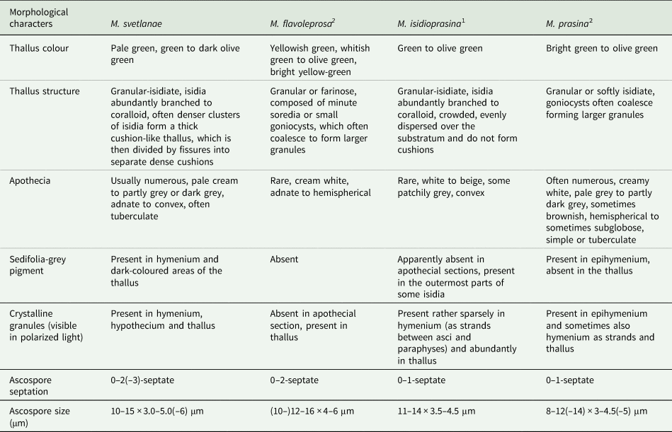
The most difficult to separate from M. svetlanae is M. isidioprasina, which also has a granular-isidiate thallus with Sedifolia-grey pigment, and crystalline granules in the hymenium and thallus. However, the isidia of M. isidioprasina are evenly dispersed over the substratum and do not form cushions, and its spores are 0–1-septate (Guzow-Krzemińska et al. Reference Guzow-Krzemińska, Sérusiaux, van den Boom, Brand, Launis, Łubek and Kukwa2019), whereas M. svetlanae has a cushion-shaped thallus, often cracked into individual cushions, and 0–2(–3)-septate spores (Table 2).
A comparison of the characteristics of Micarea svetlanae with related species having micareic acid.

Distinctive features of species according Guzow-Krzemińska et al. (Reference Guzow-Krzemińska, Sérusiaux, van den Boom, Brand, Launis, Łubek and Kukwa2019)1 and Launis et al. (Reference Launis, Malíček, Svensson, Tsurykau, Sérusiaux and Myllys2019a)2.
At the initial stages of its development, Micarea svetlanae may be similar to M. prasina with a slightly isidiate thallus, but the goniocysts of the latter species tend to merge into larger, never coralloid-branch granules. In addition, the Sedifolia-grey pigment is present in the epihymenium of M. prasina, crystalline granules visible in polarized light are present in the epihymenium and sometimes also in the hymenium as strands, but never in hypothecia, and its ascospores are 0–1-septate (Launis et al. Reference Launis, Malíček, Svensson, Tsurykau, Sérusiaux and Myllys2019a; Fig. 4A–H, Table 2).
Pale forms of Micarea svetlanae (with or without traces of Sedifolia-grey) may resemble M. levicula (Nyl.) Coppins, M. microsorediata Brand et al., M. pauli Guzow-Krzemińska et al., M. viridileprosa Coppins & van den Boom and M. xanthonica Coppins & Tønsberg, but they are easily distinguished from these species by the presence of micareic acid. Both M. microsorediata and M. pauli produce methoxymicareic acid (Guzow-Krzemińska et al. Reference Guzow-Krzemińska, Sérusiaux, van den Boom, Brand, Launis, Łubek and Kukwa2019), M. levicula and M. viridileprosa produce gyrophoric acid (van den Boom & Coppins Reference van den Boom and Coppins2001), and M. xanthonica contains thiophanic acid and other xanthones as the main secondary metabolites (Coppins & Tønsberg Reference Coppins and Tønsberg2001). In addition, M. microsorediata and M. viridileprosa form soralia, and M. levicula, M. microsorediata, M. pauli and M. viridileprosa never produce 2–3-septate ascospores.
Specimens of Micarea svetlanae with adnate apothecia and crystalline granules in the hymenium may be mistakenly identified as M. byssacea (Th. Fr.) Czarnota et al., and light forms as M. microareolata Launis et al. However, these species are distinguished by the production of methoxymicareic acid, the absence of isidia-like coralloid-branching goniocysts and 0–1-septate ascospores (Czarnota & Guzow-Krzemińska Reference Czarnota and Guzow-Krzemińska2010; Launis et al. Reference Launis, Pykälä, van den Boom, Sérusiaux and Myllys2019b).
Acknowledgements
The study was carried out in the framework of the institutional research projects ‘Cryptogamic biota of Pacific Asia: taxonomy, biodiversity, species distribution’ of the Botanical Garden-Institute of the Far Eastern Branch of the Russian Academy of Sciences (work by S. Chesnokov and L. Konoreva) and ‘Flora and taxonomy of algae, lichens and bryophytes of Russia and phytogeographically important regions of the world’ no. 121021600184-6 (work by S. Chesnokov). Ivan Frolov worked within the framework of the national project of the Botanical Garden-Institute (Russian Academy of Sciences, Ural Branch) and the project 122040800089-2 of the Botanical Garden-Institute FEB RAS. We are grateful to Curtis Björk (University of British Columbia, Canada) for the linguistic revision of the manuscript and to anonymous reviewers for valuable comments.
Author ORCIDs
Liudmila A. Konoreva, 0000-0002-4487-5154; Sergey V. Chesnokov, 0000-0001-9466-4534; Ivan V. Frolov, 0000-0003-4454-3229.
Supplementary Material
The Supplementary Material for this article can be found at https://doi.org/10.1017/S0024282924000446.