1. Introduction
Over the past few years our structural understanding of the ribosome and the initiation factors that promote formation of competent ribosomes on mRNA has increased dramatically. Although detailed analysis of eukaryotic protein synthesis has lagged behind that of bacterial systems, we are beginning to understand the underlying mechanisms of eukaryotic translation initiation despite the current lack of molecular structures of eukaryotic ribosomes.
The goal of this review is to summarize the biophysical approaches and resulting discoveries that have revealed substantial information about ribosome dynamics during the initiation of eukaryotic protein synthesis. We also discuss the challenges that lie ahead in understanding the functional states and dynamic motions of the ribosome during the initiation process. While it has been known for some time that multiple initiation factors are required for this process, our molecular understanding of how these factors promote each stage of initiation has not been clear. We have tried to present what we see as important developments in our understanding of this mechanism and direct the reader to recent specialized reviews for greater detail about specific events and the initiation factors that appear to promote them.
2. Protein synthesis and ribosome structure
Protein synthesis in all organisms is carried out by ribosomes, which consist of two ribonucleoprotein subunits that translate mRNA into protein by catalyzing the formation of peptide bonds. There are four stages to this process: initiation, elongation, termination, and recycling. During initiation, the two ribosomal subunits associate at the initiation codon of the mRNA, which is recognized by virtue of a methionyl initiator tRNA bound to the peptidyl (P) site of the small ribosomal subunit. Elongation requires the ribosome to decode the mRNA sequence by repeated cycles of three distinct steps: (1) recruitment of aminoacyl tRNAs to the aminoacyl (A) site of the small ribosomal subunit, (2) formation of a peptide bond between the incoming amino acid and the amino acid on the tRNA in the P-site, and (3) translocation of the mRNA and tRNAs so that the next codon is placed in the A-site of the ribosome. Protein synthesis terminates when a stop codon is placed in the A-site, causing the finished peptide to be released from the ribosome. Finally, ribosome recycling involves dissociation from the mRNA and the ejection of the bound deacylated tRNA so that the ribosome can enter another round of protein synthesis. Each stage of this process requires the ribosome to undergo many dynamic motions so that the mRNA is translated accurately and efficiently into protein (for details of the stages of eukaryotic protein synthesis see Kapp & Lorsch, Reference Kapp and Lorsch2004; Marintchev & Wagner, Reference Marintchev and Wagner2004; Hinnebusch et al. Reference Hinnebusch, Dever, Asano, Mathews, Sonenberg and Hershey2007; Pestova et al. Reference Pestova, Lorsch, Hellen, Mathews, Sonenberg and Hershey2007).
Ribosomes from the three kingdoms of life possess many sequence and structural similarities, indicating a common evolutionary origin. Bacterial ribosomes consist of a small (30S) subunit and a large (50S) subunit that together form the 70S ribosome. The small subunit comprises a single 1540-nt RNA (16S) and 21 proteins, while the large subunit includes a small 120-nt RNA (5S) along with a 2900-nt RNA (23S) and some 33 proteins. While eukaryotic ribosomes are larger in size than their cousins, they still possess a small (40S) subunit and a large (60S) subunit that form an 80S ribosome. The small subunit consists of a 1900-nt RNA (18S) and 33 proteins, while the large subunit comprises a 12-nt RNA (5S), a 160-nt RNA (5·8S), a 4700-nt RNA (28S) and 49 proteins. Many years of biochemical work and more recent X-ray and cryo-electron microscopic (cryo-EM) structures have helped us to understand more about the processes by which ribosomes function and are regulated during each stage of protein synthesis. Structural studies suggest that although different in size, ribosomes from all three kingdoms of life share a high degree of common structure, which likely reflects conservation in the fundamental process of protein biosynthesis (Taylor et al. Reference Taylor, Frank, Kinzy, Mathews, Sonenberg and Hershey2007). Recent X-ray crystallographic structures of the ribosome and its subunits from bacteria and archaea have indicated that the interactions between mRNA, tRNA and rRNA required for elongation primarily involve RNA–RNA contacts, and it is anticipated that this will also be true for eukaryotic ribosomes as higher-resolution structures become available (Noller, Reference Noller, Mathews, Sonenberg and Hershey2007). For all ribosomes, the small subunit has been identified as that which contacts and decodes the mRNA, whereas the large subunit contains the catalytic center that promotes peptide bond formation. The role of extra rRNA sequences in eukaryotes, called expansion segments, along with the extra proteins that they bind is currently unclear. These expansion regions are found predominantly around the periphery of the small subunit, as determined by comparing cryo-EM structures (reviewed in Taylor et al. Reference Taylor, Frank, Kinzy, Mathews, Sonenberg and Hershey2007). There are functional differences in a number of stages of protein synthesis between the kingdoms of life, particularly during the initiation of protein synthesis, which likely account for some of these eukaryotic-specific regions of ribosomes. In addition, the differences in how the ribosome functions in protein transport and folding, as well as the fact that eukaryotic ribosomes must also pass through the nuclear pore complex, may also reflect the requirement of ribosome features that are specific to eukaryotes.
3. Overview of the initiation mechanism and factors
A key regulatory step in protein synthesis is the mechanism by which ribosomes initiate protein synthesis. Some similarities and differences in this mechanism are immediately apparent between bacteria and eukaryotes and have been the subject of recent reviews (Kapp & Lorsch, Reference Kapp and Lorsch2004; Marintchev & Wagner, Reference Marintchev and Wagner2004; Merrick, Reference Merrick2004; Jackson, Reference Jackson2005; Kozak, Reference Kozak2005). A model of the steps involved is presented in Fig. 1. In each case, the ribosome must first separate into its two subunits, with the small subunit engaging a mRNA and selecting the start site for translation. Binding of a dedicated Met-tRNA to the small subunit in a GTP-dependent manner to recognize the initiation codon is also conserved, although the regulation of this step is significantly different in eukaryotes (Hinnebusch et al. Reference Hinnebusch, Dever, Asano, Mathews, Sonenberg and Hershey2007; Proud, Reference Proud2006). In bacteria, the Shine–Dalgarno (SD) sequence of the mRNA base pairs with a complementary sequence in the 16S rRNA, enabling 30S binding and initiation on polycistronic mRNAs directly at the site of the AUG codon (Shine & Dalgarno, Reference Shine and Dalgarno1974; Steitz & Jakes, Reference Steitz and Jakes1975). Importantly, the sequence located near the 3′ end of the 16S rRNA that recognizes the SD sequence is specifically deleted in the eukaryotic 18S rRNA, thereby preventing direct recognition of mRNA (Hagenbuchle et al. Reference Hagenbuchle, Santer, Steitz and Mans1978). Instead, the majority of eukaryotic mRNAs initiate by a scanning mechanism that involves 40S subunit loading at the 5′ end of the mRNA and subsequent migration to the AUG codon (Kozak, Reference Kozak1989b). The regulation of initiation codon recognition also appears to differ between bacteria and eukaryotes. Initiation in bacteria is very sensitive to the degree of secondary structure at the initiation site, while eukaryotes possess RNA helicases that reduce the effect of secondary structure on the initiation event. Interestingly, recent work has revealed mRNAs of the Flaviviridae family of viruses that appear to associate directly with the eukaryotic 40S subunit close to the AUG codon. However, these mRNAs utilize an RNA structure that allows direct binding of the 40S subunit rather than base pairing as found in prokaryotes (reviewed in Pisarev et al. Reference Pisarev, Shirokikh and Hellen2005; Fraser & Doudna, Reference Fraser and Doudna2007). Finally, once the AUG codon is recognized by the initiator tRNA, GTP is hydrolyzed and the large ribosomal subunit joins to form a competent ribosome that can decode the mRNA into protein.
Translation initiation. The proposed pathway of translation initiation in prokaryotes and eukaryotes is presented. Ribosome subunits are colored grey and labeled according to their respective sedimentation values. The functionally conserved initiation factors between prokaryotes and eukaryotes are indicated using the same colors; eIF1/IF3 are in red while eIF1A/IF1 are in yellow. In addition, eIF5B/IF2 are colored green. All other eukaryotic specific initiation factors are indicated using additional colors. The mRNA is drawn with the 5′ and 3′ ends labeled in both cases; the Shine–Dalgarno sequence (SD) in the prokaryotic mRNA is indicated as are the 5′ end cap structure and 3′ end poly(A) tail of the eukaryotic mRNA. The AUG initiation codon is also labeled. The exact timing of association and release of initiation factors are not always known, so the pathways indicate one possible model for each system. The eukaryotic pathway assumes that the 5′ end cap structure remains associated with the scanning 40S subunit, although this is currently not well understood. For additional details regarding each stage of the pathways please see the text.

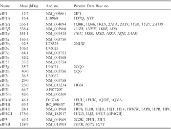
Both bacterial and eukaryotic ribosomes utilize multiple protein factors during the initiation phase. The evolution of initiation factor structure and function has recently been extensively reviewed (Marintchev & Wagner, Reference Marintchev and Wagner2004). Based on results of both structural and biochemical studies, translation initiation factors are thought to lower the energetic barriers between conformational states of the small ribosomal subunit so that each stage of initiation occurs efficiently. Three single polypeptide initiation factors, with a combined molecular weight of ∼125 kDa, bind the bacterial 30S subunit (reviewed in Boelens & Gualerzi, Reference Boelens and Gualerzi2002; Allen & Frank, Reference Allen and Frank2007; Noller, Reference Noller, Mathews, Sonenberg and Hershey2007). However, eukaryotes utilize ∼30 different initiation factor polypeptides with a molecular weight of ∼1600 kDa in order to promote efficient protein synthesis initiation. Although three of the eukaryotic initiation factors share some common structural features and functions with the bacterial initiation factors, the order of magnitude increase in molecular weight of initiation factors required by eukaryotes reflects a far more complex and highly regulated initiation process. The eukaryotic initiation factors that are known to associate with the 40S subunit are listed in Table 1. This list does not include initiation factors such as the GTP exchange factor eIF2B that does not associate with the 40S subunit, or other factors that have unclear roles at present. A full list of proteins involved during the process of eukaryotic initiation can be found in a recent review (Pestova et al. Reference Pestova, Lorsch, Hellen, Mathews, Sonenberg and Hershey2007).
Eukaryotic initiation factors

All sequences are human nucleotides; structures are all eukaryotic or archaeal (italics).
Accession numbers are taken from the National Center for Biotechnology Information database (http://www.ncbi.nlm.nih.gov).
Protein DataBase (PDB) numbers are taken from the Research Collaboratory for Structural Bioinformatics (RCSB) database (http://www.rcsb.org/pdb).
A number of model systems have been exploited to determine the eukaryotic initiation pathway. One approach has been to reconstitute the system in vitro with highly purified components, using recombinant proteins wherever possible (Schreier & Staehelin, Reference Schreier and Staehelin1973; Schreier et al. Reference Schreier, Erni and Staehelin1977; Benne & Hershey, Reference Benne and Hershey1978; Benne et al. Reference Benne, Brown-Luedi and Hershey1979; Merrick, Reference Merrick1979; Staehelin et al. Reference Staehelin, Erni and Schreier1979; Pestova et al. Reference Pestova, Borukhov and Hellen1998a, Reference Pestova, Lomakin, Lee, Choi, Dever and Hellen2000; Masutani et al. Reference Masutani, Sonenberg, Yokoyama and Imataka2007). While this approach does not allow for the true kinetics of protein synthesis to be maintained, it permits the identification of the initiation factors involved at each stage of initiation. Together with genetic approaches, it has also enabled the identification of initiation-factor domains that are important in promoting the stages of initiation (Hinnebusch et al. Reference Hinnebusch, Dever, Asano, Mathews, Sonenberg and Hershey2007). Alternative approaches have utilized the highly efficient reticulocyte lysate system that can be manipulated to study the stages of initiation, which has the advantage of maintaining kinetics close to those found in vivo while minimizing artifacts (Pelham & Jackson, Reference Pelham and Jackson1976; Jackson & Hunt, Reference Jackson and Hunt1983; Jackson, Reference Jackson2005).
In order to associate with a mRNA, a pool of 40S subunits must be available. In vivo, ribosomes tend to exist as inactive 80S particles that include the two ribosomal subunits associated with each other by virtue of inter-subunit bridge contacts. To be recycled, these ribosomes must first be separated into 40S and 60S subunits by a mechanism that is currently unclear. It is possible that at equilibrium, a small pool of free 40S subunits exists with bound initiation factors. Binding studies have shown that four initiation factors bind to free 40S subunits and thus prepare them for mRNA recruitment by remaining free of 60S subunits until AUG recognition. These initiation factors are eIF1, eIF1A, eIF3 and the eIF2⋅GTP⋅Met-tRNAi complex (Fig. 1 and Table 1). While little is known about the order of binding of these initiation factors to the 40S subunit, they appear to possess specific functions with regard to maintenance of the 40S subunit pool and the preparation of the 40S subunit for mRNA recruitment (reviewed in Marintchev & Wagner, Reference Marintchev and Wagner2004; Hinnebusch et al. Reference Hinnebusch, Dever, Asano, Mathews, Sonenberg and Hershey2007).
Prior to mRNA binding, the initiator tRNA must be recruited to the P-site of the free 40S subunit. The Met-tRNAi forms a ternary complex with eIF2 and GTP and this complex is able to associate with the 40S subunit in a manner stimulated by the small initiation factors eIF1A, eIF1 (homologs of prokaryotic IF1 and IF3) and the much larger eIF3 complex (Trachsel et al. Reference Trachsel, Erni, Schreier and Staehelin1977; Benne & Hershey, Reference Benne and Hershey1978; Peterson et al. Reference Peterson, Merrick and Safer1979; Chaudhuri et al. Reference Chaudhuri, Chowdhury and Maitra1999; Majumdar et al. Reference Majumdar, Bandyopadhyay and Maitra2003; Olsen et al. Reference Olsen, Savner, Mathew, Zhang, Krishnamoorthy, Phan and Hinnebusch2003; Passmore et al. Reference Passmore, Schmeing, Maag, Applefield, Acker, Algire, Lorsch and Ramakrishnan2007). eIF2 is a heterotrimer that has an archaeal homolog but no eubacterial homolog. Functionally, eIF2 is similar to prokaryotic IF2 in its role in stabilizing the initiator form of Met-tRNA to the small ribosomal subunit, but as we will discuss later, eIF5B is actually the structural and functional homolog of IF2 in the role of promoting subunit joining (Fig. 1). The 800 kDa eIF3 complex, unique to eukaryotes, consists of 13 proteins in humans, six of which are conserved in Saccharomyces cerevisiae. eIF3 can associate with the 40S subunit in the absence of any other initiation factors and appears to function at most of the stages of initiation (Fig. 1; reviewed in Hinnebusch, Reference Hinnebusch2006). Importantly, both eIF3 and the eIF2⋅GTP⋅Met-tRNA complex appear to be important in recruitment of the 40S subunit to mRNA. A capped mRNA is recognized by the cap-binding protein eIF4E (Sonenberg et al. Reference Sonenberg, Morgan, Merrick and Shatkin1978; Marcotrigiano et al. Reference Marcotrigiano, Gingras, Sonenberg and Burley1997), which is a component of the heterotrimeric complex eIF4F (Table 1). It has been proposed that eIF4F (eIF4E, eIF4A and eIF4G) first binds to a mRNA and recruits the 40S subunit by virtue of a direct interaction between eIF4G and eIF3 (reviewed in Kapp & Lorsch, Reference Kapp and Lorsch2004; Pestova et al. Reference Pestova, Lorsch, Hellen, Mathews, Sonenberg and Hershey2007). However, a high affinity of eIF4G for the 40S subunit appears to require not only eIF3, but also the eIF2⋅GTP⋅Met-tRNA complex, suggesting that mRNA recruitment to the 40S subunit requires more than one connection (Hui et al. Reference Hui, Terenzi, Merrick and Sen2005). It has also been speculated that eIF3 may interact directly with mRNA since it possesses strong RNA-binding characteristics (reviewed in Hinnebusch, Reference Hinnebusch2006).
Once associated with a mRNA, the 40S subunit is believed to migrate along the 5′ UTR until it reaches an AUG codon in the correct context (Kozak, Reference Kozak1986, Reference Kozak1987). The anticodon of the initiator tRNA is responsible for recognizing the AUG codon of the mRNA (Cigan et al. Reference Cigan, Feng and Donahue1988). The GTPase-activating protein (GAP) eIF5 induces the hydrolysis of eIF2 bound GTP, which commits the initiation complex to the chosen AUG codon. The association of eIF5B⋅GTP together with the 60S subunit promotes the subsequent release of initiation factors from the surface of the 40S subunit. As with IF2, its bacterial homolog, eIF5B is a ribosome-dependent GTPase, although the hydrolysis of its bound GTP appears only to be necessary for it to dissociate from the assembled ribosomes and not for its function in releasing the other initiation factors (Pestova et al. Reference Pestova, Lomakin, Lee, Choi, Dever and Hellen2000; Antoun et al. Reference Antoun, Pavlov, Andersson, Tenson and Ehrenberg2003).
4. Initiation-factor binding sites on the small ribosomal subunit
During these many stages of eukaryotic protein synthesis initiation, the small ribosomal subunit is a dynamic machine that clearly undergoes dramatic conformational changes. Identifying and understanding the molecular details of these dynamic changes continues to be a challenge, even in light of the structural framework that is currently being developed. The complexity of the ribosome, containing large rRNAs and many proteins, as well as the various ligands that must associate with it, has provided the impetus to develop new biochemical and biophysical methods to study its function and dynamics. In the following sections we will discuss some of the methods, and resulting discoveries, that have significantly added to our understanding of these events.
4.1 Chemical cross-linking approaches
Protein synthesis initiation depends on the coordinated interaction of many protein initiation factors with the 40S subunit. The growing number of initiation factors discovered in the 1970s prompted physical studies of the interaction of these factors with the 40S subunit. Initial studies of eukaryotic initiation-factor interactions with the ribosome focused on the protein components of ribosomes and to a lesser extent on the rRNA components. Standard affinity labeling and cross-linking methods were used to determine the proximity of specific ribosomal proteins to initiation factors and other initiation components such as tRNA and mRNA (Stahl et al. Reference Stahl, Bohm, Pozdnjakov and Girshovich1979, Reference Stahl, Bohm and Voderberg1981; Westermann et al. Reference Westermann, Heumann, Bommer, Bielka, Nygard and Hultin1979, Reference Westermann, Nygard and Bielka1981; Westermann & Nygard, Reference Westermann and Nygard1984). The strength of this approach allows for identification of specific positions on the ribosomal proteins, rRNA and ligands that are important for interactions. However, the generally limited efficiency of cross-linking events makes it difficult to determine if a cross-link merely reflects the transient occupancy of a highly reactive site, rather than a stable structural state. Also, cross-linked species must be verified to represent functional particles. Experiments in the 1970s and 1980s also determined positions of the ribosomal proteins within the 40S subunit by cross-linking, immuno-electron microscopy and neutron diffraction, particularly in bacterial ribosomes. These experiments established that the ribosomal proteins are mainly distributed over the outer surface of the ribosome, some distance from the predominantly RNA-containing functional interface (reviewed in Green & Noller, Reference Green and Noller1997). Although some interactions between rRNA and eukaryotic initiation factors were established in these early experiments, the specific regions of rRNA responsible for some of these interactions were not determined until much later when efficient rRNA sequencing techniques emerged (Moazed et al. Reference Moazed, Stern and Noller1986; Stern et al. Reference Stern, Moazed and Noller1988). Cross-linking initiation factors non-specifically to ribosomal proteins together with the use of immuno-electron microscopy to help determine ribosomal protein-binding sites on the emerging low-resolution electron microscopic 40S structures provided early evidence for functionally important initiation- factor-binding sites. These studies indicated that several unique eukaryotic ribosomal proteins contribute to eukaryotic initiation-factor binding. In particular, eIF2 and eIF3 were extensively studied in this way, identifying specific subunits of these complexes that associated with ribosomal proteins and rRNA (Westermann et al. Reference Westermann, Nygard and Bielka1980; Nygard & Westermann, Reference Nygard and Westermann1982).
While many of the early cross-linking studies modified ligands non-specifically, it is now common to site-specifically modify both recombinant proteins and synthesized RNA with cross-linking reagents. This approach often enables more precise structural information to be obtained than non-specific cross-linking. For example, the use of 4-thiouridine and 6-thioguanosine incorporated into an RNA sequence allows for site-specific cross-linking of these nucleotides when irradiated with 360 nm ultraviolet light. Such an approach was recently used in a detailed cross-linking study to identify functional interactions of mRNA with rRNA, the α-subunit of eIF2 and ribosomal proteins S5 and S15 within the initiation complex (Pisarev et al. Reference Pisarev, Kolupaeva, Pisareva, Merrick, Hellen and Pestova2006). The Kozak sequence that flanks the initiation codon of most cellular mRNAs is known to promote efficient protein synthesis initiation. While it had been hypothesized for many years that this sequence would likely interact with 18S rRNA (Kozak, Reference Kozak1986), it was not until this study that specific 18S nucleotides were identified to cross-link to this AUG flanking sequence. Together, these results suggest that eIF2 is the only initiation factor likely to be directly involved in AUG recognition, with most direct contacts occurring with the 40S subunit itself. However, other initiation factors may indirectly influence the conformation of the 40S subunit during initiation so that these specific regions of eIF2 and the 40S subunit recognize the AUG flanking sequence.
4.2 Footprinting
Chemical and enzymatic footprinting is a powerful technique for the study of nucleic acid complexes such as the ribosome. Footprinting refers to assays in which structural changes or ligand binding protect polymers such as nucleic acids and proteins from cleavage or modification. This experimental approach allows mapping of single residues in solution and can be done to observe thermodynamic and kinetic transitions in macromolecular complexes. Chemical footprinting is generally very reliable since only high-occupancy ligand binding results in detectable footprints. One limitation of this technique is that it does not distinguish between direct ligand–RNA contacts and the indirect effects that result from conformational changes induced by ligand binding. However, when analyzed together with data from other structural and biochemical techniques, footprinting offers valuable low-resolution structural information.
As mentioned earlier, the first stage of protein synthesis initiation requires the separation of ribosomal subunits so that the 40S subunit may associate with a mRNA (Fig. 1). The dynamics of the bridge contacts between the subunits are vital to allow the proposed ratcheting movement between subunits during the process of elongation (Frank & Agrawal, Reference Frank and Agrawal2000; Valle et al. Reference Valle, Zavialov, Sengupta, Rawat, Ehrenberg and Frank2003; Horan & Noller, Reference Horan and Noller2007). Twelve bridges have been identified in between the subunits of bacterial ribosomes, which are conserved in eukaryotes along with five additional eukaryotic-specific bridges (Cate et al. Reference Cate, Yusupov, Yusupova, Earnest and Noller1999; Spahn et al. Reference Spahn, Beckmann, Eswar, Penczek, Sali, Blobel and Frank2001a; Yusupov et al. Reference Yusupov, Yusupova, Baucom, Lieberman, Earnest, Cate and Noller2001). Early studies used footprinting to analyze these functional bridge contacts. The earliest ribosome studies examined the rRNA nucleotides important for subunit association by modifying ribosomal subunits with chemicals that prevented them from associating with each other, or determining the nucleotides that are protected during subunit joining (Chapman & Noller, Reference Chapman and Noller1977; Herr et al. Reference Herr, Chapman and Noller1979; Merryman et al. Reference Merryman, Moazed, Daubresse and Noller1999a, Reference Merryman, Moazed, McWhirter and Nollerb). Nucleotides located in single-stranded regions of the ribosomal subunits can be footprinted by modification using specific chemicals such as dimethyl sulfate (DMS), 1-cyclohexyl-3-[2-morpholinoethyl]-carbodiimide metho-p-toluenesulfate (CMCT), and ethoxy-ketobutyraldehyde (kethoxal). DMS modifies N1 of adenosine, N3 of cytosine, and N7 of guanosine; CMCT modifies N3 of uridine and N1 of guanosine; and kethoxal modifies N1 and N2 of guanosine. Alternatively, single-stranded nucleotide regions of RNA can be cleaved by nucleases such as micrococcal nuclease (Mnase). Nucleotides in double-stranded regions can be targeted by nucleases such as RNase VI (reviewed in Knapp, Reference Knapp1989). In addition, the hydroxyl radical (⋅OH) has proven to be particularly valuable as a footprinting tool that is largely independent of sequence and structural context, resulting in cleavage of the RNA backbone in single- and double-stranded regions. A number of methods can be used to produce hydroxyl radicals in order to footprint DNA, RNA and proteins. These include Fe-EDTA (Tullius & Dombroski, Reference Tullius and Dombroski1985), photolysis (Sharp et al. Reference Sharp, Becker and Hettich2004), peroxonitrite (King et al. Reference King, Jamison, Strahs, Anderson and Brenowitz1993), radiolysis (Hayes et al. Reference Hayes, Kam and Tullius1990) and synchrotron X-ray radiolysis (Sclavi et al. Reference Sclavi, Woodson, Sullivan, Chance and Brenowitz1997). Perhaps the most popular is the use of Fe-EDTA in the Fenton–Haber–Weiss reaction [Fe(II)+H2O2→Fe(III)+⋅OH+OH−]. Essentially, Fe(II) is oxidized by the addition of hydrogen peroxide to Fe(III) and can be reduced back to Fe(II) with the addition of ascorbate to the reaction. As a result, the reaction cycle produces many hydroxyl radicals from a particular site so that footprinting can occur efficiently within seconds to minute time-scales. Chelation of Fe(II) by EDTA actually prevents the transition metal ion from binding to the macromolecules being studied (Pogozelski et al. Reference Pogozelski, McNeese and Tullius1995). Over the years, determining sites of modification or cleavage of nucleotides has been simplified by using rapid primer extension techniques (Moazed et al. Reference Moazed, Stern and Noller1986; Stern et al. Reference Stern, Moazed and Noller1988). This involves purifying the modified rRNA and using it as a template in a primer extension reaction that uses a 32P-labeled primer and commercially available reverse transcriptase. The resulting DNA copy can be sequenced using standard Sanger sequencing reactions analyzed on polyacrylamide gels. Modification sites block reverse transcription at the nucleotide preceding the modified residue, so the sites are determined by stop sites compared to control reactions when separated using sequencing gels. These experiments revealed which nucleotides become more or less reactive upon subunit association, but exactly which nucleotides are directly involved in bridging the subunits was not known until cryo-EM and crystal structures of bacterial ribosomes were determined. As mentioned earlier, these contacts appear to be conserved in eukaryotes along with five additional eukaryotic-specific contacts, especially around the periphery of the subunits, based on cryo-EM studies (Spahn et al. Reference Spahn, Beckmann, Eswar, Penczek, Sali, Blobel and Frank2001a, Reference Spahn, Gomez-Lorenzo, Grassucci, Jorgensen, Andersen, Beckmann, Penczek, Ballesta and Frank2004). Changes in the chemical accessibility of rRNA during eukaryotic ribosome subunit association identified nine nucleotides that were found to have altered reactivity to chemical modification with DMS and five nucleotides with CMCT (Holmberg et al. Reference Holmberg, Melander and Nygard1994). However, this study used a magnesium concentration of 7·5 mm, which is much higher than the 1-2 mm concentration that allows efficient protein synthesis in reticulocyte lysate systems (Kozak, Reference Kozak1989a, Reference Kozak1990; Shenvi et al. Reference Shenvi, Dong, Friedman, Hanson and Cate2005). When a similar study was done at a more physiological magnesium concentration of 2·5 mm 37 nucleotides were found to have altered reactivity to DMS (Shenvi et al. Reference Shenvi, Dong, Friedman, Hanson and Cate2005). This suggests that the ribosome is more dynamic than some previous footprinting experiments suggested using high magnesium concentrations.
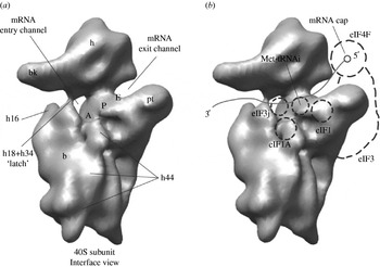
Bacterial and eukaryotic initiation factors have been shown to maintain the pool of small ribosomal subunits largely by preventing joining of the large ribosomal subunit. These discoveries were generally made by the analysis of ribosome complexes using sucrose density gradients (Subramanian & Davis, Reference Subramanian and Davis1970; Benne et al. Reference Benne, Brown-Luedi and Hershey1979; Staehelin et al. Reference Staehelin, Erni and Schreier1979; Chaudhuri et al. Reference Chaudhuri, Chowdhury and Maitra1999; Majumdar et al. Reference Majumdar, Bandyopadhyay and Maitra2003; Unbehaun et al. Reference Unbehaun, Borukhov, Hellen and Pestova2004), gel electrophoresis (Lorsch & Herschlag, Reference Lorsch and Herschlag1999; Algire et al. Reference Algire, Maag, Savio, Acker, Tarun, Sachs, Asano, Nielsen, Olsen, Phan, Hinnebusch and Lorsch2002) as well as with light scattering to observe thermodynamic and kinetic transitions in subunit joining (Grunberg-Manago et al. Reference Grunberg-Manago, Dessen, Pantaloni, Godefroy-Colburn, Wolfe and Dondon1975; Gorisch et al. Reference Gorisch, Goss and Parkhurst1976; Goss et al. Reference Goss, Parkhurst and Wahba1980, Reference Goss, Parkhurst and Wahba1982, Reference Goss, Rounds, Harrigan, Woodley and Wahba1988; Goss & Rounds, Reference Goss and Rounds1988; Antoun et al. Reference Antoun, Pavlov, Tenson and Ehrenberg2004). The conformation of the small ribosomal subunit changes upon association with initiation factors and tRNA. These conformational changes were first detected using chemical modification to observe changes in rRNA conformation. Probing of native 40S subunits purified from rabbit reticulocytes that include initiation factors identified 18 nucleotides that had altered susceptibility to chemical modification with DMS and CMCT when compared with salt-washed 40S subunits. These results suggest that these are regions with which initiation factors associate, or that change conformation upon initiation-factor binding (Melander et al. Reference Melander, Holmberg and Nygard1997). However, the footprinting data regarding how individual initiation factors may alter the conformation of the 40S subunit in preparation for mRNA binding and during the following stages of initiation is less clear. The purification of native 40S complexes relies on sucrose gradient centrifugation, which may result in the dissociation of weakly associated initiation factors. However, some of the nucleotides involved in binding to eukaryotic initiation factors appear to be similar to those involved in binding specific prokaryotic initiation factors (Moazed et al. Reference Moazed, Samaha, Gualerzi and Noller1995; Holmberg & Nygard, Reference Holmberg and Nygard1997; Melander et al. Reference Melander, Holmberg and Nygard1997; Lomakin et al. Reference Lomakin, Kolupaeva, Marintchev, Wagner and Pestova2003). In particular, the region around the platform close to the P-site of the small ribosomal subunit appears to be protected in both systems. It is possible that this may be due to the fact that the initiation factors eIF1 and the C-terminal domain of IF3 appear to be structural and functional homologs and likely bind to this region of the 40S subunit (indicated in Fig. 2). More recent footprinting data using RNase VI have shown that eIF1 localizes to a similar region on the 40S subunit to where IF3 was previously shown to bind on the 30S subunit near the P-site (Moazed et al. Reference Moazed, Samaha, Gualerzi and Noller1995; Lomakin et al. Reference Lomakin, Kolupaeva, Marintchev, Wagner and Pestova2003). Interestingly, both initiation factors appear to play a role in preventing ribosomal subunit joining (Subramanian & Davis, Reference Subramanian and Davis1970; Grunberg-Manago et al. Reference Grunberg-Manago, Dessen, Pantaloni, Godefroy-Colburn, Wolfe and Dondon1975; Kolupaeva et al. Reference Kolupaeva, Unbehaun, Lomakin, Hellen and Pestova2005). Early footprinting studies indicated that the nucleotides protected by IF3 binding, and analogously by eIF1 binding, map very close to those protected during ribosome subunit joining. Since the binding of these initiation factors likely interferes with bridge contacts involved in subunit joining, these results provide a plausible structural basis for their role in subunit dissociation (reviewed in Noller, Reference Noller, Mathews, Sonenberg and Hershey2007; Pestova et al. Reference Pestova, Lorsch, Hellen, Mathews, Sonenberg and Hershey2007). Although footprinting of other individual eukaryotic initiation factors awaits more detailed studies, it is expected that they will also associate near specific nucleotides in the rRNA, perhaps interfering with other bridge contacts.
40S subunit structure and locations of eukaryotic initiation factors, mRNA and tRNA. (a) The structure of the 40S subunit based on a cryo-EM reconstruction is presented (Spahn et al. Reference Spahn, Kieft, Grassucci, Penczek, Zhou, Doudna and Frank2001b). The 40S subunit is shown in dark grey and viewed from the subunit interface. Landmarks for the 40S subunit are indicated: A, A-site; P, P-site; E, E-site; bk, beak; b, body; pt, platform; and h, head. Helices 18, 34 and 44 of the 40S subunit are indicated as h18, h34 and h44 respectively. The latch of the 40S is formed between helix 18 and helix 34 (see text for details). In addition, the mRNA entry and exit channels are labeled. (b) The proposed positions for ligand binding are indicated by dashed lines for initiation factors and tRNA. The path of the mRNA in the 40S mRNA binding cleft is indicated as a blue line entering the 40S binding cleft through the mRNA entry channel and leaving the 40S binding cleft through the mRNA exit channel. The cap-binding complex consisting of eIF4E, eIF4G and eIF4A is labeled (eIF4F). The binding sites of eIF3j and the eIF3 complex are not presented as being physically connected, but it is expected that they are in fact associated with each other on the 40S surface. For details regarding positional information for each ligand please see the text.

Footprinting has also been used to map the functional tRNA-binding sites on the ribosome. These data determined the molecular contacts involved in tRNA binding to the A-, P- and E-sites of the ribosome. In addition, changes in footprinting patterns between specific states that the ribosome must move through provided evidence for intermediate positions that tRNA cycles through during protein synthesis, known as hybrid states (Moazed & Noller, Reference Moazed and Noller1986, Reference Moazed and Noller1989a, Reference Moazed and Nollerb, Reference Moazed and Noller1990). In addition, footprinting in the presence of either a bound A-site tRNA or IF1 revealed the same nucleotides to be protected from kethoxal modification (Moazed & Noller, Reference Moazed and Noller1986, Reference Moazed and Noller1990; Moazed et al. Reference Moazed, Samaha, Gualerzi and Noller1995). This suggests that IF1 blocks the A-site to tRNA binding during the initiation process. By analogy with its prokaryotic homolog, IF1, eIF1A is also likely to bind to a similar site on the 40S subunit, although it has not yet been investigated by footprinting assays. The proposed binding site of eIF1A close to the A-site is indicated in Fig. 2. Interestingly, the structure of these two proteins clearly indicates a conserved core, but eIF1A has significant extensions at both the N-terminus and C-terminus (reviewed in Marintchev & Wagner, Reference Marintchev and Wagner2004). Recent functional data obtained using a genetic system in yeast has indicated that these extensions in eIF1A are in fact involved in the scanning process to locate the AUG codon (Fekete et al. Reference Fekete, Mitchell, Cherkasova, Applefield, Algire, Maag, Saini, Lorsch and Hinnebusch2007). How the N- and C-terminal extensions of eIF1A may function in this way remains to be determined.
Footprinting studies have also been central to identifying nucleotides involved in the interaction of various antibiotics with ribosomal subunits. In particular, the nucleotides involved in binding antibiotics often correspond to the same, or adjacent, nucleotides directly implemented in known functional stages of protein synthesis such as initiation or translocation (reviewed in (Tenson & Mankin, Reference Tenson and Mankin2006). Many nucleotides involved in the recruitment of tRNA to the P-site of the 30S subunit also appear to be important in the association of antibiotics such as kasugamycin, pactamycin or edeine. These antibiotics all inhibit the initiation of protein synthesis, which relies on P-site function. Notably, while the footprinting evidence for kasugamycin inhibition of initiation suggests a direct competition with initiator tRNA, it has now been discovered by crystallography that it actually mimics the mRNA position on the 30S subunit (Schluenzen et al. Reference Schluenzen, Takemoto, Wilson, Kaminishi, Harms, Hanawa-Suetsugu, Szaflarski, Kawazoe, Shirouzu, Nierhaus, Yokoyama and Fucini2006; Schuwirth et al. Reference Schuwirth, Day, Hau, Janssen, Dahlberg, Cate and Vila-Sanjurjo2006). Therefore, this small molecule essentially prevents the codon–anticodon interaction from occurring from the mRNA-binding site rather than the expected binding of tRNA.
Recently, two new methods for rapid time-dependent footprinting have provided new insights into ribosome dynamics. The generation of hydroxyl radicals and incorporation of a quench flow apparatus to stop the reaction has allowed for the analysis of bridge contact formation during subunit joining of bacterial ribosomes. Importantly, this has enabled footprinting to be carried out on millisecond time-scales. High concentrations of hydroxyl radicals can be rapidly generated for these studies by using the standard Fenton reaction (Hennelly et al. Reference Hennelly, Antoun, Ehrenberg, Gualerzi, Knight, Lodmell and Hill2005) or synchrotron X-ray radiolysis (Nguyenle et al. Reference Nguyenle, Laurberg, Brenowitz and Noller2006). Together, these studies provided very similar results and have allowed a first look at the order in which bridge contacts form during subunit association. Interestingly, some nucleotides have been identified that become protected during subunit association, but are not directly involved in the subunit interface of the available crystal structures. This suggests the existence of association-dependent conformational changes in the ribosome when studied in solution compared to those observed in existing crystal structures.
The first study to use this kinetic footprinting technique to analyze initiation factor binding to the small ribosomal subunit has recently been described (Fabbretti et al. Reference Fabbretti, Pon, Hennelly, Hill, Lodmell and Gualerzi2007). The bacterial initiation factor IF3 associates with the 30S subunit and prevents 30S association with the 50S subunit. IF3 is also involved in the fidelity of initiator tRNA selection and enhances the dissociation of deacylated tRNAs from post-termination complexes. As mentioned earlier, the eukaryotic initiation factor eIF1 is a structural and functional homolog of the C-terminal domain of IF3, also playing a role in AUG recognition. Interestingly, both proteins are able to function in tRNA selection in heterologous systems (Lomakin et al. Reference Lomakin, Shirokikh, Yusupov, Hellen and Pestova2006). Hydroxyl radical probing of IF3 suggested a binding site on the platform of the 30S subunit, similar to the site footprinted by eIF1 on the 40S subunit (Lomakin et al. Reference Lomakin, Kolupaeva, Marintchev, Wagner and Pestova2003). Using time-resolved hydroxyl radical probing, the association of IF3 with the 30S subunit has now been shown to follow a specific pathway (Fabbretti et al. Reference Fabbretti, Pon, Hennelly, Hill, Lodmell and Gualerzi2007). The C-terminal domain binds first to the platform region followed by the N-terminal domain close to the E-site. Interestingly, during the association and dissociation of this initiation factor with the 30S subunit there is a transient reduction in the reactivity of specific nucleotides, suggesting that there are also conformational changes in the 30S subunit during these events. Although this novel method of time-resolved footprinting is still in its infancy we expect that it will lead to many exciting discoveries in understanding the molecular mechanisms of both bacterial and eukaryotic initiation complex dynamics.
4.3 Site-directed structural probing
Directed hydroxyl radical probing is a useful technique for studying nucleic acid environments surrounding a protein at low resolution (Culver & Noller, Reference Culver and Noller2000). In this approach, Fe(II) is covalently attached to a specific site on a recombinant protein via the linker 1(p-bromoacetamidobenzyl)-EDTA (BABE-Fe) originally devised by Rana & Meares (Reference Rana and Meares1991). Generally, a single cysteine amino acid engineered at a specific site in a recombinant protein is conjugated to BABE-Fe. Once incorporated into the desired complex, hydroxyl radicals can then be generated by the addition of hydrogen peroxide and ascorbic acid. As a result, the hydroxyl radicals cleave the phosphate backbone of nucleic acid, the sites of which are identified by primer extension, as described earlier. The degree of RNA cleavage corresponds to the distance from the Fe(II)-EDTA probe (Wang & Cech, Reference Wang and Cech1992; Han & Dervan, Reference Han and Dervan1994; Joseph & Noller, Reference Joseph and Noller2000). In general, the maximum target distance for strong cleavage is ∼18 Å, medium strength cleavage is ∼26 Å and weak cleavage is ∼42 Å (Han & Dervan, Reference Han and Dervan1994; Joseph & Noller, Reference Joseph and Noller2000). This method has a number of advantages over traditional solvent-based cross-linking and chemical probing techniques. Similar to cross-linking, tethered hydroxyl radical probing provides direct information regarding the RNA environment surrounding the Fe(II) ion. However, in general the efficiency of cross-linking is only a few percent and the reactivity of sites within the target tends to vary significantly. In contrast, since all accessible riboses are reactive to hydroxyl radicals, all targets within range are determined comprehensively. In addition, hydroxyl radical footprinting is not able to distinguish between direct and indirect effects of ligand binding, but tethering of Fe(II) restricts cleavage sites to the proximity of the tether, showing direct interactions between the protein ligand and nucleic acid. Furthermore, Fe(II) can be tethered to many positions on the protein surface, allowing probing of multiple positions of the protein in the RNA environment. As a result, mapping cleavage sites with different intensities provides sufficient constraints to allow amino-acid locations to be determined with respect to the nucleic acid structure. If the atomic structure of the protein being studied is available, it is possible to model the structure onto the RNA structure, in this case the ribosome. Directed hydroxyl radical probing has also been used to tether Fe(II) directly to positions on tRNA and to study its association with the ribosome (Han & Dervan, Reference Han and Dervan1994; Joseph et al. Reference Joseph, Weiser and Noller1997; Joseph & Noller, Reference Joseph and Noller2000).
Over the past few years, extensive use of this method has allowed many protein factors that have available atomic structures to be modeled to a high degree of accuracy onto the crystal structures of the bacterial ribosome. The interaction of many ribosomal proteins with the 16S rRNA was also determined by this method. Not surprisingly, it has also shown promise in modeling eukaryotic initiation factors onto the 40S subunit. Unfortunately, the crystal structure of the 40S subunit is not yet known. However, the high degree of homology between many conserved regions of the 18S and 16S rRNA enables detailed interaction models to be constructed based on crystal structures of the 30S subunit.
The first eukaryotic initiation factor to be studied using site directed hydroxyl radical probing was eIF1 (Lomakin et al. Reference Lomakin, Kolupaeva, Marintchev, Wagner and Pestova2003). Modeling the NMR structure of eIF1 onto the structure of the 30S subunit revealed that its position close to the P-site resembles that of the C-terminal domain of prokaryotic IF3. The proposed position of eIF1 on the 40S subunit close to the P-site is indicated in Fig. 2. This is perhaps not surprising in light of the discovery that it possesses a significant degree of structural homology to the C-terminal domain of IF3 (Lomakin et al. Reference Lomakin, Kolupaeva, Marintchev, Wagner and Pestova2003). Interestingly, the positioning of IF3, also determined by footprinting and directed hydroxyl radical probing, suggests that it directly blocks some of the bridge contacts required for 50S subunit joining, as was previously indicated (Grunberg-Manago et al. Reference Grunberg-Manago, Dessen, Pantaloni, Godefroy-Colburn, Wolfe and Dondon1975). The modeling of eIF1 suggests that it plays a similar role in directly blocking the B2b and B2d bridge contacts between eukaryotic ribosomal subunits (Spahn et al. Reference Spahn, Beckmann, Eswar, Penczek, Sali, Blobel and Frank2001a; Lomakin et al. Reference Lomakin, Kolupaeva, Marintchev, Wagner and Pestova2003). In subsequent experiments using sucrose-density gradient analysis, eIF1 was found to prevent subunit association only in the presence of its known binding partner eIF3 (Kolupaeva et al. Reference Kolupaeva, Unbehaun, Lomakin, Hellen and Pestova2005). Because eIF1 dissociates from the 40S subunit during sucrose gradient fractionation in the absence of other initiation factors, it has not been possible to determine if eIF1 possesses genuine dissociation activity on its own.
Recently, hydroxyl radical probing was used to map the interaction of the human eIF3 subunit eIF3j relative to 18S rRNA in the 40S subunit. Interestingly, this eIF3 subunit has been shown to be required for high-affinity binding of eIF3 to 40S subunits in vitro and in vivo and is able to associate with the 40S subunit in the absence of other initiation factors (Fraser et al. Reference Fraser, Lee, Mayeur, Bushell, Doudna and Hershey2004; Unbehaun et al. Reference Unbehaun, Borukhov, Hellen and Pestova2004; Nielsen et al. Reference Nielsen, Valasek, Sykes, Jivotovskaya and Hinnebusch2006). Although no atomic resolution structure is available for eIF3j, its C-terminal segment was mapped to the A-site and the mRNA entry channel of the 40S subunit, as indicated in Fig. 2 (Fraser et al. Reference Fraser, Berry, Hershey and Doudna2007). Previously, the bulk of eIF3 was found to bind on the solvent side of the 40S subunit, implying an indirect influence on mRNA and tRNA binding (reviewed in Hinnebusch, Reference Hinnebusch2006). The discovery of eIF3j in the ribosomal decoding center suggests that direct contacts by this component of eIF3 explain the observed decrease in 40S-eIF3j binding affinity in the presence of mRNA (Benne & Hershey, Reference Benne and Hershey1978; Unbehaun et al. Reference Unbehaun, Borukhov, Hellen and Pestova2004). Importantly, despite lacking a high-resolution eIF3j structure, this method provided detailed information regarding the binding of this protein to the 40S subunit.
To date, the only other eukaryotic initiation factor to be studied using site-directed hydroxyl radical probing is eIF5B. As mentioned earlier, this initiation factor is a homolog of prokaryotic IF2 and has a similar function in promoting the association of the large ribosomal subunit in a GTP-dependent fashion. Directed hydroxyl radical probing of a complex between eIF5B and the post-initiation 80S ribosome identified numerous eIF5B interactions with both the 40S subunit and the 60S subunit (Unbehaun et al. Reference Unbehaun, Marintchev, Lomakin, Didenko, Wagner, Hellen and Pestova2007). A model of eIF5B on the homologous regions of the 70S ribosome crystal structure suggests a similar placement as that determined by a previous cryo-EM structure of IF2 bound to the 70S ribosome (Allen et al. Reference Allen, Zavialov, Gursky, Ehrenberg and Frank2005). Interestingly, some of the rRNA cleavage sites determined for amino-acid positions in eIF5B were not within the expected distances when measured using the ribosome crystal structure. Since cleavages generally occur within specific distance constraints (Joseph & Noller, Reference Joseph and Noller2000), one possibility is that the ribosome conformation may in fact be different when eIF5B is bound compared to the conformation of the ribosome in the available crystal structure. These data suggest that directed hydroxyl radical probing coupled with molecular modeling can indicate whether conformational changes occur upon ligand binding. This method of structural probing is likely to enable rapid progress in our understanding of eukaryotic initiation factor interaction with the ribosome in the coming years.
4.4 Cryo-EM and X-ray crystallographic studies
Eukaryotic ribosomes were originally identified by electron microscopy in the 1950s (Palade, Reference Palade1955). Electron microscopy also provided the first low-resolution structures of bacterial and eukaryotic ribosomes as well as their individual subunits. These images were responsible for determining the general shape of the subunits and allowed the placement of ribosomal proteins using antibody labeling (Lake et al. Reference Lake, Pendergast, Kahan and Nomura1974; Tischendorf et al. Reference Tischendorf, Zeichhardt and Stoffler1975; Lutsch et al. Reference Lutsch, Noll, Theise, Enzmann and Bielka1979; Bommer et al. Reference Bommer, Noll, Lutsch and Bielka1980). In combination with cross-linking data, these low-resolution structural models revealed the general positions of initiation factors. Soon afterwards, three-dimensional reconstruction methods of negatively stained 40S subunits provided a more detailed structural model (Verschoor et al. Reference Verschoor, Zhang, Wagenknecht, Obrig, Radermacher and Frank1989). The direct visualization of eukaryotic initiation factors associated with the 40S subunit became possible, in particular the approximate placement of the large eIF3 complex facing away from the subunit interface, as indicated in Fig. 2 (Lutsch et al. Reference Lutsch, Benndorf, Westermann, Bommer and Bielka1986; Srivastava et al. Reference Srivastava, Verschoor and Frank1992). Since 1995 rapid progress has been made using single particle cryo-EM to provide higher resolution three-dimensional reconstructions of the bacterial ribosome, and more recently the eukaryotic ribosome. Using this approach, bacterial and eukaryotic ribosome structures have now been resolved to nanometer resolution. Electron microscopy is particularly well suited to the study of ribosome structure and dynamics during different stages of protein synthesis. Ribosomes are structurally stable and abundant particles with molecular weights ranging from 0·8 MDa for the 30S subunit to over 4 MDa for the 80S ribosome. Furthermore, the electron-dense RNA phosphodiester backbone of the rRNA provides for good contrast between sample and solvent during image processing and reconstruction. In addition, the requirement for only microgram quantities of material and the ability to select homogeneous individual particles allow 7–30 Å resolution models of ribosome functional states to be obtained. Many such complexes have been studied, particularly for the bacterial ribosome. These include identifying the positions of tRNAs (Agrawal et al. Reference Agrawal, Penczek, Grassucci, Li, Leith, Nierhaus and Frank1996; Stark et al. Reference Stark, Rodnina, Rinke-Appel, Brimacombe, Wintermeyer and Van Heel1997), elongation states (Stark et al. Reference Stark, Rodnina, Rinke-Appel, Brimacombe, Wintermeyer and Van Heel1997; Agrawal et al. Reference Agrawal, Penczek, Grassucci and Frank1998), termination states (Klaholz et al. Reference Klaholz, Pape, Zavialov, Myasnikov, Orlova, Vestergaard, Ehrenberg and Van Heel2003; Rawat et al. Reference Rawat, Zavialov, Sengupta, Valle, Grassucci, Linde, Vestergaard, Ehrenberg and Frank2003) and ribosome recycling (Agrawal et al. Reference Agrawal, Sharma, Kiel, Hirokawa, Booth, Spahn, Grassucci, Kaji and Frank2004; Gao et al. Reference Gao, Zavialov, Li, Sengupta, Valle, Gursky, Ehrenberg and Frank2005). In addition, recent studies also identified the positions of initiation factors bound during a late stage of the initiation step (Allen et al. Reference Allen, Zavialov, Gursky, Ehrenberg and Frank2005). Importantly, structural models of functional states of the ribosome can be combined to form a ‘movie’ to help explain ribosome dynamics during stages of protein synthesis. This has been elegantly done to help explain movements that occur during the complicated translocation stage of elongation. This ribosome ‘ratcheting’ model provides evidence for how the two ribosomal subunits rotate to open the mRNA channel when GTP and elongation factor G (EF-G) bind. Following this movement GTP is hydrolyzed and the mRNA and tRNAs translocate within the ribosome (Frank & Agrawal, Reference Frank and Agrawal2000).
During the time that cryo-EM was improving our understanding of ribosome structure and function, the emergence of bacterial ribosome crystal structures provided impressive new insight into the molecular structure of the ribosome. These structures, at resolutions of 2·4–11 Å, confirmed earlier biochemical studies indicating that the ribosome is a ribozyme, catalyzing peptide bond formation using an RNA active site (Nissen et al. Reference Nissen, Hansen, Ban, Moore and Steitz2000). These crystal structures have also provided snapshots of the ribosome bound to different components, including mRNA, tRNA and ribosome recycling factor (Berk et al. Reference Berk, Zhang, Pai and Cate2006; Korostelev et al. Reference Korostelev, Trakhanov, Laurberg and Noller2006; Selmer et al. Reference Selmer, Dunham, Murphy, Weixlbaumer, Petry, Kelley, Weir and Ramakrishnan2006; Weixlbaumer et al. Reference Weixlbaumer, Petry, Dunham, Selmer, Kelley and Ramakrishnan2007). Other structures have determined the detailed binding sites for antibiotics, explaining the molecular details of their inhibitory functions. These crystal structures together with continued advances using cryo-EM have provided a critical structural framework to help us understand the details of ribosome function. They have also helped to explain at atomic resolution many of the original studies described earlier in this review.
In contrast to the structural data available for the ribosome as a whole, the information regarding the structures of initiation events, particularly in the eukaryotic system, is quite sparse. Obtaining homogeneous samples that survive the dilution and solvent conditions required for cryo-EM analysis has been technically difficult. Atomic resolution structures of many of the initiation factors and their subunits have now been determined. A summary of links to published structures is provided in Table 1. However, the larger initiation factor complexes (eIF2, eIF2B, eIF3 and eIF4F) still pose a challenge for crystallography. Our understanding of initiation-factor binding sites on the 40S subunit has progressed in concert with structural advances for individual initiation factors. As mentioned above, the use of site-directed hydroxyl radical probing has allowed modeling of atomic structures of eIF1 and eIF5B onto the crystal structures of the ribosome and has suggested some possible conformation changes in the ribosome as a result.
Studying the association of initiation factors with the small ribosomal subunit has identified some significant conformational changes in ribosome structure. The binding of both IF1/eIF1A and IF3/eIF1 to the small ribosomal subunit induces some substantial structural rearrangements, as observed directly by crystallography and cryo-EM (Carter et al. Reference Carter, Clemons, Brodersen, Morgan-Warren, Hartsch, Wimberly and Ramakrishnan2001; Passmore et al. Reference Passmore, Schmeing, Maag, Applefield, Acker, Algire, Lorsch and Ramakrishnan2007). Originally, IF1 was localized near the A-site of the small ribosomal subunit by chemical footprinting (Moazed et al. Reference Moazed, Samaha, Gualerzi and Noller1995). Details about the binding of IF1 to the bacterial 30S subunit have now been obtained at high resolution by soaking IF1 into a 30S subunit crystal. The initiation factor clearly binds to the bottom of the A-site, interfering with tRNA binding by flipping two essential bases A1492 and A1493 out of helix 44. IF1 also prevents the intersubunit bridge B2a from forming (Carter et al. Reference Carter, Clemons, Brodersen, Morgan-Warren, Hartsch, Wimberly and Ramakrishnan2001). eIF1A has also been suggested to function in preventing subunit association, probably by interfering with the same bridge contact as that found for its homolog IF1 (Goumans et al. Reference Goumans, Thomas, Verhoeven, Voorma and Benne1980). The predicted position of eIF1A close to the A-site is shown in Fig. 2. Interestingly, the association of IF1 with the 30S subunit results in the tilting of the head and the platform regions towards the A-site. This appears to alter the mRNA binding cleft, which is formed between the head and the body of the small subunit (as indicated by the path of mRNA in Fig. 2). More recently, a cryo-EM reconstruction of eIF1A bound to the 40S subunit also induces a small conformational change in the 40S subunit around the mRNA binding cleft (Passmore et al. Reference Passmore, Schmeing, Maag, Applefield, Acker, Algire, Lorsch and Ramakrishnan2007). However, since the resolution of this complex is currently 25 Å, it will be interesting to determine if eIF1A does in fact induce the same detailed conformational changes in the 40S subunit as IF1 does in the 30S subunit.
The conformational change induced by the association of IF3 and eIF1 with the small ribosomal subunit has also been studied by cryo-EM. While eIF1 on its own does not appear to induce large changes in 40S subunit conformation (Passmore et al. Reference Passmore, Schmeing, Maag, Applefield, Acker, Algire, Lorsch and Ramakrishnan2007), IF3 appears to induce significant changes in the 30S subunit (McCutcheon et al. Reference McCutcheon, Agrawal, Philips, Grassucci, Gerchman, Clemons, Ramakrishnan and Frank1999). Even though both reconstructions were determined at ∼25 Å resolution, IF3 is observed to induce specific conformational changes in the 30S subunit that differ from the consequences of eIF1 binding to the 40S subunit. Perhaps this is understandable since IF3 possesses an N-terminal domain that is not conserved in eIF1, although at high concentrations the C-terminal domain of IF3 can function in place of full-length IF3 (Lomakin et al. Reference Lomakin, Shirokikh, Yusupov, Hellen and Pestova2006).
Cryo-EM reconstructions of the yeast 40S subunit and 40S-eIF1-eIF1A complexes, refined to ∼20 Å, clearly show significant conformational changes in the 40S subunit upon factor binding (Passmore et al. Reference Passmore, Schmeing, Maag, Applefield, Acker, Algire, Lorsch and Ramakrishnan2007). The association of eIF1 and eIF1A together appears to stabilize a 40S conformation that ‘opens’ the mRNA binding cleft. This conformational change is observed as a significant movement in the latch of the mRNA entry channel that has previously been proposed to clamp around mRNA (Frank et al. Reference Frank, Zhu, Penczek, Li, Srivastava, Verschoor, Radermacher, Grassucci, Lata and Agrawal1995). This latch is formed by helix 18 and 34 of the 18S rRNA and likely governs accessibility of mRNA to the binding cleft located between the head and body of the 40S subunit. This latch region is indicated in Fig. 2, close to the mRNA entry channel. In addition a new connection between the head and shoulder on the solvent exposed surface of the 40S subunit is formed, perhaps between helix 16 and rpS3. This connection may help to stabilize the conformation of the 40S subunit so that the open latch conformation is maintained.
Interestingly, this ‘open’ conformation of the mRNA binding cleft was first observed in a cryo-EM reconstruction that investigated the association of the hepatitis C virus (HCV) mRNA with the 40S subunit (Spahn et al. Reference Spahn, Kieft, Grassucci, Penczek, Zhou, Doudna and Frank2001b). The mRNA of HCV contains a structure in its 5′ UTR known as an internal ribosome entry site (IRES). These IRESes are believed to directly recruit the ribosome independently of the 7-methyl guanosine cap at the 5′ end of the mRNA. The HCV IRES forms a tertiary structure that binds with high affinity to the 40S subunit and promotes initiation of protein synthesis directly at the AUG codon (reviewed in Fraser & Doudna, Reference Fraser and Doudna2007; Pisarev et al. Reference Pisarev, Shirokikh and Hellen2005). Importantly, only eIF2⋅GTP⋅Met-tRNA and eIF3 are required for initiation by this IRES structure. The association of the IRES with the 40S subunit appears to promote the opening of the mRNA binding cleft in a similar way to that induced by the association of eIF1 and eIF1A. This perhaps explains why these factors are not required for correct binding of this mRNA to the 40S subunit (Pestova et al. Reference Pestova, Shatsky, Fletcher, Jackson and Hellen1998b).
4.5 Fluorescence techniques
Fluorescence spectroscopy is widely used to observe dynamics in macromolecular complexes. In the ribosome field, fluorescence-based methods have been used to detect changes in conformation during the individual stages of protein synthesis. These studies can be carried out using relatively simple steady-state emission intensity that is routinely used to measure the formation of complexes and conformational phenomena in biological molecules. In addition, including a stopped flow apparatus allows for more complicated time-resolved studies to be made. The appearance of modern fluorimeters has considerably simplified the effort needed to record and interpret the data produced by these experiments. These methods can utilize intrinsic tryptophan fluorescence of proteins or specific fluorescent tags that have been covalently attached to proteins or RNA. Changes in fluorescence intensity upon binding of a ligand or the Förster resonance energy transfer (FRET) between two fluorescent probes in close proximity can be used to quantitatively study ribosome and ligand dynamics.
Not surprisingly, fluorescence-based studies have been used to determine thermodynamic and kinetic parameters during stages of initiation, elongation, termination and recycling, particularly on bacterial ribosomes. For example, studies have indicated that the association and movement of mRNA on the ribosome can be detected by changes in fluorescence intensity of a probe attached to the 3′ end of the mRNA. During its association and movement through the ribosome's decoding center, the mRNA fluorescence increases and thereby tracks its position (Studer et al. Reference Studer, Feinberg and Joseph2003; Peske et al. Reference Peske, Rodnina and Wintermeyer2005). The association and movement of tRNA on the ribosome during elongation have been also determined by using fluorescently labeled tRNAs (Pan et al. Reference Pan, Kirillov and Cooperman2007). In addition, the association and movement between ribosomal subunits has been observed by measuring FRET efficiency between dyes attached to the small and large subunits (Peske et al. Reference Peske, Rodnina and Wintermeyer2005; Ermolenko et al. Reference Ermolenko, Majumdar, Hickerson, Spiegel, Clegg and Noller2007). These fluorescent methods have the advantage of being carried out in solution with relatively small amounts of material.
Affinities of purified cap-binding eukaryotic initiation factors and eIF3 for mRNA were determined using fluorescence intensity changes upon binding (Goss et al. Reference Goss, Carberry, Dever, Merrick and Rhoads1990a, Reference Goss, Carberry, Dever, Merrick and Rhoadsb, Reference Goss, Woodley and Wahba1987). In addition, the association of eIF3 with the 40S subunit was determined by laser light scattering, which is a method that measures the change in size during the formation of a macromolecular complex (Goss et al. Reference Goss, Rounds, Harrigan, Woodley and Wahba1988; Goss & Rounds, Reference Goss and Rounds1988). More recent studies have investigated the process of AUG recognition during eukaryotic initiation using fluorescent techniques in a reconstituted purified yeast system. eIF1 and eIF1A, two initiation factors involved in 40S subunit–mRNA binding and AUG recognition, bind cooperatively to the 40S subunit as determined by a change in the affinity of either initiation factor on the surface of the 40S subunit in the presence of the other protein (Maag & Lorsch, Reference Maag and Lorsch2003; Majumdar et al. Reference Majumdar, Bandyopadhyay and Maitra2003). This affinity change was detected using fluorescence polarization, an approach used previously for bacterial initiation factor binding to the 30S subunit (Weiel & Hershey, Reference Weiel and Hershey1981; Zucker & Hershey, Reference Zucker and Hershey1986). Fluorescence polarization, or fluorescence anisotropy, assays the rotational diffusion that occurs by Brownian motion of a molecule over time. When excited by linearly polarized light, the fluorophore attached to a macromolecule emits light and the degree of polarization of this emitted light reflects the rotation of the macromolecule during the well-defined fluorescence lifetime of the fluorophore. Experiments of this kind can determine the ligand dissociation constant, because free ligands rotate much faster than ligands bound to a larger molecule. The cooperative binding of eIF1A and eIF1 to the 40S subunit was found to be indirect, suggesting that changes occur in the conformation of the 40S subunit upon binding of each initiation factor (Maag & Lorsch, Reference Maag and Lorsch2003). Notably, although each possesses a high affinity for the empty 40S subunit, they dissociate rapidly as shown by sucrose gradient centrifugation (Majumdar et al. Reference Majumdar, Bandyopadhyay and Maitra2003). In addition, the affinity of IF1 for the 30S subunit is also enhanced by the presence of IF3, indicating a likely conformational change in the 30S subunit upon binding of each initiation factor (Zucker & Hershey, Reference Zucker and Hershey1986).
A FRET assay has been utilized to measure the interaction between eIF1 and eIF1A on the surface of the 40S subunit (Maag et al. Reference Maag, Fekete, Gryczynski and Lorsch2005). While FRET is actually a radiationless dipole–dipole transfer of energy, it is possible to think of the assay as monitoring overlap of emission and excitation of the two dyes. In this sense, a donor fluorophore is excited with a specific wavelength of light and emits light at a longer wavelength. The acceptor fluorophore is excited by the wavelength of light emitted by the donor and then emits light at a specific wavelength. However, this only occurs if the two fluorophores are in close proximity (0–10 nm). Conformational changes are observed when the distance between the two fluorophores is altered, resulting in a FRET change. Interestingly, in response to AUG codon recognition the distance between the C-termini of eIF1 and eIF1A changes in the presence of the eIF2⋅GTP⋅Met-tRNAi complex, indicating a conformational change on the surface of the 40S subunit. This was determined by modifying the C-terminus of eIF1A with fluorescein (donor) and the C-terminus of eIF1A with TAMRA (acceptor). The conformational change also reduces the affinity of eIF1 for the 40S subunit, which likely results in its dissociation from the complex (Maag et al. Reference Maag, Fekete, Gryczynski and Lorsch2005; Cheung et al. Reference Cheung, Maag, Mitchell, Fekete, Algire, Takacs, Shirokikh, Pestova, Lorsch and Hinnebusch2007).
In a separate fluorescence study, the association between mRNA and the 40S subunit was investigated using purified human components. Interestingly, a fluorescently labeled short 20 nucleotide unstructured mRNA was shown to possess a high affinity for the 40S subunit in the absence of other initiation factors (Fraser et al. Reference Fraser, Berry, Hershey and Doudna2007). This was determined by fluorescence anisotropy and suggests that the 40S subunit has high affinity for mRNA in the absence of any initiation factors. Interestingly, the association of eIF3j with the mRNA binding cleft of the 40S subunit significantly reduces the affinity of mRNA to the 40S subunit, perhaps via a change in conformation of the 40S subunit or physically blocking part of the mRNA binding cleft. High-affinity mRNA binding in the presence of eIF3j is restored upon recruitment of the eIF2⋅GTP⋅Met-tRNAi complex, even though eIF3j remains in the mRNA binding cleft (Fraser et al. Reference Fraser, Berry, Hershey and Doudna2007).
Clearly, fluorescence techniques can provide a wealth of information regarding the association of initiation factors with the 40S subunit and the dynamics that the components undergo during steps in protein synthesis. Although to date these fluorescence methods have only been used to study initiation complexes possessing a subset of initiation factors, the ability to reconstitute large multi-component factors and ribosomal particles should enable further dissection of ribosome dynamics during translation initiation. In addition, it will be important to determine the kinetics of initiation using these techniques to fully understand the sequence of events that must take place.
4.6 Single-molecule analysis
Stalling and viewing the ribosome at specific intermediate states has provided molecular ‘snapshots’ of the stages of protein synthesis. However, it is unlikely that all intermediate states can be captured in this system. Instead, a complete understanding of protein synthesis will require determining the structural and energetic landscapes of ribosomal conformations that give rise to thermodynamic and kinetic behaviors. Bulk studies measure macromolecular dynamics of a mixture of molecules. Because the molecules cannot be distinguished, only their average behavior can be determined. In contrast, single-molecule studies explore the properties of individual molecules by isolating them via chemical tethering or photon traps. Single-molecule studies have been useful in understanding many biological processes and have recently been the subject of numerous reviews (Zhuang, Reference Zhuang2005; Myong et al. Reference Myong, Stevens and Ha2006; Tinoco et al. Reference Tinoco, Li and Bustamante2006; Greenleaf et al. Reference Greenleaf, Woodside and Block2007). In general, three methods for the isolation and characterization of single molecules have become popular in these studies. Atomic force microscopy (AFM) has been applied to many biological molecules to study dynamics and the forces required to carry them out. In particular, a recent study determined the dynamics of unwinding of RNA structure by helicases involved in eukaryotic protein synthesis using AFM (Marsden et al. Reference Marsden, Nardelli, Linder and McCarthy2006). Other studies immobilize macromolecules on a surface such as a quartz cover slip and their identification is possible by the attachment of a fluorescent marker and visualization using a fluorescence microscope. Attachment of molecules to surfaces for single-molecule analysis often uses specific interactions such as a biotin-streptavidin bridge (Ha et al. Reference Ha, Zhuang, Kim, Orr, Williamson and Chu1999). Alternatively, the use of optical tweezers allows the attachment of a single molecule to a bead that is held in a tightly focused laser beam to trap the particle. Force can be measured, or applied externally during an experiment.
Early work using single-molecule spectroscopy determined that bacterial ribosomes attached to mica surfaces were functional in protein synthesis and allowed measurements of elongation rates (Jia et al. Reference Jia, Sytnik, Li, Vladimirov, Cooperman and Hochstrasser1997; Sytnik et al. Reference Sytnik, Vladimirov, Jia, Li, Cooperman and Hochstrasser1999). More recently, the attachment of mRNA to microscope coverslips allowed for tRNA dynamics to be visualized on elongating bacterial ribosomes. These studies used single-molecule FRET to determine the dynamics of tRNA as it binds and functions on the ribosome (Blanchard et al. Reference Blanchard, Gonzalez, Kim, Chu and Puglisi2004a, Reference Blanchard, Kim, Gonzalez, Puglisi and Chub). tRNAs bound to the A- and P-sites of the ribosome transitioned through different FRET states, indicating tRNA movements within the ribosome. Previously unidentified dynamic hybrid intermediate states were revealed using this method, and together with previous bulk kinetic analyses, have provided new insights into tRNA selection and kinetic proofreading (Rodnina & Wintermeyer, Reference Rodnina and Wintermeyer2001; Blanchard et al. Reference Blanchard, Gonzalez, Kim, Chu and Puglisi2004a, Reference Blanchard, Kim, Gonzalez, Puglisi and Chub; Gromadski & Rodnina, Reference Gromadski and Rodnina2004; Myong et al. Reference Myong, Stevens and Ha2006; Munro et al. Reference Munro, Altman, O'Connor and Blanchard2007).
An optical tweezer assay has been used to analyze the forces generated between the ribosome and the mRNA during the formation of the first peptide bond (Uemura et al. Reference Uemura, Dorywalska, Lee, Kim, Puglisi and Chu2007). The SD sequence anchors the 30S subunit near the start codon, via direct base paring with the 16S rRNA. This interaction is required for the stability of the initiation complex, but it must be broken as the elongation of protein synthesis commences. Recently, X-ray structures have provided snapshots of the conformational changes that occur during this event (Yusupova et al. Reference Yusupova, Jenner, Rees, Moras and Yusupov2006; Kaminishi et al. Reference Kaminishi, Wilson, Takemoto, Harms, Kawazoe, Schluenzen, Hanawa-Suetsugu, Shirouzu, Fucini and Yokoyama2007). However, analysis by single-molecule techniques suggests that it is in fact the formation of the first peptide bond that results in these conformational changes. An mRNA was attached to a coverslip via a biotin tag while the 30S subunit was attached to a bead held in a laser trap. The connection of the ribosome to the bead involved introduction of a stable RNA loop engineered into the 16S rRNA, which is subsequently hybridized to a digoxigenin-modified oligonucleotide. This study measured the force required to rupture the interaction between the mRNA and the ribosome. Interestingly, upon formation of the first peptide bond, weakening of the interaction between the SD sequence and the 30S subunit reduced the force needed to rupture it. This provided evidence that a signal from the peptidyl transferase center on the 50S subunit somehow disrupts the mRNA contacts with the 30S subunit (Uemura et al. Reference Uemura, Dorywalska, Lee, Kim, Puglisi and Chu2007).
Although no single-molecule experiments have yet been carried out with the eukaryotic ribosome, there is great potential to study the initiation of eukaryotic protein synthesis using these methods. In particular, the potential of analyzing a scanning 40S subunit is an attractive possibility.
5. Conclusions and future perspectives
Resolving the dynamic behavior of the ribosome during the stages of protein synthesis continues to be a significant challenge. The initiation of eukaryotic protein synthesis is much more complex than the initiation events observed in bacteria. The expanded set of initiation factors utilized by the eukaryotic ribosome relative to bacterial systems reflects a more highly regulated initiation process. Over the past decade, many structural details of eukaryotic initiation factors, or domains of these factors have been revealed by the appearance of crystal or solution NMR structures. However, these static structures may not indicate the conformation of the individual factors when associated with the 40S subunit. The emergence of cryo-EM reconstructions of the 40S subunit is also providing a structural framework for understanding ribosome function and dynamics at low resolution. However, high-resolution structures of initiation factor complexes and the association of these factors with the 40S subunit are still lacking. It is hoped that these will emerge in the coming years to enhance our understanding of their structure and function.
Many advances have been made in analyzing the 40S subunit conformation in stalled or blocked complexes using a combination of the methods reviewed here. Chemical modification techniques have enhanced our knowledge regarding rRNA nucleotides involved in promoting functional complexes, but without higher resolution structures it is difficult to determine whether these are direct or indirect effects. However, these data combined with data generated using site- directed hydroxyl radical probing has allowed for modeling with some confidence the placement of some factors on the 40S subunit. However, it should be remembered that such thermodynamically stable complexes might not be true initiation intermediates. In addition, it is likely that not all intermediate states may be visualized by using these techniques.
The use of fluorescent techniques is adding to our knowledge of ribosome dynamics. In particular, the attachment of fluorescent probes to allow FRET studies is uncovering important information regarding conformation changes during the stages of initiation. However, many of the studies to date have only analyzed these events in a purified system that uses a subset of initiation factors. Although a considerable challenge due to technical reasons, it is hoped that such experiments will eventually be possible in a system that utilizes a full compliment of factors. In addition, the emergence of time-resolved methods such as kinetic footprinting and single-molecule analysis are likely to enhance our understanding of the conformational changes that occur during eukaryotic protein synthesis initiation in real time.
In summary, the results obtained from the many different approaches discussed here have begun to converge and promise to yield a more detailed view of the initiation of eukaryotic protein synthesis in the future. However, although the functions of many initiation factors are now becoming clear, our molecular understanding of how eukaryotic ribosomes initiate protein synthesis is still limited. In particular, the mechanism of how the 40S subunit once bound to an mRNA migrates along the 5′ UTR during scanning has not yet been elucidated and will clearly be a significant challenge for the next decade.
6. Acknowledgements
We thank Jamie Cate, John Hershey and Katie Berry for helpful discussions and their comments on the manuscript.