Introduction
The family Potamotrygonidae (Elasmobranchii: Myliobatiformes) is the only living chondrichthyan family restricted to freshwater habitats (Lovejoy, Reference Lovejoy1996) and they are distributed throughout most of the major river systems of South America. The Amazon Basin contains the greatest number of valid species of freshwater stingrays and Brazil is the country with the highest diversity of species (approximately 18 species) (Rosa, Reference Rosa1985; Compagno & Cook, Reference Compagno and Cook1995; Charvet-Almeida et al., Reference Charvet-Almeida, Araújo, Rosa and Rincon2002; Carvalho et al., Reference Carvalho, Lovejoy, Rosa, Reis and Kullander Ferraris2003). The cururu stingray (Potamotrygon cf. histrix) is endemic to the Negro river basin (Carvalho et al., Reference Carvalho, Lovejoy, Rosa, Reis and Kullander Ferraris2003) and as a new species still deserves an appropriate taxonomic description. This high endemism has led researchers to express concern that some species may be endangered (Rosa, Reference Rosa1985; Compagno & Cook, Reference Compagno and Cook1995).
The reproductive strategy of these stingrays (Wourms, Reference Wourms1977), associated with the position they occupy in food chains, signifies that they are present in low densities in their habitats (Camhi et al., Reference Camhi, Fowler, Musick, Brauntingam and Fordham1998). Because of exploitation, environmental changes and degradation of the aquatic environment, these species might have some difficulty in maintaining their stock levels (Hoenig & Gruber, Reference Hoenig, Gruber, Pratt, Gruber and Taniuchi1990; Compagno & Cook, Reference Compagno and Cook1995; Araújo et al., Reference Araújo, Charvet-Almeida and Almeida2004; Martin, Reference Martin2005). Temperature appears to play an important role in plasma testosterone regulation, as levels in wild caught male round stingrays (Urobatis halleri) were negatively correlated with water temperature (Mull et al., Reference Mull, Lowe and Young2008). Otherwise, freshwater stingrays exhibit a very small range of tolerance to both natural and anthropogenic impacts (Compagno & Cook, Reference Compagno and Cook1995).
The reproductive cycle of stingrays of the genus Potamotrygon is directly related to the hydrological cycle, taking the seasonality (dry and rainy seasons) into account, with defined reproductive periods and differentiated periodicity for each species, although all of them present periods of gonad maturation, copulation, pregnancy, birth and resting (Charvet-Almeida et al., Reference Charvet-Almeida, Araújo and Almeida2005).
The age of sexual maturation among freshwater stingrays is highly variable. Regarding the cururu stingray, it has been estimated to be 2 years of age, with gonad maturation lasting 2 months, copulation during the rainy period, a gestational period of 3 months and parturition in the dry season. However, data on the reproductive biology of freshwater stingrays are still limited, especially concerning information on microscopic morphology (Charvet-Almeida et al., Reference Charvet-Almeida, Araújo and Almeida2005).
Freshwater stingrays have regularly been captured for ornamental purposes for decades. Currently, at least six species from the genus Potamotrygon are legally permitted to be exported as ornamental fish in Brazil (IN N. 204/08, IBAMA, 2008). Their capture represents an income source for fishermen in the northern region of Brazil (Araújo et al., Reference Araújo, Charvet-Almeida and Almeida2004; Charvet-Almeida, Reference Charvet-Almeida, Araújo and Almeida2005), as well as in other regions of South America like the Peruvian Amazonian region (Moreau & Coomes, Reference Moreau and Coomes2007). These cartilaginous fish are generally not consumed as food. However, populations of Paratrygon aiereba are receiving increasing capture pressure from the commercial fishery in Pará State (Brazil), due to its larger size and appreciable muscle mass (Charvet-Almeida & Almeida, Reference Charvet-Almeida and Almeida2007). Fishery management and conservation of freshwater stingrays is a delicate topic because of their restricted distribution and the lack of official fisheries records. Potamotrygon cf. histrix represents 50% of all freshwater stingray legally exported from Brazil (Araújo et al., Reference Araújo, Charvet-Almeida and Almeida2004). Further studies on their reproductive biology are, therefore, very much needed to help ensure sustainable management and the protection and conservation of South American stingrays. Studies on the reproduction of stingrays in captive environment are scarce and they are developed in public aquariums, universities and zoos (Oldfield, Reference Oldfield2005a, Reference Oldfieldb; Henningsen, Reference Henningsen2000).
The present study describes the testicular structure and spermatogenesis of the freshwater Amazonian cururu stingray (Potamotrygon cf. histrix), in order to provide better understanding of its reproductive biology. Data from structural analysis might contribute towards clarifying the reproductive mechanisms of this group of fish, thus providing tools for future studies on the biology and management of neotropical freshwater stingrays.
Material and methods
Ten mature male specimens of the cururu stingray were collected from the Puxurituba Igarapé, in the municipality of Barcelos, State of Amazonas, Brazil, between November and December 2005. Following capture, the body size and weight of the specimens were measured and recorded (mean total weight = 266.6 g; mean disc width = 20.6 cm and mean total length = 31.8 cm). They were then euthanized using high doses of benzocaine (1.0 g in 15 litres). The abdominal cavity was opened and the gonads were exposed for gross examination. The testes on both sides were removed and fixed in buffered 10% paraformaldehyde. Following 24 h of fixing, the samples were dehydrated in a graded series of alcohol (70–100%), cleared in xylol and infiltrated into molten paraffin. Sections of 5 μm in thickness were stained with hematoxylin–eosin and Masson's trichrome for light microscopy. Photomicrographs were produced using a Leica DM 2500 microscope and morphological measurements were made using the Leica QWin V3 software. Data were subjected to analysis of variance (ANOVA) and the means were compared using Tukey's test (5%), by means of the SAS 8.12 software.
Results
Gross structure of testes
The testes were paired, oval-shaped, reddish and elongated organs located in the abdominal cavity, ventrally to the vertebral column and suspended by a mesorchium. The dorsal surface was flattened and totally covered with the epigonial organ. Both organs were developed but the left testis was larger than the right one.
Microscopic structure of testes
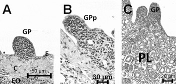
The testes were enclosed in a capsule of connective tissue, the tunica albuginea. Between the simple squamous epithelium and the collagenous fibres, germinal papillae or testicular appendages appeared as projections of the dorsal wall of the testes (Fig. 1A). The germinal papilla zone presented undifferentiated mesenchymal-like cells and primary spermatogonia surrounded by Sertoli cell precursors, thus characterizing their endodermal origin.
(A) Germinal papilla (GP), epithelium (E), collagen (C), epigonial organ (EO). (B) Germinal papilla projection (GPp). (C) Primary lobule (PL).

Proliferation of cells from the germinative lineage caused projection of the germinal papillae ventrally, with consequent formation of spermatogenic lobules, thus indicating that this was the site of origin and development of the spermatocysts (Fig. 1B).
The testicular parenchyma was defined and was filled with lobules at different phases of development. The primary lobules were covered by a thin layer of connective tissue and the parenchyma was delimited by the epigonial organ (with content resembling lymphoid tissue), with the distribution of varied cell types, according to the development stage of the seminiferous lobules (Fig. 1C). In each lobule, there were spermatocysts at different phases of spermatogenic development. As they developed, they migrated radially to the periphery of the lobule. Thus, gonad development was characterized by cell differentiation associated with changes in cell type and diameter within the spermatocysts.
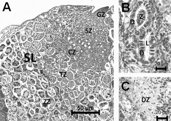
The testicular lobules were rounded, presenting zonal organization. Each zone was characterized by the presence of spermatocysts at similar phases of spermatogenesis. These zones had dorsal–ventral orientation and the upper zones were at earlier phases of spermatogenic development while the lower zones were at more advanced phases of spermatogenesis. The organization of the secondary spermatogenic lobules was characterized by the presence of germinal papillae in its upper dorsal position, followed by the germinal, spermatogonial, spermatocyte, spermatid, spermatozoid and degenerative zones (Fig. 2A).
(A) Secondary lobule (SL), germinal zone (GZ), spermatogonial zone (SZ), spermatocyte (CZ), spermatid zone (TZ), spermatozoa zone (ZZ). (B) Duct (D), lumen (L), spermatozoa (Z). (C) Degenerative zone (DZ).

The germinal zone was characterized by the presence of primary spermatogonia that were always accompanied by precursor of the Sertoli cells, encrusted in the tissue forming the spermatogenic cysts (Fig. 2A).
The spermatogonial zone was formed by cysts containing primary spermatogonia undergoing division, secondary spermatogonia, Sertoli cells and lumen. Proliferation of the spermatogonia and Sertoli cells led to an increase in the size of the spermatogenic cysts and, possibly, to disappearance of the lumen and to the process of Sertoli cell nucleus migration to the periphery of the spermatocysts (Fig. 2A).
The spermatocyte zone was totally filled by spermatocysts containing primary spermatocytes at different phases of the first meiotic division. Groups of spermatocytes surrounded by the same Sertoli cell (called spermatoblasts), gave rise to a subdivision within the spermatogenic cyst and it seemed that all the cells within each spermatoblast were at the same phase of meiotic division. There were also spermatocysts containing secondary spermatocytes with characteristics similar to the primary ones, despite being half their size. The Sertoli cells were already seen in the periphery of the spermatocysts (Fig. 2A).
The spermatid zone consisted of spermatocysts containing spermatids at different phases of spermiogenesis, in which the shape changed from oval to elongated and finally spiraled. The nuclei were turned towards the peripheral region of the spermatocysts, pointing towards the nuclei of the Sertoli cells, since their cytoplasm surrounded the spermatids (Fig. 2A).
The spermatozoid zone presented these cells in groups, initially with their heads turned towards the Sertoli cell nucleus and subsequently free within the lumen of the spermatocysts. The spermatocysts opened up in the intratesticular ducts, with release inside them. The ducts were formed by a simple ciliated columnar epithelium and were also associated with the epigonial organ, with or without spermatozoids inside it (Fig. 2A).
The degeneration zone was dispersed, including the spermatozoid zone, intratesticular ducts, spermatocysts (in the degeneration and reabsorption phase) and epigonial organ tissue (through direct contact) (Fig. 2C).
Germinative lineage
The following cells from the male germinative lineage were identified:
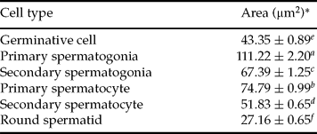
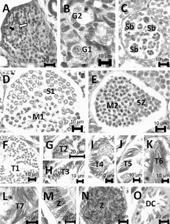
a. Primordial germinative cells were present in small number only inside the testicular papillae. They stood out from the other cell types that were also present there because of their size and rounded shape (Table 1). They presented defined regular outlines, acidophilic cytoplasm and spherical basophilic nuclei. During differentiation into spermatogonia, changes to the nuclei, such as chromatin condensation to form chromosomes, were visible. This cell type was always associated with precursors of Sertoli cells (Fig. 3A).
b. Primary spermatogonia were located within the papillae and the germinal zone and were the biggest cell type of the germinative lineage (Table 1). These cells were large, rounded and well defined, with abundant cytoplasm and low affinity for staining. The spherical nuclei contained uniform euchromatin and heterochromatin, scattered irregularly. The nucleoli were eccentric. These cells were always associated with the Sertoli cells, which had comma-shaped nuclei that were completely occupied by heterochromatin (Fig. 3B).
c. Secondary spermatogonia had characteristics similar to the primary spermatogonia. However, they were half their size and were within well defined cysts (Table 1). The cysts presented increasing sizes because of the process of spermatogonium multiplication (Fig. 3B).
d. Primary spermatocytes were present within the spermatogenic cysts and presented spherical and well-defined nuclei, with chromatin at different phases of condensation, depending on which sub-phase of prophase I they were in or, in addition, whether they were concentrated in the periphery of the nucleus, which gave them a ring appearance. These spermatocytes were also found in distinct phases of the first meiotic division, in subunits within the same spermatocyst, called spermatoblasts (Fig. 3C). Within single cysts, there were spermatoblasts that contained primary spermatocytes and others that contained secondary ones (Table 1; Fig. 3D).
e. Secondary spermatocytes were also found within the spermatogenic cysts, similar to the primary spermatocytes, but half their size (Table 1). Within single cysts, secondary spermatocytes at different phases of the second meiotic division and spermatoblasts containing round spermatids were found (Fig. 3E).
f. Spermatids were also observed within the cysts, with half the size of the preceding cell type and with spherical nuclei containing very condensed chromatin, which gave it a dark appearance with high staining affinity. As spermiogenesis progressed, the spermatids went through a process of elongation, tail formation and spiraling of the anterior part where the nucleus is located. The phases identified are described below:
f1. Round spermatids were found within the cysts, with half the size of the secondary spermatocytes (Table 1) and with spherical nuclei containing very condensed chromatin, which gave it a dark appearance with high staining affinity (Fig. 3F).
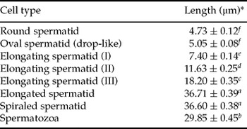
f2. Oval or drop-shaped spermatids presented a strongly colored nucleus, with a round posterior pole and a triangular anterior one, giving the appearance of a drop. The nuclei were turned towards the Sertoli cells (Table 2; Fig 3G).
f3. Spermatids going through the first stage of elongation had rod-shaped strongly basophilic nuclei, with a funneled anterior extremity that was turned towards the Sertoli cell nuclei and a rounded posterior region. There was initial formation of an acidophilic tail (Table 2; Fig. 3H).
f4. Spermatids going through the second stage of elongation had rod-shaped basophilic nuclei, with the anterior third funnelled and a point-shaped extremity that was turned towards the Sertoli cell nuclei. The posterior extremity was rounded, with an acidophilic tail that was half the size of the head, undergoing formation (Table 2; Fig 3I).
f5. Spermatids undergoing the third stage of elongation had long basophilic nuclei that were two-thirds funnelled, with a sharply pointed anterior extremity that was turned towards the Sertoli cell nuclei. There was an acidophilic tail that was undergoing formation and was smaller than the head (Table 2; Fig 3J).
f6. Long spermatids also had basophilic, long, thin and straight nuclei throughout their whole length that were grouped and turned towards the Sertoli cell nuclei. An acidophilic tail was present (Table 2; Fig. 3K).
f7. Spiraling spermatids had basophilic nuclei that were long, thin and undergoing the spiraling process around their own axis, beginning at the posterior region and heading towards the anterior region. These were in groups and were turned towards the Sertoli cell nuclei (Table 2; Fig. 3L).
g. Spermatozoids were elongated cells with an acidophilic tail and a long basophilic head, with a spiral shape around their own axis. These were found within the cysts with their anterior extremity turned towards the Sertoli cell nuclei (Fig. 3M). They were free in the lumen of the cysts (Fig. 3N), in the degeneration zone and within the intratesticular ducts (Fig. 2B).
Nuclear area of the germ cells.
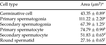
a–fMeans followed by the same letters in the column do not differ (5%). *Mean ± standard error.
Length of the cell nuclei during spermiogenesis.

a–fMeans followed by the same letters in the column do not differ (5%). *Mean ± standard error.
Table 1 presents the length of the cell nuclei during spermiogenesis and Table 2 presents the nuclear area of the germ cells.
Germinative cell lineage. (A) Germinative primordial cells  , precursor Sertoli precursor cell ➨. (B) Primary spermatogonia (G1), secondary spermatogonia (G2). (C) Spermatogenic cyst whit spermatoblast (Sb). (D) Primary spermatocyte (S1), mitosis1 (M1). (E) Secondary spermatocyte (Sc2), mitosis 2 (M2). (F) Round spermatid (T1). (G) Drop-like spermatids (T2). (H) Elongating spermatid I (T3). (I) Elongating spermatid II (T4). (J) Elongating spermatid III (T5). (K) Elongated spermatid (T6). (L) Spiraling spermatid (T7). (M, N) Spermatozoa (Z). (O) Degenerating cyst (DC). *, Sertoli cell.

Sertoli cells
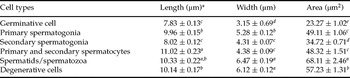
These cells accompanied the germinative lineage throughout spermatogenesis and underwent drastic modifications, with migration and morphological modification of their nuclei and increased cytoplasm (Table 3). The nuclei changed from comma-shaped (typical of precursor cells) to a triangular shape and then to an oval shape and these were larger by the end of spermatogenesis (Fig. 3B,K–M,O). After spermatozoid release, the degeneration process began. While the precursor cells were rich in heterochromatin, the Sertoli cells presented euchromatin, thus characterizing the stage of cell functionality.
Length, width and area of the Sertoli cell nuclei associated with different cell types during spermatogenic development.

a–eMeans followed by the same letters in the column do not differ (5%). *Mean ± standard error.
Intratesticular ducts
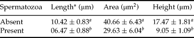
The intratesticular ducts developed simultaneously with the seminiferous lobules, interspersed with them. They were covered with a thin connective septum and were formed by simple columnar epithelium with cells presenting basophilic elongated nuclei and acidophilic cytoplasm. These ducts, which were present throughout the degeneration zone, showed elongations that connected them directly to the efferent channel, which went through the whole length of the testicle (Fig. 2B). The length and area of the nuclei and height of the duct epithelium were greater when there were no spermatozoids inside the ducts (Table 4).
Length, area and height of the duct epithelial cell nuclei, with and without presence of spermatozoa inside the duct.
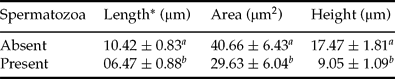
a,bMeans followed by the same letters in the column do not differ (5%). *Mean ± standard error.
Discussion
The cururu stingray is considered to be a small species in relation to the other species of the same genus and is known as the ‘dwarf freshwater stingray’, with a mean disc size of 160 mm for sexually mature males (Chavert-Almeida et al., Reference Charvet-Almeida, Araújo and Almeida2005). The stingrays of the present study presented a mean disc size of 206 mm, which is within the expected range for this species at the reproductive stage and they presented functional testicles.
Testicular structure resembling that of cururu stingray, i.e. associated with the epigonial organ, with formation of lobules proceeding radially and presence of ducts, has previously been described to Dasyatis sabina (Maruska et al., Reference Maruska, Cowie and Tricas1996), Himantura signifier (Chatchavalvanich et al., Reference Chatchavalvanich, Thogpan and Nakai2005) and also to rajids Atlantoraja cyclophora (Oddone & Vooren, Reference Oddone and Vooren2005), Leucoraja ocellata (Sulikowski et al., Reference Sulikowski, Tsang and Howell2005) and other elasmobranchs (Pratt, Reference Pratt1988). This latter author defined the seminiferous lobes as seminiferous follicles or ampullae, whereas Teshima & Takeshita (Reference Teshima and Takeshita1992), for Potamotrygon magdalenae, used the term seminiferous follicles.
A difference in size between the right and left testicle has also been described in relation to the Atlantic stingray (Dasyatis sabina) (Maruska et al., Reference Maruska, Cowie and Tricas1996) that was attributed to the size and position of the stomach and spiral intestine, which occupied the greater part of the abdominal cavity.
The differences in the cell types in the epigonial organ observed in cururu stingrays, with regard to testicles with lobules at different phases of spermatogenesis, were also described by Lutton & Callard (Reference Lutton and Callard2008) in relation to Leucoraja erinacea. These authors found greater proliferation of epigonial leukocytes and greater release into blood vessels when the gonads were at the stage of reproductive activity. This situation characterizes interaction between the immune and reproductive systems (Lutton & Callard, Reference Lutton and Callard2006). In a study on Squalus acanthias, focusing on the immunological action of the epigonial organ in the gonads, the presence of a growth inhibition factor (EGIF) was considered responsible for inhibiting spermatogenesis in that species (Piferrer & Callard, Reference Piferrer and Callard1995). In another study, changes to the immune and reproductive systems resulting in blockage of spermatogenesis was described among Atlantic stingrays that were exposed to environmental contamination due to organochlorates (Gelsleichter et al., Reference Gelsleichter, Rasmussen, Marine, Tyminki, Chang and Lombardi-Carlson2002).
Genital papillae and immature or maturing genital appendices, forming primary lobules, were identified in the testicular structure of the cururu stingrays. In addition, there were papillae that were completely developed, characterized by the presence of both primary and secondary lobules. However, among Urolophus halleri stingrays, Babel (Reference Babel1967) identified the presence of primary lobules throughout the annual reproductive cycle and secondary ones only during the reproduction period, thus characterizing the latter as temporary. Germinal papillae have also been found among other elasmobranchs, such as Squalus acanthias (McClusky, Reference McClusky2005).
The organization of the spermatogenic lobules, which was histologically characterized in the cururu stingrays by their defined development zones, followed cell differentiation and changes in spermatocyst composition and diameter that were distributed in stages equivalent to the zones described for Himantura signifier (Chatchavalvanich et al., Reference Chatchavalvanich, Thogpan and Nakai2004) and Dasyatis sabina (Maruska et al., Reference Maruska, Cowie and Tricas1996; Tricas et al., Reference Tricas, Maruska and Rasmussen2000); and for other elasmobranchs such as Mustelus canis (Conrath & Musick, Reference Conrath and Musick2002), Centroscymnus coelolepis and Centrophorus squamosus (Girard et al., Reference Girard, Rivalan and Sinquin2000) and Squalus acanthias (McClusky, Reference McClusky2005, Reference McClusky2006).
The germinative lineage found in cururu stingray was similar to that described for Dasyatis sabina (Maruska et al., Reference Maruska, Cowie and Tricas1996; Tricas et al., Reference Tricas, Maruska and Rasmussen2000), Himantura signifer (Chatchavalvanich et al., Reference Chatchavalvanich, Thogpan and Nakai2004) and Raja asterias (Barone et al., Reference Barone, De Ranieri, Fabiani, Pirone and Serena2007). At the time of cell division, it was observed that within the same spermatocyst, there were spermatoblasts containing cells at different phases of cell division, thus resulting in the presence of different cell types within the same spermatocyst, with the presence of secondary spermatocytes and some spermatoblasts and spermatids within others. Prisco et al. (Reference Prisco, Liguoro, Comitato, Cardone, D'Onghia, Ricchiari, Angelini and Andreuccetti2003) also identified different stages of cell differentiation in the same spermatogenic cyst in Torpedo marmorata and also among other elasmobranchs, such as Centroscymnus coelolepis and Centrophorus squamosus (Girard et al., Reference Girard, Rivalan and Sinquin2000).
Length measurements on the spermatids during the spermiogenesis were also made by Maruska et al. (Reference Maruska, Cowie and Tricas1996) on Dasyatis sabina stingray. The results showed that a significant difference appeared as the differentiation phenomena evolved, thus indicating that the increase in length of the reproductive cells undergoing formation is considerable. The spiraling process observed in the final events of spermiogenesis in cururu stingray is characteristic of elasmobranchs and, among these, freshwater stingrays (Maruska et al., Reference Maruska, Cowie and Tricas1996; Tricas et al., Reference Tricas, Maruska and Rasmussen2000; Chatchavalvanich et al., Reference Chatchavalvanich, Thogpan and Nakai2004, Reference Chatchavalvanich, Thogpan and Nakai2005).
Helical spermatozoids with rotation on their own axis, as identified in the present study, is characteristic of elasmobranchs in general (Wourms, Reference Wourms1977) and has also been identified in Potamotrygon magdalenae (Teshima & Takeshita, Reference Teshima and Takeshita1992) and Himantura signifer freshwater stingrays (Chatchavalvanich et al., Reference Chatchavalvanich, Thogpan and Nakai2005).
The structural modifications identified in the Sertoli cells, at different times during spermatogenesis, indicate that this cell type is directly related to the phenomena of gamete production. This is due to the greater metabolic need for their functions to be performed with the germinative lineage. The cells present in cysts in which spermatozoid release had already occurred began to degenerate (Maruska et al., Reference Maruska, Cowie and Tricas1996; Tricas et al., Reference Tricas, Maruska and Rasmussen2000; Prisco et al., Reference Prisco, Liguoro, D'Onghia, Ricchari, Andreuccetti and Angelini2002; Chatchavalvanich et al., Reference Chatchavalvanich, Thogpan and Nakai2004). This process occurs at the end of their function, when mature gametes are released and spermatocysts disappear (Prisco et al., Reference Prisco, Liguoro, Comitato, Cardone, D'Onghia, Ricchiari, Angelini and Andreuccetti2003; Liguoro et al., Reference Liguoro, Prisco, Mennella, Ricchiari, Angelini and Andreuccetti2004).
The intratesticular ducts found in cururu stingray are similar to those described in other stingrays, such as Urolophus halleri (Babel, Reference Babel1967) and also in Raja erinacea (Pratt, Reference Pratt1988). The differences in the histological structure of the intratesticular ducts that were observed in cururu stingray show the functionality of the organ, in which the cells undergo losses of cytoplasm in order to form the seminal fluid. These changes accompany the phenomena of spermatogenesis and the release of spermatozoids. Piercy et al. (Reference Piercy, Gelsleichter and Snelson2003) analysed the genital ducts of Atlantic stingrays and identified extensive histological changes in their architecture, in relation to both size and secretion activity, during the reproductive period. The information obtained from the present study will serve as a tool for gaining knowledge of the reproductive mechanism of this group of freshwater chondrichthyes and as support for further studies on reproduction in captivity or in degraded and polluted natural environments.
Acknowledgements
This work was supported by a grant from National Geographic Conservation Fund (No. C47-04). The scientific expedition was authorized by the Brazilian Ministry of Science and Technology (MCT/CNPq, process No. EXC 023/05 to J.L.M.) and by the Federal Environmental Agency (process No. 098/2005 DIFAP/IBAMA-DF).