For pathogens transmitted through the contact route, contamination on the personal protective equipment (PPE), clothing and skin of HCWs could be transferred to sites on the HCW susceptible to infection, such as the facial mucous membranes, putting HCWs at risk of infection. Contamination of PPE, clothing, and skin can also facilitate pathogen dissemination throughout the healthcare environment. Studies in intensive care units (ICUs) routinely find multidrug-resistant organisms on HCW PPE, clothing, and skin,Reference Morgan, Liang and Smith1–Reference Okamoto, Rhee and Schoeny3 but few studies of viruses have been conducted to date.
The doffing of PPE is one context in which pathogens on PPE may be transferred to the clothing and skin of HCWs. Frequently the PPE doffing practices of HCWs deviate from those recommended by the Centers for Disease Control and Prevention (CDC),Reference Phan, Maita and Mortiz4 and studies using surrogates (eg, fluorescence and bacteriophage) have found that doffing PPE can result in contamination of HCW hands and faces.Reference Casanova, Teal and Sickbert-Bennett5–Reference Kwon, Burnham and Reske7 Okamoto et alReference Okamoto, Rhee and Schoeny3 reported that the HCWs who made multiple PPE doffing errors were more likely than other HCWs to have multidrug-resistant organisms on their clothing after a patient interaction. Because of the severity of Ebola virus disease and complexity of the enhanced PPE ensembles, fear of clothing and skin contamination during doffing has heightened, and studies of doffing practices have identified numerous opportunities for contamination.Reference Casanova, Teal and Sickbert-Bennett5, Reference Mumma, Durso and Ferguson8
The primary objective of this study was to characterize the presence and magnitude of respiratory viruses on the PPE, clothing, and skin of HCWs after HCWs provided routine care for hospitalized patients having viral respiratory infections and placed under droplet and/or contact isolation. Secondary objectives were to explore the association between observed self-contacts of HCWs during the patient encounters and the magnitude of virus contamination on their PPE and to evaluate the association of HCW self-contamination and PPE doffing practices.
Methods
The study was reviewed and approved by the University of Illinois at Chicago Institutional Review Board (protocol no. 2015-0990). Care activities for hospitalized patients with viral respiratory infections were observed during a 3-hour period, typically from 8 a.m. to 12 p.m. in a 465-bed acute-care hospital in the Chicago area. The study was conducted from March to June 2017 and from September 2017 to April 2018. These periods were chosen because of the seasonally high incidence of viral respiratory illnesses. Patients infected with respiratory viral infections diagnosed within 3 days prior to observation were identified through the hospital MedMined system (Becton Dickinson, Franklin Lakes, NJ), and these patients were recruited to participate if they remained hospitalized. Patient participants provided written informed consent and authorized access to use and disclose health information for the study. A total of 52 patients participated in the study.
All patients were under isolation precautions: 30 patients were in droplet and contact isolation, 21 were in droplet isolation, and 1 was in contact isolation. Most patients had influenza A (n = 23), influenza B (n = 8), or rhinovirus (n = 15), but patients also had parainfluenza (n = 1), coronavirus (n = 1), respiratory syncytial virus (RSV, n = 3) or adenovirus (n = 1). Patients were categorized into 3 groups: (1) influenza, including influenza A and influenza B; (2) rhinovirus; and (3) other, including RSV, parainfluenza, and coronavirus. The virus grouping was based on environmental persistence characteristics.Reference Kramer, Schwebke and Kampf9 Influenza A, influenza B, and rhinovirus patients were in droplet and contact or droplet isolation. Patients infected with other viruses were in droplet and contact, droplet, or contact isolation. The PPE for each type of isolation has been reported elsewhere.Reference Phan, Maita and Mortiz4 We did not seek to evaluate whether the isolation signs were correctly chosen because some patients had coinfections. Adenovirus samples were excluded from sample processing due to the complexity of multiple virus strains.
The HCWs were recruited to participate at staff meetings and upon entry to the room of a participating patient, and they provided written informed consent. After providing care to a participating patient, Copan swabs (Copan Diagnostics, Murrieta, CA) were used to swab the following pieces of PPE: the palm of the glove on the HCW’s dominant hand, face mask, shoulders of the gown, and the cuffs of the gown. The HCWs doffed PPE using their usual practice without specific instruction from the researchers. After PPE doffing, Copan swabs were used to swab the following surfaces on the HCW’s body and clothing: the palm of the dominant hand (before hand hygiene), scrubs, and face. For face sampling, a 2-cm2 area of each cheek where the edge of the mask or respirator rested was swabbed by each side of the swab. Further details of the swabbing protocols are included in Supplemental Material 1 (online). A contact was defined as any contact made by a gloved or bare hand with a fomite. Self-contacts were observed during the patient care episode and were defined as the HCW touching his or her own mask (if used), head area other than mask, torso, hand or glove, lower body, and/or personal stethoscope (if used). Reference Phan, Maita, Mortiz, Bleasdale and Jones10
Swabs were stored on ice until transport to the DNA Services Laboratory, where they were analyzed for the virus with which the patient had a diagnosed infection. The sample processing and analysis methodology has been reported elsewhere.Reference Phan, Sweeney, Bleasdale and Jones11 Briefly, preamplification and quantitative polymerase chain reaction (qPCR) were performed using assays for each target virus on triplicates of each sample to obtain the cycle threshold values. A sample was considered positive when, having undergone preamplification, a qPCR Ct value of ≤35 was obtained for at least 2 of the triplicate results. For samples determined to not be positive, the value Ct = 40 was substituted as the limit of detection. Quantitation of each sample was based on a linear regression derived from the 5-point preamplified standard curve for each target, with correction for dilution, preamplification, volume of RNA extraction eluate, volume of reverse transcription, and double-stranded nature of the DNA standard.
Data that were recorded on paper forms, which are available in another publication,Reference Phan, Maita, Mortiz, Bleasdale and Jones10 were entered in a database using double data entry (Access, Microsoft, Redmond, WA). All data analyses were performed with the R project for Statistical Computing (The R Foundation for Statistical Computing, Vienna, Austria). Comparison of virus concentrations among 3 groups were implemented using the Kruskal-Wallis (KW) test, followed by the pairwise Wilcoxon (W) test. The Bonferroni method was used to adjust for P value of the pairwise comparison to control for type 1 error. Differences in proportions among groups were tested with the χ2 test where expected values were determined using the overall mean proportion, followed by pairwise Wilcoxon (W) test for 2-group comparisons. The χ2 test was used to test for the associations between the presence of virus and the categorical variables of HCW PPE doffing practices. Spearman’s method was used for correlation testing.
Results
A total of 59 HCWs agreed to have their PPE, clothing, and/or skin swabbed for virus measurement. Also, 11 HCWs participated more than once, resulting in 72 sets of measurements. Not all measurements were collected on all HCWs due to their preference. The samples collected on the same HCWs were treated independently in the statistical analysis because HCWs who participated more than once provided care to different patients.
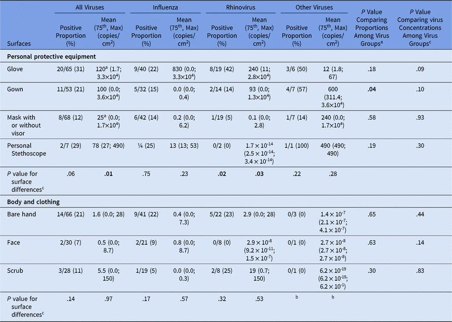
The presence and magnitude of virus contamination on the PPE are summarized in Table 1. Considering all viruses, 31% of glove samples, 21% of gown samples, 12% of face-mask samples, and 29% of personal stethoscope samples were positive for virus. All personal stethoscopes sampled had been used on patients. In these cases, the HCWs used their personal stethoscopes rather than the isolation stethoscopes available in the patient rooms. Viral contamination on the isolation stethoscopes is reported elsewhere.Reference Phan, Sweeney, Bleasdale and Jones11 The proportions of positive samples were not statistically significantly different among the sites sampled (χ2P = .06). However, the distributions of virus concentrations were significantly different (KW P = .01). Pairwise comparisons identified that the distribution of virus concentrations on gloves, which had a mean of 120 copies/cm2, differed from that on face masks, which had a mean of 25 copies/cm3 (W pairwise, P = .007). Differences in the proportion of positive samples and virus concentrations among the pieces of equipment were observed only for rhinovirus (KW P = .02), but pairwise comparisons among the equipment were not statistically significant different. Of the 68 face masks sampled, 24 HCWs had removed the visor while donning PPE, but the absence of the visor was not associated with a difference in virus concentration (W P = .95).
Virus Contamination on the Personal Protective Equipment (PPE), Clothing, and Skin of Healthcare Workers After Caring for a Patient with an Acute Respiratory Infection

Note. Bold indicates statistical significance.
a Significant pairwise comparison at the significant level of α = 0.05/3 = 0.017.
b Comparison among body parts was not tested because all samples were negative for virus presence.
c Proportions were compared using the χ2 test and virus concentrations using the Kruskal-Wallis test.
During patient care, prior to sampling, 65 of the 66 HCWs that provided bare hand samples wore gloves, 23 of 28 HCWs that provided scrub samples wore a gown, and all of the HCWs that provided face samples wore a mask or mask with visor. After PPE doffing, virus was detected on 14 of 66 (21%) of the bare dominant hands of HCWs, on 2 of 30 (7%) of the faces of HCWs, and on 3 of 28 (11%) of the scrubs sampled (Table 1). Among the 14 HCWs with positive bare hand samples, all had worn gloves. Among the 3 HCWs with positive scrub samples, 2 had worn gowns. Virus concentrations were similar on hands, faces, and scrubs, with mean values of 1.6 copies/cm2, 0.5 copies/cm2, and 5.5 copies/cm2 on the bare hands, face and scrubs of HCWs, respectively. Data in Table 1 show that the mean virus concentrations on glove, gown, and mask are higher than those on bare hand, scrub, and face, respectively. However, we did not find statistically significant differences between the distribution of virus concentrations on gown and scrub, face and mask, and glove and hand using the Wilcoxon test or in the mean difference between the surfaces using a paired t test. The proportion of samples positive for virus were similar on these 3 surfaces and for each of the virus groups (Table 1).
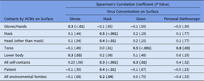
Self-contact may transfer virus between the gloves or bare hands and PPE, so we explored the association between virus concentrations on PPE and the number of self-contacts (Table 2). Self-contacts by HCWs were most frequently observed on torso and mask.Reference Phan, Maita, Mortiz, Bleasdale and Jones10 The strongest correlation was observed for the number of self-contacts with the torso and virus concentration on the stethoscope (ρ = 0.8; P = .03). The virus concentrations on HCW gloves, gown, and mask were all significantly correlated with the number of self-contact to those sites, with correlations of ρ = 0.3, ρ = 0.5, and ρ = 0.5, respectively (Table 2).
Spearman’s Correlation Between the Contacts Made by Healthcare Workers (HCWs) During Patient Care and Virus Concentrations on Personal Protective Equipment

Note. Bold indicates statistical significance.
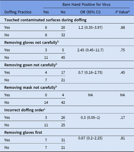
Gloves are removed during the process of doffing PPE, which means that HCWs may touch surfaces contaminated with viruses with their bare hands. As a result, we explored the influence of PPE doffing practices on the presence of virus on bare hands (Table 3). We detected a positive trend between virus presence on the hands and touching potentially contaminated surfaces during doffing (OR, 1.2; 95% CI, 0.35–3.97) and removing gloves not carefully (OR, 2.45; 95% CI, 0.45–11.7), but the odds ratios were not statistically significantly elevated. Analysis of potential determinants of virus presence on HCW bodies was limited to bare hands because of the low frequency of virus detection on the faces and scrubs of HCWs.
Association Between Personal Protective Equipment Doffing Practices and Virus Presence on the Bare Hands of Healthcare Workers After Patient Care

Note. OR, odds ratio; CI, confidence interval.
a Comparison of virus concentration between 2 categories of the characteristic of interest using Mann-Whitney test.
b PPE doffing carefulness is defined as a removal process not involving strong or vigorous movements, which can generate aerosols from infectious PPE.Reference Phan, Maita and Mortiz4
c A correct PPE doffing sequence includes removing gloves first, followed by the gown, the mask, and then hand hygiene.Reference Phan, Maita and Mortiz4
Discussion
Self-contact is one mechanism by which virus may be transferred to the PPE and bodies of HCWs from the patient or other surfaces in the patient room contaminated with virus. To our knowledge, our study is the first to evaluate the association of self-contacts by HCWs and virus contamination on their PPE and bodies. In particular, the numbers of self-contacts by HCWs with their gloves, gown (torso), and mask were moderately positively associated with virus concentrations on those pieces of PPE (Table 2). The strongest correlation identified was between self-contact with the gown (torso) and virus concentrations on a personal stethoscope, which is often draped around the neck. We were unable to discern the direction of virus transfer upon contact between the HCW hands or gloves and a surface, but our results suggest that contacts contribute to the dissemination of virus.
Personal protective equipment is intended to prevent virus from reaching the body and clothing of HCWs, including body sites susceptible to infection. The presence of virus on the bare hands under gloves (14 of 65), face (2 of 0), and scrubs under gown (2 of 23) of HCWs indicate that the PPE worn did not offer complete barrier protection, and/or that self-contamination may have occurred during PPE doffing. Two incorrect PPE doffing practices, touching contaminated surfaces during doffing and removing gloves not carefully, were positively, but not significantly, associated with virus presence on the bare hands of HCWs (Table 3). In a simulation study, contamination (fluorescein or MS2 bacteriophage) was observed on the hands and faces of 21% and 4%, respectively, of HCWs after doffing of PPE used for contact precautions.Reference Kwon, Burnham and Reske7 In an observational study, 3.2% of HCWs acquired a multidrug-resistant organism on their hand, and 3.2% acquired the organism on their clothes after PPE removal, which were associated with PPE doffing errors.Reference Okamoto, Rhee and Schoeny3 These percentages are relatively consistent with those observed in this study (Table 1) and suggests that HCW bodies are routinely contaminated after PPE doffing.
The detection of viruses on bare hands in 21% of samples collected emphasizes the significance of hand hygiene in prevention of infectious disease transmission from HCWs to patients via the contact route. In our observations of doffing practices among this population of HCWs, we found that HCWs did not perform hand hygiene before leaving the patient room in 6.6% of observations, though hygiene may have been performed later.Reference Phan, Maita and Mortiz4 The presence of viruses on the personal stethoscopes in our study highlighted the importance of stethoscope hygiene. Our study did not record personal stethoscope hygiene, but others have found that the stethoscope hygiene was rarely done by HCWs after patient encounters.Reference Holleck, Merchant, Lin and Gupta12 Others have found that even when stethoscopes were cleaned, the cleaning methods by HCWs failed to bring the contaminated stethoscopes to the level of a clean one.Reference Knecht, Mcginniss and Shankar13
Our study has several limitations. First, we were not able to measure all variables during each experiment. For example, some HCWs refused to participate in some aspects of study, such as swabbing the face. Second, the sampling efficiency of the Copan swabs used in this study has not been characterized. Previously, macrofoam swabs were found to recover <36% of norovirus applied to different environmental surfaces.Reference Park, Lee, Treffiletti, Hrsak, Shugart and Vinjé14 Therefore, virus may be present on the PPE and bodies of HCWs more often and at higher concentrations than reported in this study. Third, because we did not measure virus on HCWs prior to care delivery, we cannot rule out the possibility that contamination was not specifically the result of the observed patient care activity. This risk was minimized by measuring only the virus with which the patient was infected, but others have observed HCWs to be contaminated with multidrug-resistant organisms prior to caring for an infected or colonized patient.Reference Okamoto, Rhee and Schoeny3 Lastly, our analytical method, quantitative real-time polymerase chain reaction, only quantified the amount of viral RNA present in the swab samples and was not able to determine the amount of viable viruses in the sample. Therefore, we were not able to estimate the absolute risk of HCW exposure to viable virus.
Our study is the first to characterize the magnitude of respiratory virus contamination on the PPE and bodies of HCWs after providing care to infected patients. All 3 groups of virus were found on the PPE and bodies of HCWs with similar frequency and virus concentration distributions (Table 1). This finding, coupled with our findings that the 3 groups of virus were found in the air and on environmental fomites with similar frequency and virus concentration distributions in our other work,Reference Phan, Sweeney, Bleasdale and Jones11 suggests that these respiratory viruses follow similar exposure pathways for HCWs. Our findings of viral contamination on PPE, clothes, and skin of HCWs emphasize the significance of appropriate PPE use, PPE doffing practices, and hand hygiene in infectious transmission prevention via the contact route. Exposure modeling and quantitative microbial risk assessment should be used to evaluate the importance of the presence and magnitude of different viruses at different locations in the environment and on HCWs for occupational and patient health impact.Reference Nicas and Jones15, Reference Jones and Xia16
Supplementary material
To view supplementary material for this article, please visit https://doi.org/10.1017/ice.2019.298
Acknowledgments
We thank Charissa Fritzen-Pedicini, Rachel Weber, and Maryshe Zietsman for their helpful assistance with participant recruitment and data collection. Library preparation and sequencing were performed at the University of Illinois at Chicago Sequencing Core (UICSC), and we thank Dr Stefan Green of the UICSC facility for his advice and support. Data will be available from the corresponding author or from UIC INDIGO (https://indigo.uic.edu/) within 6 months of publication. Any opinions, findings, and conclusions expressed in this material are those of the authors and do not necessarily reflect the views for the CDC.
Financial support
This work was supported by the CDC (cooperative agreement no. 1U54CK000445-01).
Conflicts of interest
All authors report no conflicts of interest relevant to this article.