
Introduction
The possibility to remotely manipulate cellular fate and behavior in single cells has opened new frontiers for the investigation of cell signaling and gene regulation in living tissues. In this sense, physical stimuli such as electric fields and light irradiation have attracted a great deal of attention. Light offers many advantages to stimulate cell signaling, allowing high spatio-temporal resolution in a non-invasive way.[Reference Repina, Rosenbloom, Mukherjee, Schaffer and Kane1] Optogenetics refers to the control of cellular behavior when stimulating by light genetically modified cells. To date, many strategies have been developed to make specific cells responsive to light, mainly by expressing proteins containing light-responsive domains.[Reference Kushibiki, Okawa, Hirasawa and Ishihara2] In spite of many advantages, among current limitations of optogenetics is the need to engineer the cells so that they express photosensitive proteins. Other approaches rely on photoactive materials, able to convert light into an electrical, chemical or thermal stimulus. Photovoltaic materials have recently gained much attention for therapeutic purposes as drug delivery tools[Reference Tandon, Magaz, Balint, Blaker and Cartmell3] or as modulators of neuronal activity, as extensively reviewed.[Reference Antognazza, Martino, Ghezzi, Feyen, Colombo, Endeman, Benfenati and Lanzani4] Among possible alternatives, the use of organic semiconducting nanoparticles (NPs) composed of poly(3-hexyl)thiophene (P3HT) able to absorb light and to convert it into an electrical signal has been recently proposed.[Reference Zangoli, Di Maria, Zucchetti, Bossio, Antognazza, Lanzani, Mazzaro, Corticelli, Baroncini and Barbarella5] P3HT is a conjugate polymer that has been largely used for photovoltaics and photonics.[Reference Lu, Zheng, Wu, Schneider, Zhao and Yu6] Lanzani's group successfully pioneered its use to modulate cell functions by transducing light signal into cells. P3HT thin solid films, upon illumination, sustained elicitation of action potentials in primary neurons,[Reference Ghezzi, Antognazza, Dal Maschio, Lanzarini, Benfenati and Lanzani7] modulation of specific ion channels conductances in primary astrocytes[Reference Benfenati, Martino, Antognazza, Pistone, Toffanin, Ferroni, Lanzani and Muccini8] and in non-electrogenic cells,[Reference Martino, Feyen, Porro, Bossio, Zucchetti, Ghezzi, Benfenati, Lanzani and Antognazza9] electrical activity activation and inhibition in retinal and brain tissues.[Reference Feyen, Colombo, Endeman, Nova, Laudato, Martino, Antognazza, Lanzani, Benfenati and Ghezzi10, Reference Lodola, Martino, Tullii, Lanzani and Antognazza11] Moreover, they developed and tested in vivo functionally autonomous polymer-based prostheses to treat degenerative blindness,[Reference Ghezzi, Antognazza, Maccarone, Bellani, Lanzarini, Martino, Mete, Pertile, Bisti, Lanzani and Benfenati12, Reference Maya-Vetencourt, Ghezzi, Antognazza, Colombo, Mete, Feyen, Desii, Buschiazzo, Di Paolo, Di Marco, Ticconi, Emionite, Shmal, Marini, Donelli, Freddi, Maccarone, Bisti, Sambuceti, Pertile, Lanzani and Benfenati13] showing great potential of these materials to be interfaced with living cells for therapeutic purposes. The possibility to use photovoltaic materials to control cell function was further investigated in the freshwater polyp Hydra vulgaris, a small and primitive animal, structured as a living tissue, representing an amenable model to study toxicity[Reference Ambrosone, Roopin, Pelaz, Abdelmonem, Ackermann, Mattera, Allocca, Tino, Klapper, Parak, Levy and Tortiglione14] and interaction between nanomaterials and living systems,[Reference Moros, Ambrosone, Stepien, Fabozzi, Marchesano, Castaldi, Tino, de la Fuente and Tortiglione15, 16] while satisfying the 3Rs principle to reduce vertebrate experimentation. It was shown that P3HT-NPs (300 nm average size) in this eyeless animal that yet show a photobehavior, are able to enhance light sensitivity, inducing a precise behavioral response and enhancing the expression levels of a gene involved in phototransduction, opsin3-like gene.[Reference Tortiglione, Antognazza, Tino, Bossio, Marchesano, Bauduin, Zangoli, Morata and Lanzani17] The mechanisms involved in this phototransduction process remain unclear to date. In this direction, we extend here our investigations focusing on the intracellular redox disequilibrium. The change in the redox state represents an immediate consequence of the photoactivation, as reported for polythiophene drug conjugates, which under light irradiation sensitize oxygen to produce reactive oxygen species (ROS).[Reference Wang, Yuan, Zhu, Yang, Lv, Liu and Wang18–Reference Tullii, Desii, Bossio, Bellani, Colombo, Martino, Antognazza and Lanzani20] ROS are by-products of the cellular metabolism, the most relevant being singlet oxygen, hydroxyl radical, hydrogen peroxide, and superoxide anion.[Reference Dalle-Donne, Giustarini, Colombo, Rossi and Milzani21] Under physiological conditions ROS are neutralized by antioxidant systems, that are composed by endogenous enzymatic antioxidants such as catalase or superoxide dismutase (SOD), and non-enzymatic antioxidants such as α-tocopherol or ascorbic acid. ROS can have beneficial as well as deleterious effects. ROS produced at low concentrations can serve as signaling molecules to regulate different biologic and physiological processes. However, if the antioxidant mechanisms are impaired or the production of ROS is increased, oxidative stress can take place, which can induce pathology by damaging lipids, proteins, and DNA.[Reference Kannan and Jain22] Here we demonstrate that P3HT-NPs once internalized in Hydra produce an increase of oxidative stress parameters as effect of photostimulation, without, however, signs of toxicity. We showed that the animal antioxidant defense mechanisms are specifically modulated as effect of continuous exposure to a white light source. The total antioxidant defence capacity decreases already after 4 h of exposure, while after prolonged treatments (up to 24 h) both DNA and protein undergo oxidation, showing a clear effect of NP photostimulation on intracellular redox balance, and suggesting P3HT-NP as novel tool to finely tune this function for specific purposes.
Experimental
NP synthesis
P3HT-NPs were prepared from freshly synthesized P3HT by the reprecipitation method, as previously described.[Reference Tortiglione, Antognazza, Tino, Bossio, Marchesano, Bauduin, Zangoli, Morata and Lanzani17, Reference Pecher and Mecking23] Briefly, the polymer was dissolved in tetrahydrofuran, and the solution was added dropwise to distilled water in a 1:20 ratio. P3HT-NPs were separated by means of differential centrifugation into fractions of different sizes. After DLS characterization, NPs with an average hydrodynamic radius of 237 ± 82 nm and a polydispersity index of 0.12 were selected. Ultraviolet (UV)–visible (vis) spectroscopy measurements were carried out in water using a PerkinElmer Lambda 1050 spectrophotometer.
Animal culture
Hydra vulgaris (strain Zurich) was asexually cultured in Hydra medium (1 mM CaCl2 and 0.1 mM NaHCO3) at pH 7. The animals were kept at 18 ± 1 °C and fed thrice a week with freshly hatched Artemia salina nauplii.
Photostimulation of P3HT-NP-treated animals
Groups of 60 animals were placed in plastic multiwells and incubated with P3HT-NPs (50 µM) in a final volume of 300 µL of Hydra medium. Photostimulation was accomplished by using a white light source (Edmund Optics, the power density of 0.124 mW/mm2), and animals exposed for either 4 or 24 h. Then the animals were inspected by a stereomicroscope (SZX7, Olympus) and by confocal microscopy (Ti2 Eclipse, Nikon) to assess NP internalization and possible morphological changes induced by photostimulation. As control conditions, both untreated animals, and treated animals not illuminated were used.
Protein extraction
Before illumination (0 time point), and after 4 h and 24 h of continuous light exposure, groups of five animals were homogenized in detergent solution (50 mM Tris–HCl, 150 mM NaCl, 0.1% SDS, 1% NP-40) in presence of protease inhibitors, 1 mM phenylmethylsulfonyl fluoride (PMSF, Sigma) and Complete™ (Roche). Samples were centrifuged at 14,000 rpm for 10 min at 4 °C, then the soluble protein fractions were collected and stored at −20 °C.
DNA extraction
Groups of 30 Hydras were homogenized in 0.1 M sodium phosphate buffer (pH = 7.4) and incubated at 65 °C for 15 min with 10% SDS and Proteinase K (1 mg/mL). After 10 min of centrifugation at 13,000 rpm at 4 °C the supernatants were collected, and purified by adding phenol: chloroform: isoamylic alcohol = 1:1:24. Supernatants were treated with RNase (10 mg/mL) at 37 °C for 1 h, then DNA precipitated overnight with 0.7 vol of isopropanol. After centrifugation at 13,000 rpm at 4 °C for 15 min, pelleted DNA was washed with 75% ethanol, dried and resuspended in sterile water.
Total antioxidant capacity (TAC)
In brief, the fresh working solution was prepared immediately before measurements by mixing 10 volumes of acetate buffer, pH 3.6, with 1 volume of 10 mM 2,4,6–tripyridyl-S-triazine (TPTZ; Sigma) and 1 volume of 20 mM FeCl3 (Sigma). A 40 µL aliquot of protein extract was mixed with 1160 µL of the working solution and incubated at room temperature. After 20 min, the absorbance of the samples was read at 593 nm in an absorbance microplate reader against a reagent blank. The increase in absorbance is proportional to the activity of anti-oxidants in the sample. TAC was expressed as trolox equivalent anti-oxidant capacity (TEAC), that is, the concentration (amount) of trolox producing the same effect as the sample studied. Calculations were made on the basis of standard curves obtained for a trolox solution.
Western blot analysis of Cu–Zn SOD protein levels
The protein concentration was estimated with BCA assay[Reference Smith, Krohn, Hermanson, Mallia, Gartner, Provenzano, Fujimoto, Goeke, Olson and Klenk24] using bovine serum albumin (BSA) as the standard (Sigma). Then, proteins (50 µg) were resolved by 10% SDS–PAGE and electroblotted onto an Immobilon polivinylidene difluoride (PVDF) membrane (Millipore Corporation). The PVDF membrane was blocked with 1% BSA in TBST at room temperature for 1 h. As primary antibody anti-SOD1 (1:1000) (PA523245, ThermoFisher Scientific) was used. A cross-reacting band of 18 kDa [and likely corresponding to the Hydra Cu–Zn SOD (LOC100200684)] was detected after incubation with the horseradish peroxidase-conjugated secondary antibody [1:50,000 in 1% BSA in TBST (20 mM Tris–HCl, pH 7.5, 137 mM NaCl containing 0.1% Tween 20), 1 h incubation at room temperature] (Sigma Aldrich). For the loading control, mouse monoclonal anti-β-actin—peroxidase antibody was used (1:40,000 in 1% BSA in TBST, 30 min incubation at RT) (A3854, Sigma Aldrich). The chemiluminescence signal was detected with the Clarity Western Blotting Detection System (BIORAD) and a G:BOX imaging system (Syngene). The data represent the relative density normalized to β-actin.
Protein carbonylation
Stress-mediated protein carbonylation was estimated with an OxyBlot™ Protein Oxidation Detection Kit (Millipore Corporation) using the standard protocol according to the manufacturer's instructions. The protein concentration was estimated with BCA assay using BSA as the standard (Sigma). Then, proteins (20 µg) were derivatized with 2,4-dinitrophenylhydrazine (DNPH) and were resolved by 10% SDS–PAGE and electroblotted onto an Immobilon polivinylidene difluoride (PVDF) membrane (Millipore Corporation). The membrane was blocked with 1% BSA in TBST at room temperature for 1 h and protein carbonylation (proteins with DNP residues) was detected using rabbit anti-DNP antibody (diluted 1:150 in 1% BSA in TBST, overnight incubation at 4 °C) (Millipore Corporation) and goat anti-rabbit HRP-conjugated antibody (diluted 1:300 in 1% BSA in TBST, 1 h incubation at room temperature) (Millipore Corporation). For each oxyblot analysis, a negative control (no DNPH derivatization) and a positive control with a mixture of standard proteins with attached DNP residues were applied. The chemiluminescence signal was detected with ECL Plus Western Blotting Detection System (GE Healthcare) and a G:BOX imaging system (Syngene). Protein carbonylation is considered as the intensity of chemiluminescence signal of a 43 kDa band and data collected at appropriate time points for control conditions are presented as 1.0.
Protein aggregates
The concentration of protein aggregates was measured using PROTEOSTAT® Protein Aggregation kit (Enzo ENZ-51023-KP050) following the standard protocol according to the manufacturer's instructions. Briefly, protein extract from Hydra cells (50 µg) was subjected to protein aggregation content analysis. Fluorescence was measured after 15 min incubation of samples with PROTEOSTAT® detection dye using microplate reader with an excitation setting of 550 nm and an emission filter of 600 nm. The calculation was made on the basis of a positive control.
DNA damage
Oxidative DNA damage as a level of 8-hydroxy-2′-deoxyguanosine (8-OHdG, 8-oxo-dG) was measured using Epigentek EpiQuik 8-OHdG DNA Damage Quantification Direct Kit, following the standard protocol according to the manufacturer's instructions. Briefly, DNA extracted from Hydra polyps in each experimental condition (100 ng) was subjected to 8-OHdG content analysis. Absorbance was read at 450 nm using a Tecan Infinite® M200 absorbance mode microplate reader. The calculation was made on the basis of positive control solutions and 8-OHdG content is presented as ng 8-OHdG per 100 ng DNA.
Photo-induced absorption spectroscopy
P3HT-NP water dispersion has been excited by a solid-state laser source (Oxxius-561-100-COL-PP) with an optical power of about 100 mW, modulated by a mechanical chopper at a frequency of 235 Hz. The probe beam is given by a 100 W tungsten halogen lamp focused on the P3HT-NP dispersion by two spherical mirrors, in order to avoid chromatic aberrations. Sample transmission is spectrally analyzed by a monochromator and collected by a silicon photodetector (Thorlabs FDS100) and the signal is analyzed with a lock-in amplifier (Stanford Research Systems SR830), locked in frequency to the modulation of the chopper, with an integration time of 10 s. The measured signal ΔT/T is the modulation in the transmitted light (ΔT) at a certain wavelength, due to the presence of the pump excitation, and normalized to the sample transmission (T) in the absence of the pump. In particular, a negative signal (ΔT/T < 0) indicates the occurrence of photo-induced absorption processes.
Results and discussion
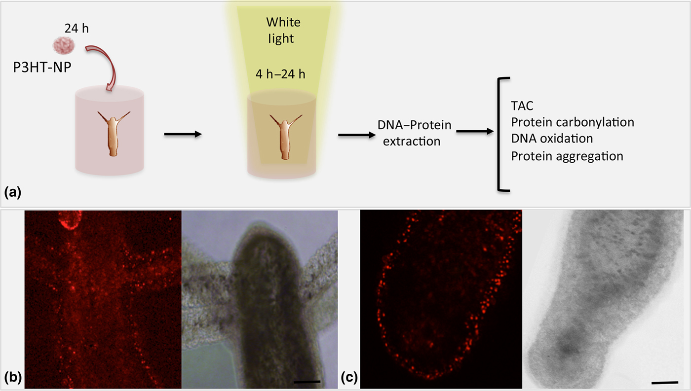
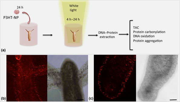
P3HT-NPs were prepared by a reprecipitation method, using P3HT polymer that was previously synthesized by oxidative polymerization.[Reference Zucchetti, Zangoli, Bargigia, Bossio, Di Maria, Barbarella, D'Andrea, Lanzani and Antognazza25] The P3HT-NP display a spherical shape, they have an average hydrodynamic radius of 237 ± 82 nm and a polydispersity index of 0.12, as measured by dynamic light scattering (Fig. S1). The UV–vis spectrum shows a broad optical absorbance in the visible range 400–600 nm, peaking at 500 nm (Fig. S1). In vivo assays were accomplished on groups of 60 animals, soaked with P3HT-NPs (50 µM) for 24 h, to ensure efficient NP uptake into all animal tissues. After extensive washes living animals were observed by confocal microscopy. As shown in Fig. 1, P3HT-NPs appeared thoroughly distributed along tentacles and body length. The animals were then exposed to a light-emitting diode (LED) white light source (0.124 mW/mm2) for 4 or 24 h. After illumination, the toxicity of the P3HT-NPs and/or the photoexcitation protocol was assessed by analyzing animal morphology, that may be seriously affected (tentacle and body contractions, tissue disintegration, and general damages up to complete death) in presence of organic or inorganic toxicants, as reported.[Reference Ambrosone, Mattera, Marchesano, Quarta, Susha, Tino, Rogach and Tortiglione26–Reference Ambrosone and Tortiglione28] No morphological changes such as tissue alterations were observed, indicating the absence of toxicity after long-term photostimulation. As it has been reported that in P3HT solid thin films exposed to aqueous electrolytes net charge–transfer phenomena can give rise to efficient oxygen reduction,[Reference Mosconi, Salvatori, Saba, Mattoni, Bellani, Bruni, Santiago Gonzalez, Antognazza, Brovelli, Lanzani, Li, Brédas and De Angelis29] we aimed to evaluate the changes in intracellular redox equilibrium by analyzing oxidative stress endpoints in the animals. Production of ROS can be detected by using fluorescent compounds that change their intensity or emission peak upon oxidation by the ROS molecules. However, these probes are difficult to use for live imaging in Hydra, due to artifacts caused by animal movements. As it is known that ROS can cause damage by attacking carbohydrate, lipids, proteins, DNA and RNA, several oxidative stress endpoints were considered, namely the TAC, the rate of both DNA and protein oxidation, the amount of defensive enzymes such as Cu–Zn SOD and protein aggregation. As for the toxicity analysis, animals either untreated or treated with P3HT-NPs were exposed to continuous white light LED illumination (4 or 24 h) or kept in the dark prior to DNA and protein extraction.
Photostimulation of living animals with P3HT-NPs. (a) Living Hydras were incubated with 50 µM P3HT-NPs for 24 h, and then exposed to white light for 4 or 24 h before processing for DNA and protein extraction. (b, c) Laser scanning confocal microscopy shows efficient internalization of nanoparticles into Hydra tissue, both in (b) the head of the animals, and (c) in the lower body including the foot. In each panel, both fluorescence and bright field images are reported, respectively, on the left and right part. Scale bar, 200 µm.

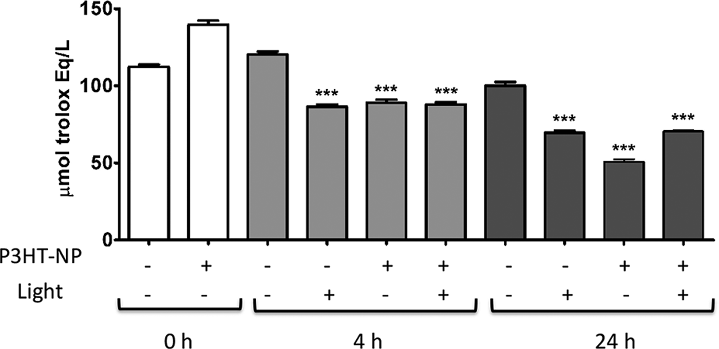
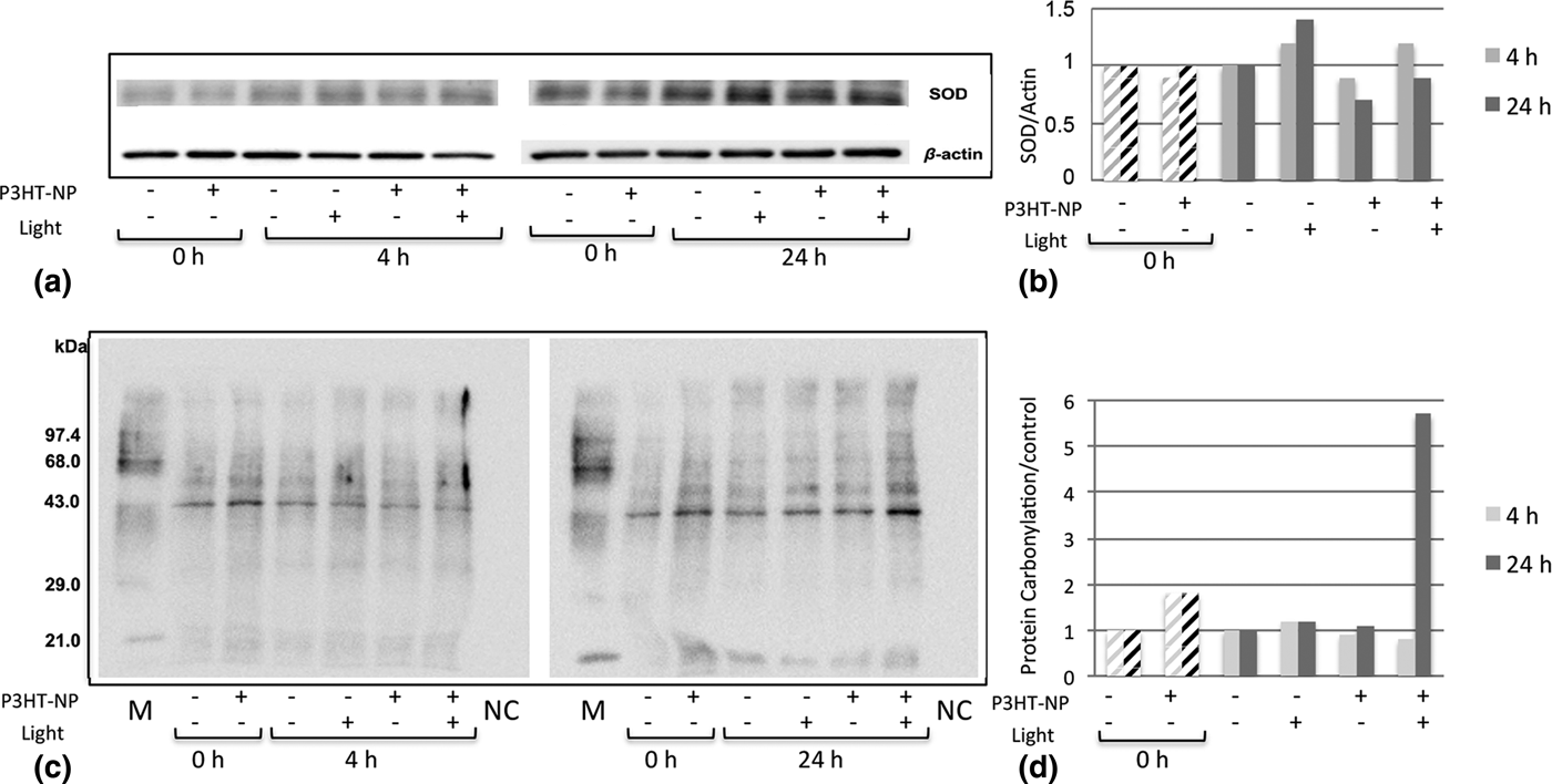
TAC is a useful technique to estimate the antioxidant status of biologic samples and provides an indication of the overall capability to counteract ROS. For quantifying TAC, we performed the Ferric Reducing Antioxidant Power (FRAP) assay using a ferric ion complex containing tripyridyltriazine (TPTZ).[Reference Lewinska, Wnuk, Slota and Bartosz30] An intense blue color is formed when Fe(III) species are reduced to Fe(II) by the electron-donating antioxidants of the sample, which can be measured at 593 nm. The presence of P3HT-NPs combined with continuous illumination significantly decreases TAC when compared with non-treated animals, as determined using the ferric ion reducing anti-oxidant power assay (Fig. 2). After 4 and 24 h of illumination comparable results were found, indicating a relatively short dynamics of induction of oxidative stress. In general, TAC is decreased in conditions associated with oxidative stress or increased susceptibility to oxidative damage.[Reference Young31] Interestingly, P3HT-NPs in the dark, continuous illumination, and a combination of both led to the similar outcome. As a complementary approach to measure the expression of antioxidant enzymes, a Western blot analysis was performed to assess possible changes in the levels of Cu–Zn SOD [Fig. 3(a)]. No changes in protein expression levels were detectable at both time points unless a slight decrease at 24 h in P3HT-NP treated animals, however uncorrelated to light exposure [Fig. 3(b)]. As a decrease in TAC can impact on protein redox state, the possibility that P3HT-NP photostimulation may lead to imbalanced ROS and thus change total protein redox state was next evaluated. Protein carbonylation is an irreversible oxidative modification of the proteins that can be considered a final result of long or strong oxidative stress, and it has been related to different pathological conditions and aging. The carbonyl groups of total protein extracts prepared from animals in the four experimental conditions as above were detected by immunodetection (Oxyblot). Results shown in Fig. 3 display no differences in the protein carbonylation levels after 4 h of continuous irradiation, while after 24 h a strong increase was detected in protein samples extracted from treated and illuminated animals [Figs. 3(c) and 3(d)].
Total anti-oxidant capacity (TAC) of animals after P3HT-NP photostimulation. Comparison of TAC among protein extracts prepared from groups of animals in the four experimental conditions relative to the exposure of P3HT-NP and Light, indicated by the sign + and − below the graph, i.e. NP/Light −/−, −/+,+/−, and +/+. Animals were illuminated under a continuous white light LED for 4 h (light gray bars) or 24 h (dark gray bars) before extracting the proteins. Proteins were also extracted from animal before starting the illumination (time zero, white bars). TAC was expressed as trolox equivalent anti-oxidant capacity. Bars indicate SD, n = 3, ***P < 0.001 compared with control (−/−) at appropriate time point (ANOVA and Dunnett's a posteriori test).

Impact of P3HT-NP photostimulation on Cu–Zn SOD protein expression and protein oxidative damage. (a) Modulation of Cu–Zn SOD levels as the effect of P3HT-NP photostimulation—Protein extracts were prepared from groups of animals in the four experimental conditions as above and compared to control animals. (b) Cu–Zn SOD expression levels were quantified by densitometry analysis relative to β-actin expression. (c) Oxyblot analysis of total protein carbonylation. M and NC have been included as a positive and negative controls, respectively. (d) Densitometry quantification of the 43 kDa band was performed considering controls (NP/Light, −/−) at appropriate time points as 1.0.

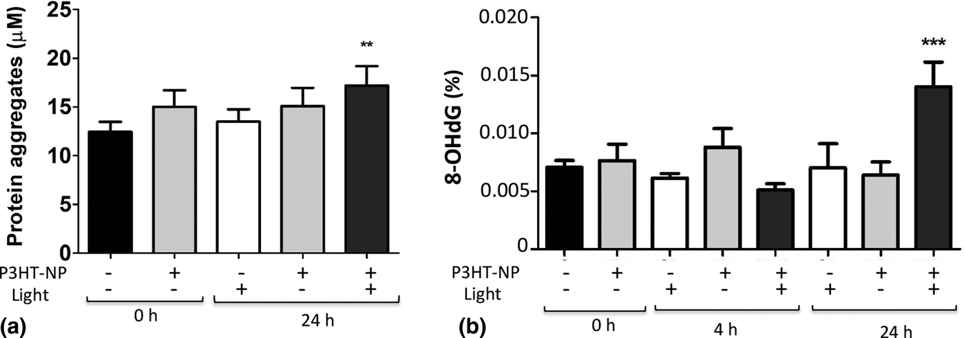
The estimation of protein aggregation represents an additional indication of the protein modification induced by the oxidative stress since oxidized proteins are thermodynamically unstable and may aggregate. To this aim, the protein aggregation states were assessed in all experimental conditions above analyzed. As expected, significant protein aggregation was detected in protein extracts from an animal treated with P3HT-NPs and exposed for 24 h to white light [Fig. 4(a)]. Conversely, neither light nor NPs by themselves induced protein aggregation, confirming the specific effect played by P3HT-NPs photoexcitation. Finally, the possible DNA damage was analyzed by detecting 8-hydroxy-2′-deoxyguanosine (8-OHdG) a critical biomarker for oxidative stress.[Reference Valavanidis, Vlachogianni and Fiotakis32] 8-OHdG results when hydroxyl radical attacks deoxyguanosine, and unless repaired prior to DNA replication, it may lead to mutagenesis. As in the case of the protein aggregates, only animals exposed to P3HT-NPs and illuminated for 24 h showed a significant increase in the levels of 8-OHdG [Fig. 4(b)]. We can therefore conclude that although both the P3HT-NPs and light alone have an impact on the TAC of the animal (Fig. 2), they have a negligible effect on the rate of biomolecule oxidation. Importantly, they are not able to produce a high enough oxidative stress to damage DNA or give rise to protein aggregation. On the other hand, long periods of illumination (24 h) in animals treated with P3HT-NPs produce higher levels of protein carbonylation and aggregation, as well as DNA damage. We notice that, at fixed photoexcitation density, the extent of illumination period is critical in term of the promotion of oxidative stress, as no significant effects are detected at shorter times both in protein carbonylation or 8-OHdG levels.
Effect of P3HT-NP photostimulation on protein and DNA. Protein and DNA extracts were prepared from groups of animals treated with either P3HT-NP and/or Light, as indicated by the sign + and − below the graphs, i.e. NP/Light: −/−,−/+,+/−, and +/+. (a) Protein aggregation was estimated by using PROTEOSTAT® Protein Aggregation kit according to the manufacturer's instructions. Protein extracts at 24 h were compared with experimental control condition at time 0. (b) Detection of DNA damage by means of 8-OHdG. DNA extracts were prepared from animals treated as above at 4 and 24 h time points, and compared with controls at time 0. Bars indicate SD, n = 3, **P < 0.01, ***P < 0.001 compared with control at the appropriate time point (ANOVA and Dunnett's a posteriori test).

The formation of ROS after light excitation of inorganic semiconductor NPs has been previously described.[Reference Rajendran, Lehnig and Niemeyer33] Upon irradiation with UV light cadmium-based quantum dots generate superoxide- and hydroxyl radicals, that have been exploited to manipulate enzymatic activity or for photodynamic therapy, as demonstrated for metal and graphene-based QD.[Reference Waiskopf, Ben-Shahar, Galchenko, Carmel, Moshitzky, Soreq and Banin34, Reference Ge, Lan, Zhou, Liu, Guo, Wang, Jia, Niu, Huang, Zhou, Meng, Wang, Lee, Zhang and Han35] Although an excess of ROS production is a deleterious effect, their controlled production on demand using light can rise many opportunities in numerous biologic functions. Some ROS play key roles in many cellular signaling pathways, by modulating cell proliferation, apoptosis, and gene expression through activation of transcription factors, like NFkappa-B and hypoxia-inducible-factor-1α.[Reference Poljsak, Suput and Milisav36] Cell homeostasis is indeed guaranteed by a tightly controlled balance between the production and scavenging of ROS. The antioxidant defense system must thus minimize the levels of most harmful ROS on one side while still permitting enough ROS concentration for their useful purposes (e.g., cell signaling and redox regulation).
Many approaches based on exogenous inorganic semiconductors have been developed to modulate the intracellular redox equilibrium at non-toxic levels. In the large majority of cases, however, optically-controlled ROS modulation is determined by Near-Infrared photothermal heating, leading to ROS enhancement as a secondary step. By contrast, very little is known about the behavior of NPs entirely composed of P3HT polymer,[Reference Zucchetti, Zangoli, Bargigia, Bossio, Di Maria, Barbarella, D'Andrea, Lanzani and Antognazza25] simply dissolved in an electrolyte medium and without external bias. In particular, for the present work, it is key to verify that P3HT-NPs maintain the capability to create free charges, prone to undergo oxygen reduction processes and thus to efficiently sustain ROS formation. To this goal, we carried out photoinduced absorption (PIA) spectroscopy experiments on a ms time regime, compatible with slow activation kinetics typical of a liquid, ionic environment. Figure S2 shows the PIA spectrum of P3HT-NPs in the electrolyte solution. Photoinduced absorption in the spectral range 900–1100 nm confirms the presence of long-lived charged states. The latter can react with the oxygen dissolved in the aqueous biologic environment, according to the scheme[Reference Tullii, Desii, Bossio, Bellani, Colombo, Martino, Antognazza and Lanzani20]:
 $${\rm P3HT} + hv\, \to \,{\rm P3HT}^{\ast}$$
 $${\rm P3HT}^{\ast}\, \to \,{\rm P3H}{\rm T}^{+} + e^- $$
 $$e^- + {\rm O}_2\, \to \,{\rm O}_2^{\bullet -} $$
The superoxide, which is an anion radical, can further evolve leading to the generation of different ROS within the living animal cells. Superoxide can be converted into hydrogen peroxide (H2O2) by the action of cytosolic and mitochondrial SODs.[Reference Schieber and Chandel37] Hydrogen peroxide may then oxidize cysteine residues on proteins to initiate redox biology or be converted to water by antioxidant proteins, such as peroxiredoxins (PRx), glutathione peroxidase (GPx), and catalase (CAT). However, when hydrogen peroxide levels are elevated, hydroxyl radicals (OH•) are formed via reactions with metal cations (Fe2+) that promote irreversible biomolecule damage, e.g., oxidative protein and DNA damage.[Reference Schieber and Chandel37]
These results are in agreement with a direct photocatalytic generation of ROS species, sustained by photoexcitation of the P3HT-NPs. On the other hand, photothermal effects can be safely disregarded, because the estimated temperature increase at the NP surface caused by photoexcitation at the operating conditions of this work is well below 10−3 K.
In conclusion, we have demonstrated that photoexcitation of P3HT-NPs with low-intensity white light can modulate the intracellular redox balance without causing macroscopic damage to the animals. This function can be further investigated, i.e., by using other illuminating regimes for instance, or other illuminating periods, to finely tune the amount of ROS species produced and achieve controlled manipulation of cell function for specific purposes.
Supplementary material
The supplementary material for this article can be found at https://doi.org/10.1557/mrc.2018.104.
Acknowledgment
Technical assistance of Giuseppe Marino and Ilaria Panico (ISASI-CNR, Pozzuoli) for animal culturing is gratefully acknowledged. We also thank for technical assistance in animal irradiation using white light LED source Dr. Massimo Rippa (ISASI-CNR, Pozzuoli). MM acknowledges the European Union´s Horizon 2020 research and innovation programme (Marie Skłodowska-Curie grant agreement No. 660228).