INTRODUCTION
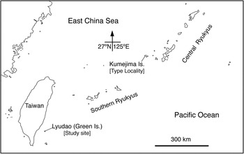
Lissoclinum midui Hirose & Hirose, Reference Hirose and Hirose2011 is a colonial ascidian harbouring the cyanobacterial symbiont Prochloron in the tunic. The colonies investigated in the original description were found on non-geniculate coraline algae in the reef crest (0.5–1 m deep) off Shinri-hama (Kumejima Island, Ryukyu Archipelago, Japan) (Figure 1). This species has two character states that are unique among didemnid ascidians: a cloacal aperture in each zooid; and an unknown organ in the bottom wall of the thorax (Hirose & Hirose, Reference Hirose and Hirose2011). Despite these unique features, L. midui has never been recorded from any site other than the type locality. In contrast, we have observed photosymbiotic ascidian fauna at various sites across Taiwan–Ryukyus (Hirose, Reference Hirose2013 and references therein). However, it is unlikely that the distribution range of this species is strictly limited to the type locality. We found multiple colonies of L. midui in the reef crest off Gongguan in Lyudao (Green Island), Tawain (Figure 1). A molecular phylogeny inferred from the cytochrome oxidase subunit I (COI) gene sequences supported the monophyly of this species as well as of the genus Lissoclinum. It is possible that the range of this species is broad but has merely not been recorded.
A map of Taiwan and the Central–Southern Ryukyus showing the type locality of Lissoclinum midui and the study site.

MATERIALS AND METHODS
Animals
Colonies of Lissoclinum midui were collected from the reef crest off Gongguan (22°38′42″N 121°30′5″E, Lyudao, Taiwan) on 24 June 2013. The colonies were found as an aggregate, and those from the two aggregates were sampled for this study. The first author found four aggregates of colonies within a 1-hour survey by snorkelling. The colonies were photographed using a digital camera before they were scraped off the substrate using a steel knife. We fixed the colonies in 10% formalin–seawater after anaesthetizing them with menthol and 0.37 M MgCl2. Some specimens were deposited in the National Museum of Natural Science, Taichung, Taiwan (NMNS-7254-001). Some fresh colonies were preserved in 99% ethanol at −30°C for DNA analyses.
The zooids were dissected from the fixed colonies and observed under a binocular stereomicroscope. In some photomicrographs, several images were combined to increase the depth of field using the post-processing image software Helicon Focus Pro 4.2.8 (Helicon Soft, Ltd., Kharkov, Ukraine).
DNA extraction, amplification and sequencing
Tissue samples were preserved in ethanol at −30°C. Three preserved colonies from a colony aggregate were dissected under a stereo microscope, and genomic DNA was extracted from about 40 zooids using a DNeasy Tissue Kit (Qiagen K.K., Tokyo, Japan) following the manufacturer's protocol. A segment of the mitochondrial COI gene was amplified using the primers UroCox1-244F (5′–CATTTWTTTTGATTWTTTRGWCATCCNGA–3′) and UroCox1-387R (5′–GCWCYTATWSWWAAWACATAATGAAARTG–3′), following the method of Hirose et al. (Reference Hirose, Yokobori and Hirose2009) for the amplification and sequencing of the partial COI fragments. Five clones were randomly selected and sequenced. Polymerase chain reaction products were treated with ExoSAP-IT (GE Healthcare) prior to the sequencing reactions. Sequencing reactions were performed using Operon Biotechnology (Tokyo). The sequences determined in this study have been deposited in the DNA databases of GenBank, European Molecular Biology Laboratory (EMBL) and DNA Data Bank of Japan (DDBJ) under the accession number AB899783.
Phylogenetic analyses
Partial COI gene sequences of 14 didemnid ascidian specimens (14 species from five genera) and Ciona savignyi (Cionidae) were obtained from GenBank and aligned with the present sequence for molecular analysis. Ciona savignyi was used as the outgroup. Initial alignments were performed using MUSCLE (Edgar, Reference Edgar2004), which were then inspected by eye and edited manually. The following analyses were performed on the aligned DNA sequences of the partial COI gene: maximum likelihood (ML) using the October 2008 version of TREEFINDER (Jobb, Reference Jobb2008) and phylogenetic analysis based on Bayesian inference (BI) using MrBayes 3.12 (Ronquist & Huelsenbeck, Reference Ronquist and Huelsenbeck2003). jModeltest 0.1.1 (Posada, Reference Posada2008) was used to select an appropriate nucleotide substitution model. For the BI analysis, a Markov chain Monte Carlo (MCMC) analysis was run for 1,000,000 generations, and trees were built at 100-generation intervals (burn-in = 2500). Statistical support for the ML tree was evaluated using a non-parametric bootstrap test with 1000 resampling events.
RESULTS
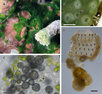
The colonies of Lissoclinum midui investigated were found on non-geniculate coraline algae growing on coral limestones in the reef crest open to the ocean (0.5–1 m deep) (Figure 2A). The approximately 1 mm thick irregularly shaped colonies (up to 3 cm in the longest axis) were attached to the substratum via filiform extensions of the tunic from the colonial margin (arrowheads in Figure 2B). Several zooids shared a common cloacal aperture (arrows in Figure 2B). The colonies were entirely green due to the presence of a symbiotic cyanophyte, Prochloron sp., in the tunic (Figure 2C). The area of the colonies closest to the substratum was white due to clusters of 10–20 µm diameter globular spicules (Figure 2C). Zooids were ~1.0 mm long (0.6 mm thorax, 0.4 mm abdomen, excluding the testis). Each branchial siphon was six-lobed, with the wide atrial aperture exposing most of the branchial sac. The branchial sac contained four rows of stigmata, and the right and left halves of the branchial sac respectively had seven stigmata in the first row, six or seven in the second row, six in the third row and five in the fourth row (Figure 2D). An unknown organ was observed in the bottom floor of the branchial sac of each zooid (Figure 2D). An undivided spherical testis was located under the abdomen, with an uncoiled vas deferens. Oocytes and embryos were not found in the specimens examined to date.
Lissoclinum midui from Lyudao: (A) colonies in situ; (B) fixed colony. Arrowheads indicate filiform extensions of the tunic. Arrow indicates common cloacal aperture, while the others are branchial apertures; (C) spicules and Prochloron cells in the tunic; (D) a zooid. Numbers indicate the stigmata in each row. en, endostyle; st, stomach; ts, testis; un, unknown organ. Scale bars: 0.5 mm in B, 20 µm in C, 0.1 mm in D.

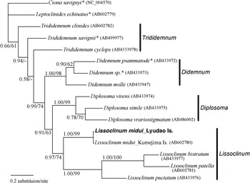
The partial mtCOI DNA sequences of L. midui from Shinri-hama (Kumejima Island, Japan) and Gongguan (Lyudao, Taiwan) showed a 3/401 base pair difference (0.75%). Since we sequenced only one specimen (40 zooids from several colonies) from Shinri-hama (Hirose & Hirose, Reference Hirose and Hirose2011) and one specimen from Gongguan (this study), the genetic variability within each site is unknown. Figure 3 shows the BI tree generated using a general time-reversible model with an invariant rate and a gamma distribution substitution (GTR + I + G). The topology of the ML phylogenetic tree was nearly identical. The phylogenetic trees inferred from the partial COI sequences supported the monophyly of L. midui and the genera Didemnum, Diplosoma, and Lissoclinum with high PI (i.e. Bayes posterior probability) and BP (i.e. bootstrap probabilities), but they did not support the monophyly of Trididemnum.
Bayesian inference consensus tree of selected didemnid species based on partial COI sequences. Ciona savignyi was used as the outgroup. The GTR + I + G model was used for the analysis. Bayes posterior probability and bootstrap probabilities greater than 50% for the maximum likelihood analyses are noted. New sequences from this study are shown in bold.

DISCUSSION
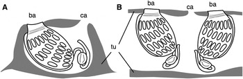
Many of the morphological features observed in the present specimens were consistent with the original descriptions of L. midui (Hirose & Hirose, Reference Hirose and Hirose2011). In particular, the specimens from Kumejima (the type locality) and Lyudao shared the same stigma number pattern, and their zooids always had an unknown organ in the bottom floor of the branchial sac. Stigma number pattern is sometimes a useful key for species identification in some didemnids (Nagaya & Hirose, Reference Nagaya and Hirose2013 and references therein), and the unknown organ is exclusive to this species among didemnid ascidians (Hirose & Hirose, Reference Hirose and Hirose2011). In contrast, the zooids in the colonies from Lyudao shared a common cloacal aperture, whereas each zooid in the type and topotype colonies of L. midui had a separate cloacal aperture (Figure 4). While the absence of the common cloacal system in L. midui is exceptional among didemnid ascidians, the presence of common cloacal apertures in the present specimens indicates a plasticity of this feature in this species. The differences in cloacal aperture morphology are thought to depend on the density of zooids within the colonies. The zooids in the colonies from Lyudao are more crowded than those from Kumejima Island (the type locality), and each zooid stands almost vertical within the colony (Figure 4B). In the topotype specimens, each zooid usually lies on its ventral side in the colony, with the oesophagus curving dorsally at the mid-point, and the alimentary tract looping perpendicularly to the anterior–posterior axis of the zooid (Hirose & Hirose, Reference Hirose and Hirose2011) (Figure 4A).
Schematic drawing of the section of Lissoclinum midui colonies: (A) a colony from Shinrihama, Kumejima Island (A, type locality); (B) a colony from Gongguan, Lyudao (this study). ba, branchial aperture; ca, cloacal aperture; tu, tunic.

In the partial mtCOI sequences of Diplosoma species, the intraspecific divergences were lower (0–11.0%) than the interspecific divergences among congeners (10.2–22.4%) (Hirose & Hirose, Reference Hirose and Hirose2013). In L. midui, the intraspecific divergence between the two collection sites (0.75%) was much smaller than the interspecific divergences among four Lissoclinum species (18.0–26.7%). This demonstrates the utility of using the partial mtCOI sequences as DNA barcodes in Lissoclinum species, as has been shown for other didemnid ascidians (Hirose et al., Reference Hirose, Tochikubo and Hirose2010; Hirose & Hirose, Reference Hirose and Hirose2013).
Although we carried out the faunal survey for photosymbiotic ascidians in Lyudao from October–November 2009 (Hirose & Nozawa, Reference Hirose and Nozawa2010), we did not find colonies of L. midui, likely because we were unable to dive off the eastern coast (including Gongguan) due to seasonal wave conditions at that time. To date, L. midui has only been recorded from Shinri-hama (Kumejima Island: the type locality) and Gongguan (Lyudao). The colonies were found on non-geniculate coraline algae in the reef crests (0.5–1 m deep) at both collection sites, indicating that this habitat is suitable for this species. Whereas we have not surveyed at −3 m or deeper at the both sites, the amount and species number of photosymbiotic ascidians generally decrease at deeper sites because of the shortage of solar radiation for photosymbionts. To date, no colonies have been found in similar habitats in Taiwan and the Ryukyu Archipelago. The crucial factor(s) controlling the occurrence of L. midui remains uncertain. If L. midui requires specific conditions for survival, this species could serve as an indicator for a particular environment. It is possible that this species has a broad range that has not been recorded. Since shallow, active wave conditions are inconvenient for SCUBA divers, such areas tend to be less investigated for small benthic invertebrates than more accessible habitats.
ACKNOWLEDGEMENTS
The present study was supported by a Grant-in-Aid for Scientific Research (C) no. 23510296 from the Japan Society for the Promotion of Science and by the International Research Hub Project for Climate Change and Coral Reef/Island Dynamics from University of the Ryukyus. This paper includes a contribution from Green Island Marine Research Station (GIMRS), Academia Sinica.