INTRODUCTION
The cabbage whitefly, Bemisia tabaci (Homoptera: Aleyrodidae), is an important pest that inflicts damage in temperate and tropical regions worldwide. The B-biotype and Q-biotype are the two most damaging sibling species of B. tabaci. The B-biotype is one member of a complex of biotypes that vary greatly in characteristics such as host range, fecundity, insecticide resistance, ability to transmit plant viruses and ability to induce plant disorders (Perring Reference Perring2001; Dinsdale et al. Reference Dinsdale, Cook, Riginos, Buckley and De Barro2010; De Barro et al. Reference De Barro, Liu, Boykin and Dinsdale2011; Sun et al. Reference Sun, Xu, Luan and Liu2011). This biotype exhibits a strong ability to develop resistance to many insecticides including organophosphates, carbamates, pyrethroids, neonicotinoids and juvenile hormone mimics (Brown et al. Reference Brown, Frohlich and Rosell1995; Horowitz et al. Reference Horowitz, Kontsedalov and Ishaaya2004; Nauen & Denholm Reference Nauen and Denholm2005). Furthermore, the mechanisms of resistance to these insecticides have been studied widely (Nauen et al. Reference Nauen, Stumpf and Elbert2002; Rauch & Nauen Reference Rauch and Nauen2003; Karunker et al. Reference Karunker, Benting, Lueke, Ponge, Nauen, Roditakis, Vontas, Gorman, Denholm and Morin2008, Reference Karunker, Morou, Nikou, Nauen, Sertchook, Stevenson, Paine, Morin and Vontas2009; Feng et al. Reference Feng, Wu, Xu, Wang, Chang, Xie and Zhang2009; Wang et al. Reference Wang, Yan, Yang and Wu2010; Yang et al. Reference Yang, Xie, Jones, Bass, Jiao, Yang, Liu, Li and Zhang2013; Zhang et al. Reference Zhang, Kong, Wang, Gao, Zeng and Shi2015a). However, the resistance of B. tabaci to diafenthiuron and the mechanisms of this resistance have not been reported. Diafenthiuron is a thiourea derivative and is a very useful chemical insecticide against B. tabaci. Diafenthiuron has been reported to be safe for parasitoids and predators (Ismail Reference Ismail1997; Shi & Liu Reference Shi and Liu1998; Torres et al. Reference Torres, Silva-Torres, Silva and Ferreira2002; Hameed et al. Reference Hameed, Aziz and Aheer2010) and it can be incorporated in integrated pest management strategies (Delbeke et al. Reference Delbeke, Vercruysse, Tirry, De Clercq and Degheele1997; Yoo & Kim Reference Yoo and Kim2000).
The most common causes of resistance to insecticides are enhanced metabolic detoxification and target site insensitivity. It has been reported that enhanced metabolism mediated by cytochrome P450 monooxygenases (P450s) and glutathione S-transferase (GST) is involved in abamectin resistance (Wang & Wu Reference Wang and Wu2007). Resistance to neonicotinoid and thiamethoxam is mediated in part by P450s (Nauen et al. Reference Nauen, Stumpf and Elbert2002; Rauch & Nauen Reference Rauch and Nauen2003; Feng et al. Reference Feng, Wu, Xu, Wang, Chang, Xie and Zhang2009, Reference Feng, Wu, Wang, Chang, Xie, Xu and Zhang2010). Furthermore, increased levels of P450s resulted in resistance to both imidacloprid and thiamethoxam (Bass et al. Reference Bass, Puinean, Andrews, Cutler, Daniels, Elias, Paul, Crossthwaitte, Denholm, Field, Foster, Lind, Williamson and Slater2011). Alon et al. (Reference Alon, Alon, Nauen and Morin2008) found that organophosphate resistance in B. tabaci is related to a point mutation in an ace1-type acetylcholinesterase and also to over-expression of carboxylesterases (COEs). A specific P450 from B. tabaci, CYP6CM1, was shown to be involved in resistance to imidacloprid and was capable of metabolizing this insecticide in vitro (Karunker et al. Reference Karunker, Benting, Lueke, Ponge, Nauen, Roditakis, Vontas, Gorman, Denholm and Morin2008, Reference Karunker, Morou, Nikou, Nauen, Sertchook, Stevenson, Paine, Morin and Vontas2009; Jones et al. Reference Jones, Daniels, Andrews, Slater, Lind, Gorman, Williamson and Denholm2011). Another P450 gene, CYP6CX1, was found to have a putative role in imidacloprid resistance in a field population of B. tabaci (Zhuang et al. Reference Zhuang, Wang, Zheng, Wu, Miyata and Wu2011). In previous studies by the present authors (Zhang et al. Reference Zhang, Kong and Zeng2015b), a diafenthiuron-resistant strain (R-DfWf) was reared that was 24·7-fold more resistant than the susceptible strain (S-Lab) after selection for 20 generations. The biochemical characteristics and gene expression profiles of metabolic detoxification enzymes were studied in these strains, including COE, GST and P450. These studies revealed increased enzyme activity and upregulation of COE, GST and P450 genes in the R-DfWf strain, which may be responsible for the development of resistance to diafenthiuron (Zhang et al. Reference Zhang, Kong and Zeng2015b).
In the present study, the induction profiles of several detoxification genes found to be upregulated in the R-DfWf strain have been examined to further test whether both the induction and constitutive over-expression of detoxification genes are involved in insecticide resistance by increasing the overall expression levels of detoxification genes.
MATERIALS AND METHODS
Insect materials
In the present experiments, two B-type B. tabaci strains, R-DfWf and S-Lab, were used and reared as previously described (Feng et al. Reference Feng, Wu, Xu, Wang, Chang, Xie and Zhang2009; Xie et al. Reference Xie, Wang, Wu, Feng, Pan, Jiao, Zhou, Yang, Fu, Teng, Xu and Zhang2011). The R-DfWf strain exhibited approximately 24·7-fold greater resistance to diafenthiuron compared with the S-Lab strain. Samples of each strain were composed of unsexed whitefly adults (within 5 days after eclosion). The samples were frozen at −80 °C and stored for later use in RNA experiments.
Bioassays and sample preparation
A leaf-dip bioassay was performed according to the methods of Feng et al. (Reference Feng, Wu, Xu, Wang, Chang, Xie and Zhang2009, Reference Feng, Wu, Wang, Chang, Xie, Xu and Zhang2010) with slight modifications. Diafenthiuron was dissolved in and diluted with distilled water. Leaf discs (22 mm in diameter) obtained from medium-sized leaves of cabbage plants were dipped in insecticide solution for 10 s. After drying, leaf discs were placed onto a bed of agar (2 ml of 15 g/l) in a flat-bottomed glass tube (78 mm long) with their adaxial surface downwards. Control groups were treated with distilled water that was applied to the leaf discs in the same manner as the diafenthiuron. Each tube containing approximately 30 unsexed whitefly adults (within 5 days after eclosion) was placed in an incubator at 25 °C with a 14 : 10 h light : dark photoperiod. Mortality was recorded after 48 h. Each treatment included 3–4 replicates with 5–7 concentrations based on preliminary data.
In the present study, the dose range and time course were first established in preliminary experiments. Induction assays of detoxification genes were then performed on the adults using a range of concentrations (LC10 and LC50) and a time course of 24, 48 and 72 h. When detoxification genes were induced in both the R-DfWf and S-Lab strains, there were clear concentration (LC10) and time (48 h) dependent responses. Based on these preliminary results, two different diafenthiuron treatment experiments were conducted to assess the expression of the detoxification genes: (1) 300 adults of each of the two B. tabaci strains were treated with diafenthiuron at the LC10 for each strain, as shown in Table 1, and the surviving adults were collected for RNA extraction at 24, 48 and 72 h after treatment; (2) the two B. tabaci strains were treated with diafenthiuron at the LC10 and LC50 for each strain, as shown in Table 1, and the surviving adults were collected for RNA extraction at 48 h after diafenthiuron challenge. Control groups that were treated with distilled water were collected at the same time points as their diafenthiuron-treated counterparts. The experiments were conducted at least three times.
Diafenthiuron treatment of Bemisia tabaci adults

Insecticides and chemicals
Diafenthiuron (98·1% purity, technical grade, crystal) was obtained from Foshan Entry-Exit Inspection and Quarantine Bureau (Foshan, China). TRIzol reagent was purchased from Invitrogen (Shanghai, China). Taq DNA polymerase and DNA Marker DL 2000 were purchased from Sangon Company (Shanghai, China). Agarose, DNase I and SYBR Green I were purchased from TaKaRa (Dalian, China). All other chemicals used were of reagent grade.
Primer design
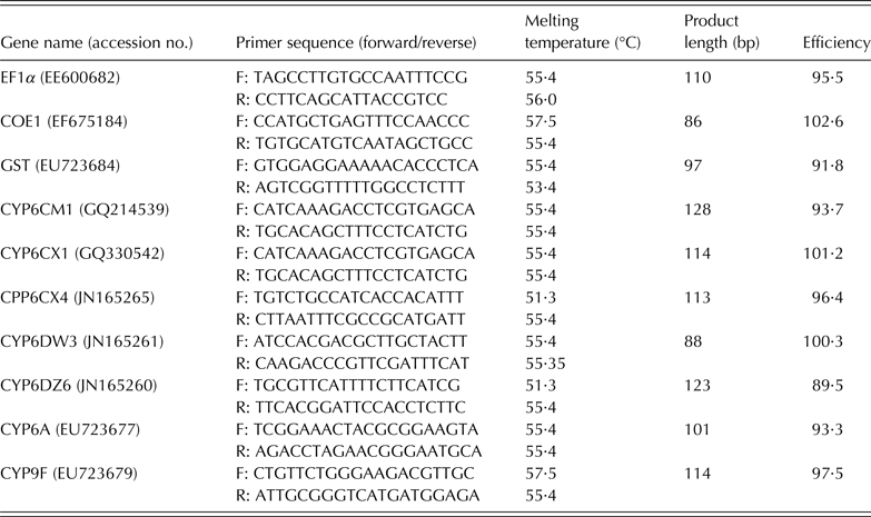
The primers for quantitative real-time polymerase chain reaction (PCR) (qRT-PCR) were designed using Primer3 (http://primer3.ut.ee/). The primers were designed on the basis of sequences published by the National Center for Biotechnology Information (NCBI) and are listed in Table 2.
Sequences of primers used to determine the relative expression of target genes in Bemisia tabaci

Total RNA isolation and reverse transcription
Total RNAs were extracted from approximately 30 B. tabaci adults using TRIzol reagent (Invitrogen, Shanghai, China) following the manufacturer's protocol. The resulting total RNA was re-suspended in nuclease-free water and was quantified on a NanoDrop 2000 spectrophotometer (Thermo Scientific, Wilmington, DE). First-strand cDNAs were synthesized with 1·0 µg/μl total RNA using the PrimeScript® RT reagent kit (Takara Biotech, Dalian, China) according to the manufacturer's protocol.
Quantitative real-time polymerase chain reaction
The relative expression levels of CYP6CM1, CYP6CX1, CYP6CX4, CYP6DW3, CYP6DZ6, CYP6A, CYP9F, COE1 and GST in B. tabaci adults were examined using qRT-PCR. Each reaction was performed in a 20 µl mixture containing 1 µl of cDNA, 10 µl of SYBR Green qRT-PCR SuperMix-UDG, 0·15 µl of each primer and 8·7 µl of H2O. All samples, including a ‘no-template’ negative control, were tested in duplicate. The amplification efficiency of the target genes and housekeeping gene (EF − 1α) were estimated using E = 10−1/slope − 1, where the slope was derived from the plot of the cycle threshold value v. the log of the serially diluted template concentration. The optimized qRT-PCR programme consisted of initial steps at 50 °C for 2 min and 94 °C for 2 min, followed by 50 cycles of 94 °C for 15 s and 60 °C for 30 s. After running the cycling protocol, melting curves were obtained by increasing the temperature from 60 to 95 °C (0·2 °C/s) to denature the double-stranded DNA. The qRT-PCR amplifications were carried out in 96-well plates. The assays were run in an ABI 7500 system using the sequence detection software (SDS) Version.1.4 application software (Applied Biosystems, Foster City, CA, USA). Quantification of the mRNA transcript level of nine detoxification genes was performed using the comparative 2−ΔΔCT method (Pfaffl Reference Pfaffl2001).
Statistical analysis
Statistical and bioassay analyses were performed using Microsoft Excel (2010) and Polo (Probit and Logit Analysis) (LeOra Software Company, Petaluma, CA). The gene expression data are expressed as means ± standard error (s.e.). Statistical analysis was performed using one-way analysis of variance and Tukey's test (P < 0·05) to compare the expression levels of P450 genes with InStat Version.3.0 software (GraphPad Software, San Diego, CA).
RESULTS
Dose response of the expression of carboxylesterase (COE1), glutathione S-transferase (GST) and cytochrome P450 monooxygenases (P450) genes after diafenthiuron challenge in both the S-Lab and R-DfWf strains
Nine putative detoxification genes (COE1, GST and 7 P450s) in the S-Lab and R-DfWf strains were tested in the present study. The results of an earlier study revealed that six detoxification genes – GST, CYP6CX1, CYP6CX4, CYP6DW3, CYP6DZ6 and CYP9F – were all over-expressed in the R-DfWf strain, while the other three detoxification genes, COE1, CYP6CM1 and CYP6A, were not over-expressed in the R-DfWf strain (Zhang et al. Reference Zhang, Kong and Zeng2015b). Based on these data, the LC10 and LC50 of diafenthiuron for each strain were chosen for use in the present induction study and the expression of nine detoxification genes in response to diafenthiuron challenge at 48 h was characterized in each strain. Significant (P < 0·05) induction of the expression of GST was detected in both the S-Lab and R-DfWf strains treated with both concentrations of diafenthiuron at 48 h compared with their respective controls (Fig. 1(a)). In the S-Lab strain, GST was readily induced by diafenthiuron at LC10 and reached a maximum (2·4-fold), with no further significant induction at LC50 (1·7-fold). Similarly, in the R-DfWf strain, GST was readily induced by diafenthiuron at LC10 and reached a maximum (2·3-fold), with no further significant induction at LC50 (1·4-fold). Glutathione S-transferase was induced readily in the S-Lab and R-DfWf strains by diafenthiuron at LC10 and reached a maximum. Significant (P < 0·05) induction of GST was more evident in the R-DfWf strain than in the S-Lab strain. It was found that four of the five P450 genes were upregulated in both the S-Lab and R-DfWf strains treated with diafenthiuron at both concentrations at 48 h compared with the controls. The expression levels of CYP6CX4, CYP6DW3, CYP6DZ6 and CYP9F were significantly (P < 0·05) increased by diafenthiuron at both the LC10 and LC50, showing 1·5-fold increased expression compared with the control (Figs 1(c)–(f)). No induction of CYP6CX1 was detected in the S-Lab or R-DfWf strains treated with diafenthiuron at LC10 or LC50, respectively (Fig. 1(b)). Although CYP6CX4 was readily induced in the S-Lab strain by diafenthiuron at LC10 and reached a maximum (1·5-fold increase) with no further significant induction up to LC50, the induction was more pronounced in the R-DfWf strain than in the S-Lab strain, with an induction peak of 2·4-fold at LC10 (Fig. 1(c)). A similar induction pattern was also observed for CYP9F in both the S-Lab and R-DfWf strains (Fig. 1(f)). Diafenthiuron at LC10 readily induced CYP9F in both the S-Lab and R-DfWf strains and reached a maximum in each strain (1·9- and 1·9-fold, respectively) with no further significant induction up to LC50. However, this induction was more pronounced in the R-DfWf strain than in the S-Lab strain at LC50. Diafenthiuron at LC10 also significantly (P < 0·05) induced CYP6DW3 in both the S-Lab and R-DfWf strains by diafenthiuron at LC10, with further significant (P < 0·05) induction up to LC50 (1·80- and 2·00-fold, respectively). The induction was also more evident in the R-DfWf strain than in the S-Lab strain at both LC10 and LC50 (Fig. 1(d)). A similar induction pattern was also found for CYP6DZ6 in both the S-Lab and R-DfWf strains (Fig. 1(e)). However, CYP6DZ6 was not induced significantly in either the S-Lab or R-DfWf strain by diafenthiuron at LC10, but there was a significant (P < 0·05) induction up to LC50 (1·5- and 1·7-fold, respectively). This induction was also more evident in the R-DfWf strain than in the S-Lab strain at LC50.
Dose response of the expression of detoxification genes known to be constitutively over-expressed in the resistant strain (R-DfWf) after diafenthiuron exposure. The expression of detoxification genes in each strain of B. tabaci at 48 h after treatment with diafenthiuron at LC10 and LC50 (4 µg/ml and 100 µg/ml for the susceptible strain (S-Lab) and 100 µg/ml and 300 µg/ml for the R-DfWf strain, respectively). The relative level of gene expression shown along the Y-axis represents the ratio of gene expression in each treatment compared with the control. The experiments were conducted three times. The results are shown as the mean ± s.e. (Colour online).
Interestingly, the other three detoxification genes measured, COE1, CYP6CM1 and CYP6A, were not over-expressed in the R-DfWf strain. In response to diafenthiuron, COE1 was induced in both the S-Lab and R-DfWf strains by diafenthiuron at LC10, with further significant (P < 0·05) induction up to LC50 (2·20- and 1·60-fold, respectively). The induction was more evident in the S-Lab strain than in the R-DfWf strain at both LC10 and LC50 (Fig. 2(a)). A similar induction pattern was also found for CYP6A in both the S-Lab and R-DfWf strains, such that CYP6A was readily induced in both the S-Lab and R-DfWf strains by LC10 and reached maximum values (4·2- and 2·8-fold, respectively), with no further significant induction when exposed to diafenthiuron at LC50. The induction was also more apparent in the S-Lab strain than in the R-DfWf strain at LC50 (Fig. 2(c)). No significant induction of CYP6CM1 expression was detected in the S-Lab strain treated with diafenthiuron at either concentration (Fig. 2(b)); however, in the R-DfWf strain, the expression of CYP6CM1 was readily induced by LC10 and reached a maximum (2·1-fold) with no further significant induction after exposure to LC50. These results are slightly different from those observed in the R-DfWf and S-Lab strains, where most of the up-regulated P450s peaked at LC10 and some of the up-regulated P450s peaked at LC50.
Dose response of the expression of detoxification genes that are not constitutively over-expressed in the resistant strain (R-DfWf) after diafenthiuron exposure. The expression of detoxification genes in each strain of B. tabaci at 48 h after treatment with diafenthiuron at LC10 and LC50 (4 µg/ml and 100 µg/ml for the susceptible strain (S-Lab) and 100 µg/ml and 300 µg/ml for the R-DfWf strain, respectively). The relative level of gene expression shown along the Y-axis represents the ratio of gene expression in each treatment compared with the control. The experiments were conducted three times. The results are shown as the mean ± s.e. (Colour online).
Based on the above data and data from previous studies, CYP6CX1, CYP6CX4, CYP6DW3, CYP6DZ6, CYP9F and GST were all up-regulated in the R-DfWf strain, and CYP6CX4, CYP6DW3, CYP6DZ6, CYP9F and GST were all significantly (P < 0·05) upregulated in both the R-DfWf and S-Lab strains after diafenthiuron treatment compared with the control. The up-regulation of CYP6CX4, CYP6DW3, CYP6DZ6, CYP9F and GST was also more pronounced in the R-DfWf strain than in the S-Lab strain when the strains were treated with diafenthiuron at LC10 or LC50. However, CYP6CX1 was not significantly upregulated in either the R-DfWf strain or the S-Lab strain after diafenthiuron treatment (LC10 or LC50) compared with the control. In contrast, CYP6CM1, CYP6A and COE1 were not upregulated in the R-DfWf strain. The levels of CYP6A and COE1 were significantly (P < 0·05) higher in both the R-DfWf and S-Lab strains after diafenthiuron treatment compared with the control. However, the induction of CYP6A and COE1 was more pronounced in the S-Lab strain than in the R-DfWf strain after exposure to both of the diafenthiuron concentrations. Interestingly, CYP6CM1 was significantly (P < 0·05) higher in the R-DfWf strain but not in the S-Lab strain after treatment with diafenthiuron at both concentrations. So there are some differences in the expression of different detoxification genes in different treatments.
Time-dependent response of carboxylesterase (COE1), glutathione S-transferase (GST) and cytochrome P450 monooxygenases (P450) expression after diafenthiuron challenge in both the S-Lab and R-DfWf strains
To further study the effects of diafenthiuron on the induction of putative detoxification genes (COE1, GST and 7 P450s), gene expression in B. tabaci adults challenged with diafenthiuron was also measured at the LC10 for various durations (24, 48 and 72 h). The evaluation of GST gene expression following diafenthiuron treatment at the LC10 revealed a significant (P < 0·05) induction in both the S-Lab and R-DfWf strains at 48 and 72 h (Fig. 3(a)). In the S-Lab strain, the initial induction (1·3-fold) occurred 24 h after exposure to LC10 (4 µg/ml) treatment and reached a peak at 48 h (1·9-fold increase), with a decrease observed at 72 h after treatment. In the R-DfWf strain, the initial induction (2·2-fold) occurred at 24 h after the exposure to LC10 (100 µg/ml) treatment and increased at 48 h (1·3-fold increase), with a decrease observed at 72 h after treatment. However, CYP6CX4 expression in both the R-DfWf and S-Lab strains reached maximum levels (2·6- and 1·7-fold) at 72 h after exposure to diafenthiuron at LC10 (Fig. 3(c)). The expression of CYP6DW3 and CYP9F in the R-DfWf strain reached maximum levels (1·8- and 1·9-fold, respectively) at 48 h after exposure to LC10 and declined at 72 h after treatment (Figs 3(d) and (f)). Similarly, the expression of CYP9F in the S-Lab strain also reached a maximum level (1·4- and 1·9-fold, respectively) at 48 h after exposure to LC10 and declined at 72 h after the treatment (Fig. 3(f)). Nevertheless, no significant induction of CYP6CX1 or CYP6DZ6 was identified in either of the two strains tested at any time points after treatment with diafenthiuron at LC10 (Figs 3(a) and (d)).
Time course of the expression of detoxification genes known to be constitutively over-expressed in the resistant strain (R-DfWf) after diafenthiuron exposure. The relative expression of detoxification genes in B. tabaci following treatment with diafenthiuron at LC10 (4 µg/ml and 100 µg/ml for the susceptible and resistant (S-Lab and R-DfWf) strains, respectively) was analysed at 24, 48 and 72 h after treatment. The relative level of gene expression shown along the Y-axis represents the ratio of gene expression in each treatment compared with control. The experiments were conducted three times. The results are shown as the mean ± s.e. (Colour online).
The other three detoxification genes, COE1, CYP6CM1 and CYP6A, were not over-expressed in the R-DfWf strain. Exposure to diafenthiuron at the LC10 led to a significant (P < 0·05) induction of COE1 expression in both the S-Lab and R-DfWf strains at 48 and 72 h (Fig. 4(a)). In the S-Lab strain, the initial induction (1·3-fold) occurred at 24 h after exposure to diafenthiuron at LC10 (4 µg/ml), reached a peak at 48 h (1·7-fold increase in expression), and declined after 72 h. In the R-DfWf strain, the initial induction (1·1-fold) occurred at 24 h after exposure to diafenthiuron at LC10 (100 µg/ml), increased further at 48 h (1·3-fold increase in expression) and reached a peak at 72 h with an induction level of 1·5-fold. Interestingly, CYP6A expression in the R-DfWf strain reached a maximum (2·7-fold increase) at 24 h after exposure to diafenthiuron at LC10 and then declined 48 h after treatment, while the expression of CYP6A in the S-Lab strain reached a maximum (4·3-fold increase) at 48 h after exposure to LC10 and then declined 72 h after treatment (Fig. 4(c)).
Time course of the expression of detoxification genes that are not constitutively over-expressed in the resistant strain (R-DfWf) after diafenthiuron exposure. The relative expression of COE1 in B. tabaci following treatment with diafenthiuron at LC10 (4 µg ml/ and 100 µg/ml for the S susceptible and resistant (S-Lab and R-DfWf) strains, respectively) was analysed at 24, 48 and 72 h after treatment. The relative level of gene expression shown along the Y-axis represents the ratio of gene expression in each treatment compared with control. The experiments were conducted three times. The results are shown as the mean ± s.e. (Colour online).
The expression of CYP6CM1 in the R-DfWf strain reached a maximum (2·1-fold increase) at 48 h after exposure to LC10 and declined at 72 h after treatment (Fig. 4(b)). CYP6CM1 expression was not significantly induced in the S-Lab strain from 24 to 72 h after treatment with diafenthiuron at LC10. These results are slightly different from those observed in the R-DfWf and S-Lab strains, where the expression of most of the up-regulated P450 genes peaked at 48 h after diafenthiuron treatment, while the expression of some of these genes peaked at 24 or 72 h after diafenthiuron treatment. So, there are some differences in the expression of different detoxification genes at different times after treatment.
DISCUSSION
Typically, enhanced metabolic detoxification in insects can contribute to insecticide resistance (Li et al. Reference Li, Schuler and Berenbaum2007; Low et al. Reference Low, Ng, Morton, Parker, Batterham and Robin2007). The metabolic detoxification system in insects consists of three major groups of enzymes. Phase I detoxification enzymes, which act directly on a broad range of substrates to reduce their toxicity, are P450s. Phase II enzymes, including GST, uridine diphosphate glucose-glucuronosyltransferases and COE, facilitate the excretion of hydrophobic toxic compounds by improving their hydrophilicity. Many studies have indicated that increased expression of detoxification genes results in increased levels of detoxification enzymes and that the activity of detoxification enzymes is responsible for insecticide resistance (Liu & Scott Reference Liu and Scott1998; Liu et al. Reference Liu, Li, Reid, Yang and Zhang2011; Schuler Reference Schuler2011; Gong et al. Reference Gong, Li, Zhang, Gao and Liu2013; Zhang et al. Reference Zhang, Kong, Wang, Gao, Zeng and Shi2015a). Constitutive increases in gene expression and induction of detoxification enzymes are thought to be responsible for increased detoxification of insecticides (Pavek & Dvorak Reference Pavek and Dvorak2008). Previous research by the present authors indicated that six detoxification genes – GST, CYP6CX1, CYP6CX4, CYP6DW3, CYP6DZ6 and CYP9F – were over-expressed in the R-DfWf strain (Zhang et al. Reference Zhang, Kong and Zeng2015b), whereas COE1, CYP6CM1 and CYP6A were not. In the present study, it was found that five of the detoxification genes that were constitutively over-expressed in the resistant strain were significantly induced in both the R-DfWf and S-Lab strains in response to diafenthiuron; however, CYP6CX1 was not significantly induced. Similar results have been reported in Drosophila melanogaster and Culex quinquefasciatus (Brandt et al. Reference Brandt, Scharf, Pedra, Holmes, Dean, Kreitman and Pittendrigh2002; Zhu et al. Reference Zhu, Li, Zhang and Liu2008; Liu et al. Reference Liu, Li, Reid, Yang and Zhang2011; Gong et al. Reference Gong, Li, Zhang, Gao and Liu2013). These results indicate that the induction and constitutive over-expression of these detoxification genes play an important role in the detoxification of insecticides and that the development of insecticide resistance results from the increased expression of multiple detoxification genes in the resistant strain. The coincidence of induction and constitutive over-expression of GST, CYP6CX4, CYP6DW3, CYP6DZ6 and CYP9F suggests that these five P450 genes have a common function in the diafenthiuron-resistant strain. In addition, the other three detoxification genes (COE1, CYP6CM1 and CYP6A) that were not constitutively over-expressed in the resistant strain were significantly induced in either the R-DfWf strain or the S-Lab strain in response to diafenthiuron, suggesting that some detoxification genes were only constitutively over-expressed (e.g. CYP6CX1) and some were only induced (e.g. COE1, CYP6CM1 and CYP6A). It was also found that seven detoxification genes – GST, COE1, CYP6CX4, CYP6DW3, CYP6DZ6, CYP6A and CYP9F – were induced in both the R-DfWf and S-Lab strains. However, significant induction of CYP6CM1 after diafenthiuron treatment was found in the R-DfWf strain but not in the S-Lab strain. Interestingly, the induction of COE1 and CYP6A was more pronounced in the S-Lab strain than in the R-DfWf strain after diafenthiuron treatment. These results may indicate that some detoxification genes are common to the response of both susceptible and resistant strains to diafenthiuron, while others, such as CYP6CM1, are more specific, with roles only in the response of the R-DfWf strain to insecticides, and CYP6CX1 is only constitutively over-expressed in the R-DfWf strain. In previous studies, Rauch & Nauen (Reference Rauch and Nauen2003) and Cahill et al. (Reference Cahill, Gorman, Day, Denholm, Elbert and Nauen1996) reported that enhanced metabolic detoxification resulting from increased expression of P450 genes is a major factor resulting in pest tolerance to neonicotinoid insecticides. The P450 genes metabolize endogenous compounds and of all the P450 families, the CYP6 family has been most often implicated in insecticide resistance (Pavek & Dvorak Reference Pavek and Dvorak2008; Scott Reference Scott and Liu2008; Puinean et al. Reference Puinean, Foster, Oliphant, Denholm, Field, Millar, Williamson and Bass2010). For example, CYP6CM1 and CYP9F have been reported to be linked with imidacloprid resistance in whitefly (Karunker et al. Reference Karunker, Benting, Lueke, Ponge, Nauen, Roditakis, Vontas, Gorman, Denholm and Morin2008; Qiu et al. Reference Qiu, Liu, Li, Mathur, Qin and Ren2009; Jones et al. Reference Jones, Daniels, Andrews, Slater, Lind, Gorman, Williamson and Denholm2011), and CYP6CX1 reportedly exhibited significantly higher mRNA levels in the context of imidacloprid and thiamethoxam resistance in whitefly (Zhuang et al. Reference Zhuang, Wang, Zheng, Wu, Miyata and Wu2011; Yang et al. Reference Yang, Xie, Jones, Bass, Jiao, Yang, Liu, Li and Zhang2013). This finding may indicate that CYP6CM1, CYP6CX1 and CYP9F are involved not only in imidacloprid and thiamethoxam resistance but also in diafenthiuron resistance in whitefly.
In addition to the P450s, GST and COE could also be involved in the detoxification of allelochemicals (Lindroth & Weisbrod Reference Lindroth and Weisbrod1991; Feng et al. Reference Feng, Davey, Pang, Ladd, Retnakaran, Tomkins and Palli2001; Yoshiyama & Shukle Reference Yoshiyama and Shukle2004). Enhanced expression of GSTs has been shown to be a mechanism of resistance to DDT and organophosphates and has also been implicated in resistance to pyrethroids in certain insects (Huang et al. Reference Huang, Hu, Yao, Wu, Chiang and Sun1998; Ranson et al. Reference Ranson, Rossiter, Ortelli, Jensen, Wang, Roth, Collins and Hemingway2001; Vontas et al. Reference Vontas, Small and Hemingway2001). These findings are somewhat consistent with the current finding that a single GST was highly over-expressed and induced by diafenthiuron in the resistant strain. Organophosphate resistance in the B-biotype of B. tabaci is associated with a point mutation in an ace1-type acetylcholinesterase and with over-expression of COEs (Alon et al. Reference Alon, Alon, Nauen and Morin2008). Yang et al. (Reference Yang, Xie, Jones, Bass, Jiao, Yang, Liu, Li and Zhang2013) observed a higher level of COE1 expression. In the present study, although COE1 was not differentially expressed between the R-DfWf and S-Lab strains, significant induction of COE1 was observed in both the strains treated with diafenthiuron. This finding may indicate that GST and COE1 are also involved in diafenthiuron resistance in whitefly.
In conclusion, the present study strongly suggests a common mechanism for induction of GST, COE and the suite of P450 genes in response to detoxification-mediated insecticide resistance in whitefly. Enhanced metabolic detoxification by GST, COE and the suite of P450 genes may be an important mechanism by which the B-biotype has attained a high level of resistance to commonly used insecticides in China.
This work was funded by the Special Fund for Agro-scientific Research in the Public Interest (grant no. 201303019-01).