Introduction
It has long been acknowledged that intrauterine growth retardation and preterm birth constitute risk factors for the development of future adverse neurological, nephrological and ophthalmological events.Reference Mercuro, Bassareo and Flore 1
However, more recent studies have demonstrated that subjects born preterm and/or with a low birth weight also display an increased cardiovascular risk, being more frequently affected by coronary artery disease, stroke, hyperlipidaemia, high blood pressure, diabetes, atherosclerosis, cardio-renal syndrome, QT tract prolongation at basal electrocardiogram, and potentially emboligenic interatrial septal aneurysms than their peers born at term.Reference Mercuro, Bassareo and Flore 1 , Reference Bassareo, Fanos, Puddu, Flore and Mercuro 2
Epicardial fat tissue (EFT) is the large visceral fat storage located between the epicardium and pericardium and surrounding the large coronary arteries.Reference Payne, Kohr and Tune 3 This tissue exerts an important endocrine and paracrine activity, being involved in the development of atherosclerosis through the cytokine pathway.Reference Lau, Dhillon, Yan, Szmitko and Verma 4 Furthermore, EFT is an emerging cardiometabolic risk factor capable of predicting the future development of high blood pressure, dislipidaemia, overweight/obesity, metabolic syndrome and coronary artery disease. EFT can be easily and accurately measured by means of echocardiography.Reference Iacobellis and Willens 5
The aim of this study was to measure EFT in a group of young subjects born extremely preterm (i.e. with a birth weight of <1000 g) in comparison with a group of controls born at term. A possible relationship with patient characteristics, including echocardiographic measurements, gestational age, birth weight and duration of stay in neonatal intensive care unit (NICU) was also investigated.
Materials and methods
In total, 30 young adults born with an ex-extremely low birth weight (ex-ELBW; 10 males and 20 females) ranging between the ages of 17 and 28 (mean age±s.d.: 20.1±2.5 years) were enrolled in the study and compared with a control group (C) comprising 30 healthy subjects born at term from a healthy mother in the nursery of our University and matched for sex, age and body mass index (BMI). All ex-ELBW subjects were contacted in alphabetical order from the Records of the NICU of the University of Cagliari, Italy. These subjects were the first surviving ex-ELBW assisted in the sole NICU present in Cagliari (Italy). Cagliari is the capital of Sardinia, a region with a relatively small number of inhabitants, the majority of which reside in Cagliari and surrounding areas.
Exclusion criteria were as follows: secondary hypertension, cerebrovascular disease, severe valvular heart disease, atrial fibrillation, substance use, severe renal insufficiency (serum creatinine: ⩾1.5 mg/dl in men and ⩾1.4 mg/dl in women), major non-cardiovascular diseases and known diabetes or fasting glucose ⩾126 mg/dl.
All study participants underwent cardiological assessment, including echocardiographic measurements.
Echocardiographic imaging study
Transthoracic echocardiographic procedures were performed on all study subjects by the same trained physician.
Examination was conducted in left lateral decubitus position using acommercially available echocardiographic Toshiba Artida (Toshiba Medical Systems Corporation, Tochigi, Japan) machine fitted with a 2.5–3.5 MHz probe. Measurements were taken during normal breathing at end expiration. Left ventricular end-systolic (Lv.d) and end-diastolic diameters (LVID), end-diastolic interventricular septal thickness (Iv.) and end-diastolic LV posterior wall thickness (PW) were measured at end-diastole according to established standards of the American Society of Echocardiography.Reference Lang, Bierig and Devereux 6
Left ventricular ejection fraction was calculated by means of the biplane Simpson’s method. Left ventricular mass (LVM) was calculated using the Devereux formula: LVM=(1.04[(LVDd+IVSth+PWth)3−(LVDd)3]−13.6).Reference Devereux and Reichek 7 Subsequently, left ventricular mass index (LVMI) was obtained by the following formula: LVM/body surface area. Left ventricular hypertrophy was defined as LVMI values exceeding 125 g/m2 in men and 110 g/m2 in women. 8
Measurement of EFT
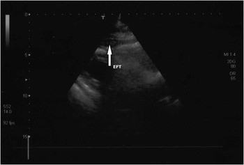
EFT was measured in individuals in left lateral decubitus position using standard echocardiographic parasternal long-axis and short-axis views from bi-dimensional images. Images from standard parasternal long- and short-axis views were digitally stored and reviewed by a single cardiologist blind to patients’ clinical data. At ultrasound imaging EFT is generally identified as the relatively echo-free space between the outer wall of the myocardium and the visceral layer of pericardium. EFT is measured perpendicularly on the free wall of the right ventricle at end-systole in three cardiac cycles (Fig. 1).Reference Iacobellis, Assael and Ribaudo 9
Epicardial fat thickness (EFT) (white arrow) detected by transthoracic echocardiography at free wall of right ventricle (parasternal long view).

The average value of three cardiac cycles from each echocardiographic examination was determined. The same trained cardiologist scanned 13 randomly selected subjects on a second occasion 1 week after the initial scan. The coefficient of intra-observer variation was 1.6%.
Statistical analysis
The results of the study population (n=30) were first analysed, and ex-ELBW subsequently compared with subjects born at term (n=30) using the Student’s t-test. The relationship between EFT and the previously mentioned variables were assessed by Pearson’s correlation coefficients and by plotting corresponding regression lines. Partial correlation analysis was also applied. Multivariate analysis was not applied due to small sample size. Statistical significance was set at P<0.05 throughout the study. Commercially available computer software (SPSS version 22.0 SPSS Inc, Chicago, IL, USA) was used for all analyses.
Results
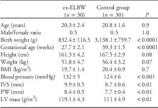
The characteristics of the population studied (ex-ELBW v. C) are summarized in Table 1. Statistically significant differences (ex-ELBW v. C) were detected for interventricular septum thickness, posterior wall thickness and LVM. Two male ex-ELBW subjects had a LVM exceeding 125 g/m2.
Characteristics of the population in the study

ex-ELBW, ex-extremely low birth weight; BMI, body mass index; IVS, interventricular septum; PW, posterior wall; LV, left ventricular.
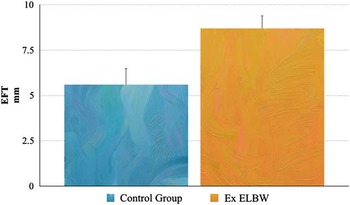
In addition, EFT was significantly higher (8.7±0.7 v. 5.6±0.9; P<0.001; Fig. 2) in former preterm individuals than in controls, and was correlated with birth weight (r=−0.47, P=0.0009), gestational age (r=−0.39, P=0.03) and LVM (r=0.51, P=0.004). When excluding the influence of BMI, birth weight was the sole remaining determinant of EFT, irrespective of gestational age (r=−0.37, P=0.04). Furthermore, even when excluding the possible influence of blood pressure values on the cardiac structures, the correlation between birth weight and EFT remained statistically significant (r=−0.40, P=0.028).
Comparison of epicardial fat thickness (EFT) between the two groups [ex-extremely low birth weight (ex-ELBW) v. controls: 8.7±0.7 v. 5.6±0.9 mm].

Discussion
To the best of our knowledge, this is the first study to investigate the thickness of adipose tissue in former preterm newborns and report an association between EFT and intrauterine growth restriction.
During embryogenesis, EFT originates from the splanchnopleuric mesoderm, with vascularization being supplied by branches of the coronary arteries. Under physiological conditions, EFT represents ~20% of the heart mass.Reference Iacobellis and Bianco 10
Although somewhat controversial, there is a general consensus that epicardial fat is lower in younger subjects.Reference Tansey, Aly and Sheppard 11
Conversely, in our study population of young ex-ELBW, the thickness of pericardial adipose tissue appears to be greater than in age-matched controls.
EFT has been increasingly recognized as a marker for cardiovascular risk factors, including glucose intolerance, hypertension, dyslipidaemia, hyperinsulinaemia and atherosclerosis. A potential explanation may be provided by the activity carried by EFT as an endocrine and paracrine organ capable of releasing a series of atherogenic inflammatory cytokines.Reference Funahashi, Nakamura and Shimomura 12 Indeed, EFT produces several biologically active molecules (adipocytokines), which may be involved in the development of atherosclerosis. Taken together, these findings suggest that, in addition to the conventional coronary risk factors, EFT is also likely implicated in the development of coronary artery disease by means of its direct endocrine and paracrine effects. This hypothesis was first put forward many years ago, based on the absence of atherosclerosis inhuman intramyocardial, but not epicardial, coronary arteries.Reference Robicsek and Thubrikar 13 Segments of coronary arteries lacking EFT or separated from the latter by a bridge of myocardial tissue appear to be protected against the development of atherosclerosis, whilst coronary atherosclerotic plaques tend to be more prominent on the arterial side in contact with EFT deposits.Reference Prati, Arbustini and Labellarte 14
Based on these premises, former preterm newborns in our study appear to be at higher risk of developing a number of adverse cardiovascular events in the future.
To the best of our knowledge, this is the first study to report an association between EFT and intrauterine growth restriction, expressed as birth weight. This finding implies an adverse influence of birth weight on EFT development. In addition, this study, carried out in a population of former preterm newborns, has demonstrated for the first time a relationship between EFT and LVM in these subjects. This finding is in agreement with previous reports focusing on the above-stated association in patients affected by hypertension and metabolic syndrome.Reference Pucci, Battista and de Vuono 15
Conversely, increased EFT has been found to be associated with left ventricular hypertrophy irrespective of the concomitant presence of hypertension.Reference Iacobellis, Ribaudo, Zappaterreno, Iannucci and Leonetti 16
A number of mechanisms may play a role in the relationship between EFT and increased LVM.
The first of these involves a direct effect of EFT on the myocardium through the secretion of several proinflammatory mediators, proatherogenic cytokines and growth factors.Reference Sacks and Fain 17
In this respect, in cell culture models, it has been demonstrated that interleukin 6, transforming growth factor β and macrophage chemotactic factor 1, secreted from EFT, are capable of inducing myocardial hypertrophy.Reference Meléndez, McLarty and Levick 18
Further, adipocytes in EFT release free fatty acids, which may diffuse directly into the myocardium, together with myocardial uptake of plasma-free fatty acids, thus exacerbating myocardial steatosis. Intracardiac steatosis and extracardiac fat lead to increased heart weight and mechanical pumping effort, subsequently resulting in left ventricular hypertrophy, diastolic dysfunction, cardiac failure and increased risk of arrhythmias.Reference Poirier, Giles and Bray 19
With regard to the indirect mechanisms that induce left ventricular hypertrophy, high plasma levels of fatty acid levels may stimulate activity of the cardiac autonomic nervous system, with a consequent increase in plasma catecholamine concentrations.Reference Manzella, Barbieri and Rizzo 20 The latter, in turn, induce an increase in arterial stiffness, with consequent augmented heart pumping effort and secondary left ventricular hypertrophy.Reference Natale, Tedesco and Mocerino 21
The present study undoubtedly features several limitations: (a) the small number of patients, that should consequently be enlarged in future studies. This is an objective limitation, due to the small number of subjects with the required characteristics available at our University; (b) the possibility of better defining the amount of pericardial fat by means of cardiac magnetic resonance. In fact this imaging technique, in view of its ability to yield three-dimensional images, appears to be the gold standard in calculating EFT compared with other imaging procedures such as echocardiography;Reference Sicari, Sironi and Petz 22 (c) the need to follow these patients to verify the possible onset of severe cardiovascular diseases over time (i.e. coronary artery disease, stroke, etc.). In fact, even if significant changes in the cardiac morphology and function of subjects born with intrauterine growth restriction have been previously described, mostly during childhood, uncertainty exist about their real impact on the cardiovascular risk in these individuals;Reference Crispi, Bijnens and Figueras 23 – Reference Arnott, Skilton and Ruohonen 26 (d) it would be argued that some of our findings might be influenced by the different gestational age between the patients in the study and controls. However, as above stated, when excluding the possible influence of BMI, the relationship between intrauterine growth restriction and EFT has remained statistically significant, whilst that between the latter and gestational age has lost its significance; (e) the available case records underlined a marked discrepancy between the two genders, with a clear prevalence of females. This disparity may be due to the fact that in the initial stages of neonatal intensive care, a considerably higher number of preterm female ELBW infants survived for pathophysiological reasons never fully clarified.Reference Mercuro, Bassareo and Flore 1
Conclusions
As previously hypothesized in numerous animal models, intrauterine growth restriction is capable of influencing fat distribution in later life.Reference Sarr, Yang and Regnault 27
EFT is significantly increased in former preterm subjects compared with controls and is associated with an increase in cardiac LVM in these subjects. As the latter is known to be correlated with a higher incidence of clinical events attributable to cardiovascular disease, EFT appears to be an easy-to-measure tool for the early detection of subclinical target organ damage and capable of predicting the likely development of future adverse cardiovascular events in these subjects, irrespective of traditional cardiovascular risk factors.Reference Thornburg 28 – Reference Wang, Shu and Sheng 30
Acknowledgements
The authors wish to thank the subjects who took part in the study.
Conflicts of Interest
None.
Ethical Standards
The study was approved by the Internal Ethics Board in accordance with the Declaration of Helsinki. Informed written consent was obtained from all ex-ELBW and C or their parents.