Vascular access-related complications following cardiac catheterisation can cause significant morbidity if not recognised and treated. We describe a patient who developed an arteriovenous fistula and pseudoaneurysm in an accessory arterial branch following femoral access for placement of a Harmony TPV 25, which were treated endovascularly.
Case report
A 17-year-old male with repaired Tetralogy of Fallot and severe pulmonary insufficiency was referred for placement of a Harmony TPV 25. Percutaneous access was obtained in the right femoral vein using ultrasound guidance, and the valve delivery system was advanced through a 26 French Gore DrySeal sheath. Arterial access was obtained in the contralateral femoral artery. One month later, a palpable thrill and audible bruit were noted over the right inguinal access site. Vascular ultrasound showed a femoral arteriovenous fistula. CT angiogram demonstrated the right common femoral artery branched into a superficial femoral artery, deep femoral artery, and an additional medial profunda femoris artery arising below the inguinal ligament. There was an arteriovenous fistula from the medial profundal femoris to the common femoral vein, and an arterial pseudoaneurysm adjacent to the fistula. After discussion with our vascular surgeon, we felt both lesions could be treated with percutaneous implantation of a covered endovascular stent.
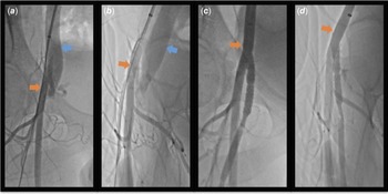
The patient returned to the catheterisation laboratory, and a delivery sheath was advanced from the left common femoral artery around the iliac artery bifurcation to the right common femoral artery. Angiography demonstrated a fistula arising from the medial profundal femoris artery 11 mm inferior to its origin, with contrast opacifying the common femoral vein. A pseudoaneurysm was seen on the anterior wall of the artery adjacent to the origin of the arteriovenous fistula. The common femoral artery branched into the superficial and deep femoral artery branches 14 mm below the level of the arteriovenous fistula. The medial profundal femoris was carefully engaged, and a 6x39 GORE VIABAHN VBX (W.L. Gore and Associates Inc., Flagstaff, AZ) stent was deployed. Follow-up angiography showed appropriate stent-positioning within the medial profundal femoris with occlusion of the arteriovenous fistula and exclusion of the pseudoaneurysm (Fig. 1). Repeat ultrasounds prior to discharge and after 1 month showed no residual arteriovenous fistula or pseudoaneurysm and a widely patent medial profundal femoris artery. No thrills or bruits were appreciated on exam.
Angiograms in panels a and b were performed immediately following access and demonstrate the arteriovenous fistula. The orange arrow represents the common femoral artery, and the blue arrow represents the common femoral vein. Contrast injection in the femoral artery opacifies the common femoral vein due to the fistulous connection. Angiograms in panels c and d were performed following covered stent placement in the profunda femoris artery. The common femoral artery is represented by the orange arrow. Contrast injection in the common femoral artery opacifies the deep femoral, superficial femoral, and profundal femoris arteries without residual fistulous connection.

Discussion
Femoral arterial complications following cardiac catheterisation occur in 5–8% of patients. Risk factors include patient size, vessel diameter, number of access attempts, sheath size, interventional procedures performed, and procedure duration. Haematomas, arterial thrombosis, persistent bleeding, pseudoaneurysm formation, and arteriovenous fistulas are among the most common complications.Reference Siracuse, Farber and Cheng1–Reference Glatz, Shah and McCarthy7 Most morbidity is due to bleeding and thrombosis, with pseudoaneurysms and fistulas representing an estimated incidence of less than 1%,Reference Kawano, Tamura and Kadota8–Reference Glaser, McKellar and Scher10 but if untreated, it can lead to high-output heart failure, arterial degeneration, and reduced blood flow to distal vasculature.Reference Ohlow, Secknus and von Korn6,Reference Tsetis11–Reference Russu, Mureşan and Kaller13 Known risk factors for the development of femoral arteriovenous fistula include baseline anticoagulation therapy, female gender, systemic hypertension, and low groin puncture.Reference Wang, al Jabri, Jewell and Jellison12–Reference Altin, Flicker and Naidech14 Size and number of sheaths used during catheterisation have not been associated with an increased risk of fistula formation.Reference Perings, Kelm, Jax and Strauer9 One study noted the incidence of iatrogenic fistula is increased when ipsilateral femoral artery and venous access were obtained for the procedure;Reference Altin, Flicker and Naidech14 however, arterial access was obtained in the contralateral femoral artery for valve implantation in our patient.
Ultrasound is widely used for femoral access in children, decreases the number of attempts and frequency of inadvertent arterial sticks,Reference Iwashima, Ishikawa and Ohzeki15,Reference Law, Borasino, McMahon and Alten16 and reduces the risk of arterial complications by almost 50%.Reference Rashid and Hughes17 Additionally, ultrasound often identifies the presence of deep or smaller calibre vessels that may contribute to complications such as pseudoaneurysms.Reference Gabriel, Pawlaczyk and Waliszewski18 We achieved right femoral vein access in one attempt using ultrasound guidance with single wall vessel puncture. The common femoral artery bifurcation was identified below the inguinal ligament, and venous access was obtained superior to this. The medial profundal femoris artery, which had a high takeoff near the inguinal ligament, was not identified by ultrasound and was likely traumatised during femoral venous access, resulting in the fistula and pseudoaneurysm.
Though anatomic variants in femoral artery branches have been reported, most reports describe variation in the medial and lateral circumflex arteries and their branch points.Reference Chun19–Reference Tomaszewski, Henry and Vikse21 This is recognised as an important consideration for interventionalists and surgeons, and one such variant has also been associated with iatrogenic fistula formation.Reference Yamamoto, Uchiyama and Onuki22 To our knowledge, our case is the first reported of an iatrogenic arteriovenous fistula from an accessory arterial branch to the femoral vein. While CT imaging is recommended prior to transcatheter aortic valve replacement to assess iliac artery diameter and calcification, this is not routinely performed in pulmonary valve replacement.Reference Blanke, Weir-McCall and Achenbach23 This is likely multifactorial, as patients requiring transcatheter aortic valve replacement are typically older with significant peripheral artery disease. Imaging is required in high-risk patients to assess candidacy for percutaneous intervention based upon ilio-femoral vessel sizing. Emphasis is placed on characterising atherosclerosis, calcification, and tortuosity, all of which are leading contributors to vascular complications in elderly patients.Reference Perry, George and Lee24 However, patients with CHD undergoing transcatheter aortic valve replacement often have less baseline risk factors, and no guidelines exist for routine anatomic imaging prior to intervention. While imaging would identify accessory arterial branches and may decrease the chances of arteriovenous access complications, the overall incidence of such complications is low and should be weighed when considering extensive imaging and radiation exposure prior to intervention.
Surgical repair has been the first-line management of femoral access complications,Reference Franco, Goldsmith and Veith25,Reference Kufner, Cassese and Groha26 but recent studies have reported successful transcatheter approaches for treating arteriovenous fistulas.Reference Lee27–Reference Sarac, Vargas and Kashyap31 While the success rate of surgical management is nearly 100%, post-operative morbidity remains up to 25%,Reference Tsetis11 largely attributable to pre-existing co-morbidities such as calcification and arterial stenosis.Reference Tsetis11,Reference Franco, Goldsmith and Veith25 However, patients with significant vascular co-morbidities may not be suitable candidates for endovascular treatment and require resection of the damaged arterial section and vein repair.Reference Tsetis11 Younger patients without significant peripheral vasculature lesions may be suitable candidates for endovascular approaches. Additionally, while it is unclear to what extent anatomic variants play a role in the development of fistulas and pseudoaneurysms (due to their relative infrequency), such variants may also lend themselves to percutaneous closure. Both self-expanding and balloon expandable stents have been used to treat various acquired vascular lesions in the iliac and femoral arteries, and further imaging and anatomic assessment can be used to guide this decision.
Once a fistula is identified, complete anatomic assessment with contrast CT should be performed to evaluate the lesion and vascular anatomy. Given the fistula arose from an accessory medial profundal femoris artery, we felt percutaneous approach was a reasonable alternative to surgery and would allow for simultaneous fistula and pseudoaneurysm occlusion. Additionally, the medial profundal femoris artery was an accessory branch and occlusion would be of low haemodynamic significance, justifying a less invasive approach. While the medial profundal femoris branch has remained patent at 1 month follow-up on ultrasound, if it were to ultimately become occluded, it would not lead to vascular compromise as noted above.
Conclusion
We demonstrated endovascular treatment for an arteriovenous fistula from the right medial profundal artery to the common femoral vein and arterial pseudoaneurysm, which developed following Harmony TPV placement in a patient with an accessory arterial branch. Thorough assessment of the vascular anatomy should be performed to determine if percutaneous therapy is appropriate.
Author contribution
All authors contributed equally to the writing of this manuscript. Gary Stapleton served as the senior author.
Financial support
This research received no specific grant from any funding agency, commercial or not-for-profit sectors.
Competing interests
None.