Introduction
White light endoscopy uses reflected light in the visible spectrum (400–700 nm) to inspect the nasal, pharyngeal and laryngeal surface for gross mucosal changes, and is the most common tool used by otolaryngologists. Narrow-band imaging constitutes an improvement over white light endoscopy because it uses only the blue (415 nm) and green (540 nm) light absorbed by haemoglobin; the blue light penetrates deeply and enables visualisation of the vasculature beneath the mucosal layer, whereas the green light is reflected by the mucosal layer, highlighting the mucosal surface and the superficial capillary network. Narrow-band imaging can be used to analyse both mucosal abnormalities and alteration of the surrounding vasculature,Reference Zhou, Wu, Huang, Jin, Qin and Chen1 to obtain better characterisation and demarcation of the investigated lesion.
Narrow-band imaging is used by many physicians to inspect mucosal surfaces,Reference Sasanuma, Ohtsuka, Kudo, Ogata, Maeda and Misawa2 but interest in the possibility of extending its use to other fields of application is increasing.Reference Ma, Chowdary, Eskander, Ellett, McIlwaine and Manwaring3–Reference Prada, Pipolo, Del Bene, Maccari and Saibene5 For example, Prada et al.Reference Prada, Pipolo, Del Bene, Maccari and Saibene5 applied narrow-band imaging during video-assisted trans-sphenoidal giant pituitary adenoma surgery. Adenomas are characterised by hypercellularity demanding high blood support, which is satisfied by the development of a new irregular and fragile vasculature.Reference Verdelli, Avagliano, Creo, Guarnieri, Scillitani and Vicentini6 The capability of narrow-band imaging to enhance the visualisation of adenomas facilitates their complete removal.
Our team is currently testing the potential of narrow-band imaging in parathyroid surgery to assist with the visualisation of the different vascular architectures exhibited by the thyroid, normal parathyroid and pathological parathyroid. Currently, parathyroidectomy can be executed with minimally invasive approaches, using both an endoscopic view (purely endoscopic or minimally invasive video-assisted parathyroidectomy, in accordance with the endoscopic technique initially introduced by Gagner et al. in 2001Reference Gagner and Inabnet7,Reference Miccoli, Pinchera, Cecchini, Conte, Bendinelli and Vignali8 ) and direct vision.Reference Sessa, Lombardi, De Crea, Raffaelli and Bellantone9 It must be underlined that the endoscope is normally used only to achieve magnification of the surgical field.Reference Sessa, Lombardi, De Crea, Raffaelli and Bellantone9 Our team tested narrow-band imaging applicability in parathyroid surgery to evaluate whether it could be of assistance in distinguishing parathyroid adenomas from normal or hyperplastic parathyroids (in case of primary hyperparathyroidism due to multi-glandular disease), as well as the thyroid, peri-tracheal fat and lymph nodes.
Our study aimed to show that narrow-band imaging provides much additional information to the surgeon, and is a useful instrument during endoscopic interventions. This paper represents a preliminary study of this approach on a small series of patients.
Materials and methods
The study was conducted respecting the Declaration of Helsinki. Indication and technique of surgery were not modified, nor was the scheduling of surgical interventions delayed. All patients gave informed consent to undergo the procedure.
A preliminary evaluation was conducted on five potential patients for selective, minimally invasive video-assisted parathyroidectomy. Three were female and two male, with a mean age of 54.4 years (range, 35–77 years).
The patients were referred to our Endocrine Surgery Unit following biochemical diagnosis of primary hyperparathyroidism. Total calcium, parathyroid hormone (PTH), phosphate, 25-hydroxycholecalciferol (25-OH-D) and calciuria on 24 hours’ urine test results were verified in all patients. Females were diagnosed as a result of osteoporosis, one male was diagnosed because of symptomatic nephrolithiasis, and the other male with hypercalcemia was detected by chance during a check-up, with serum calcium persistently 1 mg over the upper normal level.
Both neck ultrasonography and sestamibi scintigraphy were performed to check for an enlarged parathyroid and determine features such as location, and to exclude the presence of further lesions.
Two-dimensional, minimally invasive video-assisted parathyroidectomy was carried out in an operating theatre using a high-definition endoscopic system (Video Processor CV-170 (Olympus, Tokyo, Japan) and 8 mm Forward-Oblique Telescope 30° (Karl Storz, Tuttlingen, Germany)). All minimally invasive video-assisted parathyroidectomy procedures were performed by the same expert surgeon (LDP).
The pathological gland suspected as being adenoma was initially identified by visual inspection. After recognition, the vascular pattern of the gland was checked using narrow-band imaging light. The neoplastic vascular pattern was evaluated by a different surgeon (AMS), who had already been trained in narrow-band imaging endoscopy for head and neck malignancies. This author compared the vasculature supporting the surrounding fat, thyroid and normal parathyroid tissue. Narrow-band imaging was not used as a decision tool during these procedures.
After checking the vascular pattern, minimally invasive video-assisted parathyroidectomy was carried out in accordance with our standard protocol. Patients underwent intra-operative PTH monitoring. Blood samples for PTH level were collected in the operating theatre from a peripheral vein (in accordance with the Vienna criterion) before skin incision (baseline measurement) and 10 minutes after removal of the suspicious gland. Intra-operative PTH decline was defined according to Carter and Howanitz.Reference Carter and Howanitz10 Patients were discharged the day after surgery, with oral calcium supplementation if found to be hypocalcaemic.
Results
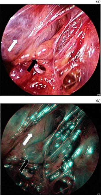
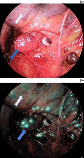
Narrow-band imaging uses the physical phenomena of scattering and absorption to efficiently reveal capillary networks showing distinctive vascularisation of neoplastic or inflamed tissues. Parathyroid tissue indicative of adenomas exhibited a dark-spotted, homogeneous and vessel-dense pattern. This results from irregular neo-angiogenesis (Figures 1 and 2), sharing the same pattern of a variety of neoplastic lesions such as oral or gastric neoplasia.Reference Zhou, Wu, Huang, Jin, Qin and Chen1–Reference Ma, Chowdary, Eskander, Ellett, McIlwaine and Manwaring3
(a) Image obtained during minimally invasive video-assisted parathyroidectomy shows a normal parathyroid gland. The gland (black arrow) presents a yellowish appearance, in good contrast with the normal thyroid gland (white arrow). The gland can be more difficult to distinguish from peri-thyroid fat, which appears less vascularised. (b) Image shows a normal parathyroid gland as visualised with narrow-band imaging. The gland presents a slightly darker appearance than the normal thyroid gland (white arrow).

(a) Image obtained during minimally invasive video-assisted parathyroidectomy shows a parathyroid adenoma. The adenoma (blue arrow) presents a reddish appearance, in moderate contrast with the normal thyroid gland (white arrow). (b) Image shows a parathyroid adenoma as visualised with narrow-band imaging. The adenoma presents a much darker appearance than the normal thyroid gland (blue arrow), as a result of narrow-band imaging's capacity to reveal capillary networks.

In four of five cases in this study, narrow-band imaging integrated the common white light endoscopic vision. The enlarged parathyroid typical of adenomas was easily identified, and diagnosis was promptly confirmed by histology and by a significant intra-operative PTH drop. In all cases, narrow-band imaging revealed a dark neoplastic tissue, which was more vascularised than thyroid, fat and all the surrounding tissues.
In one of the five cases, narrow-band imaging was crucial in discriminating between a pathological and normal parathyroid. A preliminary neck ultrasound revealed a suspect nodule behind the right superior thyroid lobe. During surgery, the nodule was identified using white light and inspected using narrow-band imaging, but no vascular abnormalities were identified. The histological analysis conducted on frozen sections confirmed that it comprised normal thyroid tissue. Exploration of the whole thyroid tissue with the narrow-band imaging revealed a further dark and hyper-vascularised lesion located just behind the right inferior thyroid lobe, which was removed. Histological analysis performed on a frozen section confirmed the removal of pathological parathyroid tissue indicative of adenoma; moreover, PTH serum level dropped from 158 pg/ml to 32 pg/ml approximately 10 minutes after removal.
For all patients, we observed serum calcium normalisation in the first post-operative and histological diagnosis of adenoma. At six months’ follow up, the serum calcium and PTH levels of these patients were normal.
Discussion
In this preliminary study, narrow-band imaging helped distinguish between the hyperfunctioning parathyroid tissue and the surrounding structures by visualising the different vascular patterns. Pathological parathyroid glands are usually well distinguishable from normal thyroid structures. Nevertheless, the distinction between a single adenoma and multiple gland disease (hyperplasia) remains difficult, and surgeons may not be completely sure of surgical radicality.Reference Sessa, Lombardi, De Crea, Raffaelli and Bellantone9 The most diffuse technique used to evaluate successful removal is intra-operative PTH monitoring; however, many researchers have recently questioned the validity of this method because of bias linked to the measurement of serum PTH levels.Reference Carter and Howanitz10 Another useful tool to define parathyroid adenomas is histological analysis of frozen sections, which inevitably leads to increased procedure duration and cost.
In our experience, narrow-band imaging is a valid and helpful instrument for the detection and removal of a pathological parathyroid resulting in adenoma. Furthermore, greater confidence in tumour recognition minimises the accidental removal of healthy parathyroid or thyroid tissue. We consider narrow-band imaging as valuable for achieving this objective and providing the best treatment. Of course, these benefits will come at a cost: training a surgeon in the interpretation of narrow-band imaging data is mandatory, and requires significant time and effort.
Conclusion
Although our patient sample size is small, this preliminary study on narrow-band imaging applied to parathyroid surgery is promising. Prospective validation on a larger scale is needed, but we believe that narrow-band imaging represents a powerful tool in parathyroid surgery. Its application may lead to a reduction in surgical duration and to increased surgeon confidence regarding the validity of the selected intervention. We hope that our initial observation will inspire more colleagues to experiment with this technique in parathyroid surgery and to gather more data on this potential application.
Acknowledgement
We thank Dr Susanna Diamanti for the careful linguistic revision of this work.
Competing interests
None declared