Introduction
Echinococcosis is a zoonotic parasitic disease caused by adult or larval stages of dog tapeworm belonging to the genus Echinococcus (family Taeniidae) (McManus et al., Reference McManus, Zhang, Li and Bartley2003; Umur, Reference Umur2003). The disease has a worldwide geographical distribution and occurs in all continents, including circumpolar, temperate, subtropical and tropical zones, especially in undeveloped and developing countries. The highest prevalence of the parasite is found in parts of Euroasia, Africa, Australia and South America (Schantz et al., Reference Schantz, Chai, Craig, Eckert, Jenkins, McPherson, Thakur, Thompson and Lymbery1995; Craig et al., Reference Craig, Rogan and Allan1996; Umur, Reference Umur2003).
Echinococcosis is a major public health and economic problem in Turkey, as in many other parts of the world. The disease can cause severe clinical signs and possible death in humans, and results in economic losses due to treatment costs, lost wages and livestock-associated production losses (Altintas, Reference Altintas2003; Budke et al., Reference Budke, Deplazes and Torgerson2006). Survey reports indicate that Turkey has a high prevalence of E. granulosus in both dogs and livestock animals as well as a high incidence of cystic echinococcosis (CE) in humans (Altintas, Reference Altintas1997; Economides & Thrasou, Reference Economides and Thrasou1999). The prevalence of E. granulosus in dogs in Turkey varies between 0.32% and 44%, and the prevalence of CE infection in livestock animals varies between 1.6% and 79.6%, depending upon the geographical location (Altintas, Reference Altintas2003; Akyol, Reference Akyol, Altintas, Tinar and Coker2004). The Ministry of Health has recorded 21,303 hospital cases of human CE, with an annual average of 2663 between 1987 and 1994 (Altintas, Reference Altintas1997). Related to a total population of approximately 61 million, the average annual incidence rate is 4.4 per 100,000 inhabitants (Altintas, Reference Altintas1997; Economides & Thrasou, Reference Economides and Thrasou1999; Altintas, Reference Altintas2003). Major reasons for the high prevalence of echinococcosis in Turkey are thought to be uncontrolled slaughter, insufficient abattoir/slaughterhouse facilities and abundance of stray dogs (Altintas, Reference Altintas1997; Umur, Reference Umur2003).
To establish effective control programmes, it is epidemiologically important to obtain an estimation of how long the protoscolices remain infective to carnivore hosts. Although several authors have studied the survival of protoscolices in storage (Andersen & Loveless, Reference Andersen and Loveless1978; Ohnishi et al., Reference Ohnishi, Nakao and Inaoka1984), until now there has been no study of the infectivity of E. granulosus protoscolices towards definitive hosts. In this study, conducted in Bursa, the aim was to examine the infectivity of larval E. granulosus to definitive hosts under different conditions and at various levels of viability.
Materials and methods
Dogs
Eighteen male and female cross-bred dogs, 6 months to 2 years of age, were divided into six groups (n = 3 each), and were raised separately in captivity under helminth-free conditions. For this purpose, dogs were treated with a praziquantel+pyrantel pomate+oxantel pomate combination upon arrival, and subsequent faecal examinations were performed until they revealed negative results for helminth eggs prior to infection. The lack of infection was proved by means of a flotation technique with saturated NaCl solution (specific gravity 1.20) according to Hendrix (Reference Hendrix1998). Furthermore, dogs were vaccinated against distemper, hepatitis contagiosa canis, parvoviral enteritis, leptospirosis and rabies. All animals were fed with commercial dog food and clean, drinkable water ad libitum.
Protoscolices
Echinococcus granulosus protoscolices were obtained from liver hydatid cysts of naturally infected sheep slaughtered at three different abattoirs located in Bursa. Livers were taken to the laboratory within 3 h and transferred into the incubation cabinet adjusted to temperatures of − 10°C, 0°C, +10°C, +30°C or +40°C with 50%, 60%, 65%, 75% or 80% relative humidity (RH), respectively (table 1). The incubation cabinet used in the study was specially produced, including an electronic climatic control unit, to obtain the relative humidity and temperature conditions. The conditions inside the cabinet were monitored digitally by means of thermometer and hygrometer which were calibrated before the study.
The number of protoscolices of E. granulosus given to dogs, viability rates of protoscolices and necropsy days of dog groups under different conditions of temperature and humidity.

Viability assessment
Livers with hydatid cysts were taken from the incubation cabinet for observing the viability of protoscolices and their ability to exclude 0.1% aqueous eosin. Protoscolices stained with eosin (pink-red) were counted as non-viable and the unstained protoscolices (clear) were counted as viable, and the percentages of viable protoscolices were calculated.
Experimental infection and necropsy
After fasting for 24 h before infection, dogs were fed with offal mince harbouring approximately 20,000 protoscolices of E. granulosus at different viabilities (table 1).
Infected animals were fasted for 24 h before being euthanized on 29–49 days post-infection. Intestines were removed within 40 min after the death of the dogs, tied at both ends and placed in numbered plastic bags. Materials were deep frozen for 5 days at − 80°C in order to avoid the infection risk from E. granulosus eggs (Eckert et al., Reference Eckert, Gottstein, Heath, Liu, Eckert, Gemmel, Meslin and Pawlowski2002).
Examination of the intestine
The intestine of each dog was placed on a large metal tray, opened for its full length with scissors and examined macroscopically, and then cut into 20-cm long segments. Segments of the intestine were immersed in a large beaker in water at 37°C. The beaker was kept in an incubator for 45 min. The mucosal wall of the intestine was stripped between two pressed fingers three times. All intestinal contents were sieved through a 75 μm metal mesh to obtain mature E. granulosus. Sieved material was examined in small portions of 10–15 ml in glass dishes under a stereomicroscope to collect worms. Collected worms for each dog were fixed in 10% formalin in separate small jars to be counted afterwards.
Ethics and biosafety
The experimental procedure complied with the Uludag University Ethical Committee Regulations and biosafety regulations were considered in all phases of the study (Eckert et al., Reference Eckert, Gottstein, Heath, Liu, Eckert, Gemmel, Meslin and Pawlowski2002).
Statistical analysis
Viability values of protoscolices and infectivity results were analysed by chi-square (χ2) test (Sumbuloglu & Sumbuloglu, Reference Sumbuloglu and Sumbuloglu1995), using Minitab® (V-13.20) software. (Minitab Inc., State College, Pennsylvania, USA). The value of P < 0.05 was considered to be significant.
Results
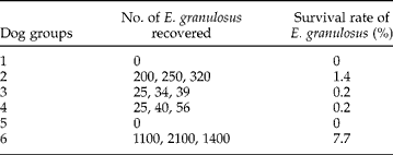
Table 2 shows the number and survival rates (%) of E. granulosus recovered from the small intestine of each group of dogs.
The number and survival rate (%) of E. granulosus recovered from the small intestine of each group of dogs.

No E. granulosus was recovered from the first and fifth groups of dogs, necropsied on days 48 and 45 post-infection, respectively. Dogs in the second group were euthanized on the 35th day of infection and mean burden of E. granulosus was found to be 256.7 ± 60.3 (range 200–320). The number of worms ranged from 25 to 39 and mean intensity was 32.7 ± 7.1 in the third group on the 49th day after the infection. In the fourth group, two dogs on 33rd day and one dog on day 36 post-infection were euthanized and the mean worm burden was found to be 40.3 ± 15.5 (range 25–56). In the control group, dogs were euthanized on days 29, 32 and 39 post-infection and the mean worm burden was found to be much greater than those obtained from the other groups: 1533 ± 513, ranging from 1100 to 1400.
The difference between the first group and the fifth group was not significant (P = 0.4964); differences among other groups were significant (P < 0.001).
Discussion
The transmission dynamics of E. granulosus are influenced by several biotic and abiotic factors. Within the abiotic factors, the most important ones are environmental temperature and humidity. Therefore the biotic potential may vary widely in different ecological situations and climatic zones (Torgerson & Heat, Reference Torgerson and Heat2003; Jenkins, Reference Jenkins2005).
Various results have been reported on the viability of Echinococcus eggs in different climatic conditions around the world (Sweatman & Williams, Reference Sweatman and Williams1963; Veit et al., Reference Veit, Bilger, Schad, Schafer, Frank and Lucius1995; Thevenet et al., Reference Thevenet, Jensen, Drut, Cerrone, Grenovero, Alvarez, Targovnik and Basualdo2005). Early studies by Sweatman & Williams (Reference Sweatman and Williams1963) have shown that E. granulosus eggs may remain viable for a maximum of 120 days under the climatic conditions of New Zealand. Veit et al. (Reference Veit, Bilger, Schad, Schafer, Frank and Lucius1995) reported that E. multilocularis eggs survived for 240 days in autumn and winter and 78 days in summer in Germany. Thevenet et al. (Reference Thevenet, Jensen, Drut, Cerrone, Grenovero, Alvarez, Targovnik and Basualdo2005) reported that eggs of E. granulosus were able to generate infection in sheep after 41 months under the environmental conditions of an arid climate (Patagonia, Argentina).
Also, the viability of metacestodes under different environmental conditions influences the parasite's biotic potential (Torgerson & Heat, Reference Torgerson and Heat2003). The effect of temperature on the viability of E. granulosus protoscolices has been well studied (Andersen & Loveless, Reference Andersen and Loveless1978; Ohnishi et al., Reference Ohnishi, Nakao and Inaoka1984). In these studies cysts from different animal species were used, methods for assessing viability differed and the temperature ranges and periods of time of observation were varied. Several differences were found between the results of these studies on the viability of protoscolices. The protoscolices of E. granulosus were reported to survive for 2 h to 70 days between the temperatures of − 20°C and+40°C. These differences are probably due to differences in the methods of storage, in viability assessment and/or genetic variation and diversity of E. granulosus (Andersen & Loveless, Reference Andersen and Loveless1978; Ohnishi et al., Reference Ohnishi, Nakao and Inaoka1984).
Although several authors studied intensively the infectivity of E. granulosus eggs and the survival of protoscolices in storage under different conditions with different methods, the infectivity of E. granulosus protoscolices towards definitive hosts has not hitherto been investigated (Andersen & Loveless, Reference Andersen and Loveless1978; Ohnishi et al., Reference Ohnishi, Nakao and Inaoka1984). The present study is the first observation on the infectivity of E. granulosus protoscolices under different conditions of temperature and humidity. Therefore, it is difficult to discuss and compare results of other researchers with those of our own study.
In our study, infectivity of E. granulosus protoscolices under the effect of different temperatures and humidities was demonstrated by recovering worms from the intestine of dogs which were experimentally infected. For this purpose approximately 20,000 protoscolices with different viabilities were fed to each dog, within the offal mince, resembling the natural infection. No E. granulosus was recovered from the small intestines of dogs of the first and fifth groups, which were infected with protoscolices of 5% and 11% viability. According to these findings protoscolices of E. granulosus ≤ 11% viability do not seem to have the ability to infect definitive hosts. Percentages of developing worms increased in parallel with the viability rate during the infection.
Bursa is located in the southern part of Marmara region, where sheep raising is intensive. In this region stray and roving dogs are numerous and prevention or treatment of the parasitic infections in these dogs is difficult. The prevalence of E. granulosus in stray dogs in Bursa was reported to be 36% in 1988 (Tinar et al., Reference Tinar, Coskun, Dogan, Demir, Akyol and Aydin1989). Stray and roving dogs that scavenge for food around the slaughterhouses can easily access dead or discarded carcasses of infected animals, and they feed not only on freshly killed animals but also on decayed carcasses. The food habits of dogs and the presence of protoscolices in decayed offal, viscera and carcasses play an important role in the transmission of echinococcosis. Results obtained in the present study indicate that the protoscolices in decayed carcasses keep their infectivity for more than 3 weeks, and suggest that the saprophagy of canines may be epidemiologically important for the transmission of E. granulosus.
Gemmel (Reference Gemmel1958) investigated the effect of weather conditions, particularly rainfall, on the transmission and distribution of E. granulosus, and reported that 25 mm rainfall per month throughout half the year provides the optimal conditions for transmission. Under natural conditions, the mean temperature in Bursa region is about 0°C to +10°C with 60% to 70% RH between November and March, and strong rainfall periods are observed in autumn and winter months. High rainfall and mild winters provide favourable conditions for E. granulosus eggs and larval metacestodes. According to the results of our study, this period is risky for the transmission of the disease. Our study suggests that, under natural conditions within our geographical area, hydatid cysts within improperly discarded sheep carcasses might represent a potential source of infection to stray or roving carnivores.
In conclusion, the persistence of protoscolices in decayed carcasses suggests that canines are infected with E. granulosus not only by predation but also by saprophagy. Considering the epidemiological importance of the disease, dogs should be properly controlled; stringent and adequate preventive control programmes, based on education, combinations of dog de-worming, stray dog culling and vaccination, must be established in areas where the disease is endemic. Cold-storage depots in slaughterhouses and abattoirs where sheep carcasses might be discarded should be kept at − 20°C for 2–3 days. Animal pits should be enclosed or adequately fenced and slaughterhouses should be renovated.
Acknowledgements
This work was supported by the Research Fund of the University of Uludag (Project number V-2003\1).