Introduction
Mastoid surgery is indicated for a number of conditions. The most common reasons for this surgery are chronic suppurative otitis media (CSOM) that does not respond to clinical treatment and cholesteatomas.Reference Prata, Antunes, de Abreu, Frazatto and Lima1 Local destruction of the middle- and inner-ear structures resulting from cholesteatomas can have profound consequences for the individual, such as hearing loss.
The choice of pre-operative imaging in presumed uncomplicated mastoid surgery remains controversial.Reference Walshe, McConn Walsh, Brennan and Walsh2 Pre-operative computed tomography (CT) scans may aid the operator in identifying the extent of disease, and any existing or underlying anatomical abnormality, and can assist the otologist in the planning of the procedure. Other imaging techniques for mastoid pathology are in use, such as magnetic resonance imaging (MRI). However, CT scans have certain benefits over other modalities, such as improved visualisation of bony detail, which can provide information on potential asymptomatic complications.Reference Yildirim-Baylan, Ozmen, Gun, Yorgancilar, Akkus and Topcu3
This study aimed to determine the correlation between pre-operative CT findings and intra-operative findings.
Materials and methods
This study was designed as a service evaluation and therefore did not require formal ethical approval. A retrospective cohort study was conducted in a district general hospital in South Wales. Consecutive case notes were analysed over a 12-month period (1 August 2016 to 31 July 2017). Pre-operative CT imaging reports were compared to the operation notes. Eligible patient records were captured using Operating Room Information Management System (‘ORMIS’) software.
The codes included in the patient records search represented: mastoidectomy, mastoid exploration, revision mastoidectomy and atticotomies. This generated a provisional cohort of 139 patients. Thirty-three patients did not undergo pre-operative imaging, or there were no Picture Archive and Communication System (‘PACS’) data available; these patients were therefore excluded (final cohort, n = 106).
A Medline and PubMed literature search was conducted, using the terms ‘preoperative’, ‘computed tomography’ and ‘cholesteatoma’ within all fields. This generated a number of studies evaluating pre-operative imaging in relation to intra-operative findings, which were directly comparable to our study.Reference Prata, Antunes, de Abreu, Frazatto and Lima1–Reference O'Reilly, Chevretton, Wylie, Thakkar, Butler and Sathanathan19 Abstract-only papers and studies concerning MRI were excluded. The search revealed a mixture of both retrospective and prospective analyses. The publication dates of the studies ranged from 1984 to 2018, and cohort numbers ranged from 20 to 80.
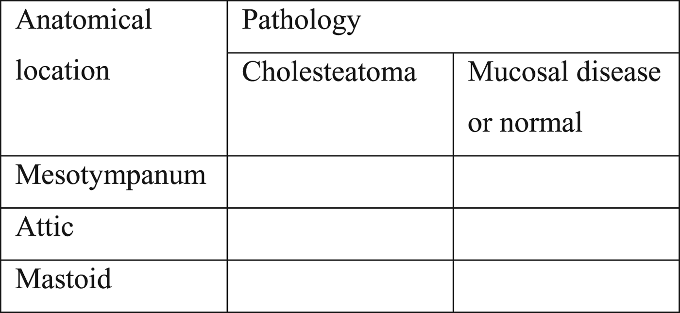
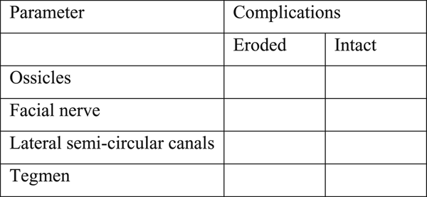
Data were evaluated based on a modified, existing proforma obtained from the literature.Reference Badran, Ansari, Al Sam, Al Husami and Iyer20 Patient age, and information on laterality obtained from both the CT scan and the operation, were collected. Results were categorised into two broad subset groups: pathology and complications. Figures 1 and 2 show examples of the data collection forms.
Data-collecting proforma for identifying cholesteatoma based on anatomical location (mesotympanum, attic and mastoid).

Data-collecting proforma for identifying complications of cholesteatoma involving the ossicles, facial nerve, lateral semi-circular canals and tegmen.

Under the category of pathology, findings concerning the mesotympanum, attic and mastoid were grouped into two separate variables: ‘cholesteatoma’ and ‘mucosal disease or normal’. Complications were classed as either ‘intact’ or ‘eroded’ (or ‘removed’ if absent at revision surgery). The structures included in the complications subset group were: ossicles, facial nerve, lateral semi-circular canals and tegmen.
Results
Naturally, there is some degree of variability in nomenclature between radiologists on their reporting of structures on CT scans, and indeed between ENT surgeons in their operation notes. We therefore have had to make certain assumptions and generalisations regarding the terms used. The full list of assumed structures and terms can be found in Appendix 1.
A total of 106 patients underwent mastoid surgery and had pre-operative CT scans performed at the single site within the 12-month period. Patients’ median age at the time of the operation was 30.5 years.
Fifty-seven operations were performed on the left mastoid and 49 were performed on the right mastoid. Incidentally, there was a discrepancy between the laterality of the pathology reported on CT and the side that was operated on in 5.7 per cent of the cases. This was either part of a two-stage operation or an erroneous operation note; the operation note error was later rectified in subsequent follow-up clinic letters.
The median time delay or ‘lag’ between the CT scan and the operation was 215 days. However, some patients may have been under ‘active surveillance’ in out-patient clinics before they were ultimately scheduled for operative treatment, and so this may have positively skewed the lag.
To ensure standardisation, both the radiology reports and the operation notes had to explicitly state whether disease was present or absent in order to be included in the final data analysis. If only one of these (the radiology report or the operation note) commented on the independent variable, the patient was excluded, as no valid comparisons could be made.
Pathology
Mesotympanum
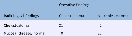
In 62 out of 106 cases, the mesotympanum was commented on by both the radiologist and surgeon (Table 1). The CT scans predicted 33 cholesteatomas, 31 of which were confirmed at operation. Radiology predicted 29 cases where there was mucosal disease or no evidence of cholesteatoma, 21 of which were found to be normal intra-operatively. The sensitivity of CT scans in identifying cholesteatoma affecting the mesotympanum was 80 per cent, with a specificity of 91 per cent. The positive predictive value and negative predictive value were calculated at 94 per cent and 72 per cent respectively.
Presence of cholesteatoma affecting the mesotympanum, identified on pre-operative CT and intra-operatively

Data represent numbers of cases. CT = computed tomography
Attic
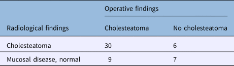
In 52 cases, both specialties (radiology and ENT) commented on the attic in their respective reports (Table 2). There were 39 cholesteatomas identified intra-operatively, 30 of which were correctly predicted by pre-operative radiology (sensitivity of 77 per cent). Specificity was lower at 54 per cent. The positive predictive value was 83 per cent and the negative predictive value was 56 per cent.
Presence of cholesteatoma affecting the attic, identified on pre-operative CT and intra-operatively

Data represent numbers of cases. CT = computed tomography
Mastoid
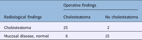
In 48 out of 106 cases, comments on the appearances of the mastoid were eligible for interpretation (Table 3). Sensitivity was 81 per cent and specificity was 88 per cent. There was a high positive predictive value of 93 per cent and a negative predictive value of 71 per cent.
Presence of cholesteatoma affecting the mastoid, identified on pre-operative CT and intra-operatively

Data represent numbers of cases. CT = computed tomography
Total
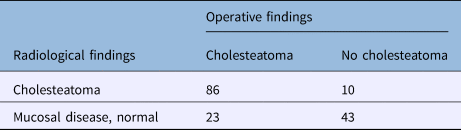
The prediction for cholesteatoma in all cases was calculated by determining the total number of cases implicating the mesotympanum, attic or mastoid (Table 4). The sensitivity for predicting cholesteatoma in any of the three anatomical locations was 79 per cent, and the specificity was 81 per cent. The positive predictive value was 90 per cent, but the negative predictive value was lower, at 65 per cent.
Total presence of cholesteatoma identified on pre-operative CT and intra-operatively

Data represent numbers of cases. CT = computed tomography
Complications
The number of cases where both radiology and ENT specialists commented on the involvement of the middle-ear structures was noticeably fewer than when commenting on the presence of cholesteatoma. It may be inferred that if there was no comment on complications of the disease process, then it is unlikely to be present. For the purposes of data analysis, however, this was not assumed and cases were excluded. Given the paucity of data, the significance of results for individual subsites can be called into question. Therefore, in the analysis, we have included the grouped results, incorporating the data from comments on the ossicles, facial nerve, lateral semi-circular canals and tegmen. The data from individual subsites are wholly presented in Appendix 2.
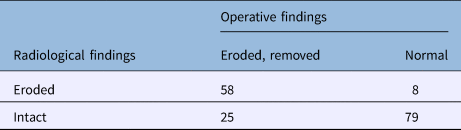
Regarding the grouped results, the sensitivity was fairly poor at 70 per cent, whereas the specificity for predicting complications was 91 per cent. The positive predictive value and negative predictive value were calculated as 88 per cent and 76 per cent respectively (Table 5).
Total presence of complications identified on pre-operative CT and intra-operatively

Data represent numbers of cases. CT = computed tomography
A positive indicator for diagnosing cholesteatomas from CT is often erosion of the ossicles.Reference Khavasi, Malashetti and Chandrashekarayya4 Our study demonstrated this, as our positive predictive value for ossicular chain involvement was 88 per cent. We had a low sensitivity for identifying complications associated with the facial nerve, largely because of a low number of operation notes commenting on whether there was dehiscence or not. This gave us a sensitivity of 25 per cent, specificity of 96 per cent, positive predictive value of 67 per cent and negative predictive value of 79 per cent. In terms of identifying complications associated with the lateral semi-circular canals, the sensitivity, specificity, positive predictive value and negative predictive value were 60 per cent, 96 per cent, 86 per cent and 85 per cent respectively.
Discussion
Based on the results of this cohort, pre-operative imaging of the mastoid has good positive predictive values for cholesteatomas and associated complications (90 per cent and 88 per cent respectively). These high values are comparable to those of other studies.Reference Prata, Antunes, de Abreu, Frazatto and Lima1,Reference Yildirim-Baylan, Ozmen, Gun, Yorgancilar, Akkus and Topcu3,Reference Khavasi, Malashetti and Chandrashekarayya4,Reference Mohamed, Yousif and Mehnab7,Reference Jackler, Dillon and Schindler10,Reference Alzoubi, Odat, Al-Balas and Saeed11,Reference Payal, Pranjal, Gul, Mittal and Rai15,Reference Park, Park, Kwon, Kim, Park and Sung16,Reference O'Donoghue, Bates, Anslow and Rothera18 Imaging was not as accurate when predicting a non-diseased ear (65 per cent negative predictive value for cholesteatomas and 76 per cent negative predictive value for complications). There were, however, a large proportion of ‘undecided’ reports (49 per cent) that were not included, as either the radiology report or the operation notes failed to comment on one of the independent variables, thereby precluding valid comparison. This is a limitation of retrospective analysis. Similar results from the literature show that sensitivity values are not as reliable, suggesting that the role of CT in predicting complications of CSOM is not absolute.Reference Faramarzi and Roosta21
Our cohort was representative of the population, as several studies had mean patient ages ranging from 24.02 years to 38.2 years.Reference Prata, Antunes, de Abreu, Frazatto and Lima1,Reference Yildirim-Baylan, Ozmen, Gun, Yorgancilar, Akkus and Topcu3,Reference Karki, Pokharel, Suwal and Poudel6,Reference Mohamed, Yousif and Mehnab7,Reference Alzoubi, Odat, Al-Balas and Saeed11,Reference Gerami, Naghavi, Wahabi-Moghadam, Forghanparast and Akbar13,Reference Rogha, Hashemi, Mokhtarinejad, Eshaghian and Dadgostar14 Das et al.Reference Das, Ranjan Rout, Shilpa Sri, Mohanty and Vittal Prasad5 and Chintale et al.Reference Chintale, Kirdak, Jatale and Shaikh8 had age ranges that reflected our cohort. Khavasi et al.Reference Khavasi, Malashetti and Chandrashekarayya4 had a younger cohort, with 45 per cent of their patients with cholesteatomas being between 11 and 20 years.
We had a large number of patients in our cohort. The largest number of patients found in comparative literature was 80.
Invariably, disease progresses chronologically. It can therefore be theorised that a longer time delay between a CT scan and an operation can influence the discrepancy between the two findings. Our median time lag was over seven months. This duration could quite easily allow changes to occur between what is reported on a CT scan and what is found at operation. This is what was found in our cohort, as the prediction of a non-diseased middle ear was lower than that for predicting a cholesteatoma (65 per cent vs 90 per cent respectively). What is not taken into account in our calculations, but is appreciated as a limitation, is that a proportion of the CT scans were used for surveillance or diagnosis, and not as a pre-operative planning adjunct.
It is also worth noting that CSOM and cholesteatomas rarely affect the anatomical locations described in our study in isolation.Reference Sethi, Rafferty and Kayarkar22 Variations in the reported frequencies of individual anatomical locations of cholesteatomas must therefore be considered, as well as the calculations of sensitivity and positive predictive value.
A further limitation of this study is the assumptions made associated with the nomenclature used in the reporting of the findings in order to categorise the data effectively. If this was not done, direct comparisons between verbatim reports would be unfeasible and lead to multiple, substantially smaller cohorts.
The sensitivity for predicting cholesteatoma based on pre-operative CT imaging varies in the literature. Our results are most comparable with those of Prata et al.,Reference Prata, Antunes, de Abreu, Frazatto and Lima1 Jackler et al.Reference Jackler, Dillon and Schindler10 and Alzoubi et al.Reference Alzoubi, Odat, Al-Balas and Saeed11 (who reported sensitivity values of 72.73–80 per cent). Other sensitivity values reported ranged from 61.8 per cent to 100 per cent.Reference Walshe, McConn Walsh, Brennan and Walsh2–Reference Khavasi, Malashetti and Chandrashekarayya4,Reference Mohamed, Yousif and Mehnab7,Reference Tatlipinar, Tuncel, Öğredik, Gökçeer and Uslu12,Reference Gerami, Naghavi, Wahabi-Moghadam, Forghanparast and Akbar13,Reference Payal, Pranjal, Gul, Mittal and Rai15,Reference Park, Park, Kwon, Kim, Park and Sung16,Reference O'Donoghue, Bates, Anslow and Rothera18
Much like our sensitivity findings, our specificity was similar to Prata et al.Reference Prata, Antunes, de Abreu, Frazatto and Lima1 (81 per cent vs 82.5 per cent respectively). However, specificity ranged from 48 per cent to 94.7 per cent in studies by Yildirim-Baylan et al.Reference Yildirim-Baylan, Ozmen, Gun, Yorgancilar, Akkus and Topcu3 and Alzoubi et al.Reference Alzoubi, Odat, Al-Balas and Saeed11
There was variability in both the positive predictive value and negative predictive value within the literature, ranging from 60.6 per cent to 97.1 per cent and from 66.67 per cent to 85.7 per cent, respectively. Multiple studies have demonstrated positive predictive values comparable to our results. The positive predictive values were 77.27 per cent in the study by Prata et al.,Reference Prata, Antunes, de Abreu, Frazatto and Lima1 85.2 per cent in that by Yildirim-Baylan et al.,Reference Yildirim-Baylan, Ozmen, Gun, Yorgancilar, Akkus and Topcu3 and 94.3 per cent in the study by Kumaresan and Nirmala.Reference Kumaresan and Nirmala9 Payal et al.Reference Payal, Pranjal, Gul, Mittal and Rai15 had a considerably lower positive predictive value of 68.95 per cent.
There was a big discrepancy in the sensitivity values for erosion or dehiscence of the facial nerve in the literature. A low sensitivity was also found by Khavasi et al.,Reference Khavasi, Malashetti and Chandrashekarayya4 Das et al.Reference Das, Ranjan Rout, Shilpa Sri, Mohanty and Vittal Prasad5 and Payal et al.Reference Payal, Pranjal, Gul, Mittal and Rai15 (42 per cent, 42.9 per cent and 40 per cent, respectively). These values were considerably lower than in some other studies, such as those by Prata et al.,Reference Prata, Antunes, de Abreu, Frazatto and Lima1 Yildirim-Baylan et al.Reference Yildirim-Baylan, Ozmen, Gun, Yorgancilar, Akkus and Topcu3 and Karki et al.,Reference Karki, Pokharel, Suwal and Poudel6 which reported sensitivity values for facial nerve compromise of over 98 per cent.
Again, there was variability in each of the parameters measured (complications of the ossicles, facial nerve, lateral semi-circular canals and tegmen). Sensitivity values ranged from 33 per cent to 100 per cent.Reference Prata, Antunes, de Abreu, Frazatto and Lima1–Reference Khavasi, Malashetti and Chandrashekarayya4,Reference Karki, Pokharel, Suwal and Poudel6,Reference Chintale, Kirdak, Jatale and Shaikh8,Reference Kumaresan and Nirmala9,Reference Tatlipinar, Tuncel, Öğredik, Gökçeer and Uslu12,Reference Payal, Pranjal, Gul, Mittal and Rai15,Reference Park, Park, Kwon, Kim, Park and Sung16,Reference O'Donoghue, Bates, Anslow and Rothera18 Specificities were also widely spread, ranging from 48 per cent to 100 per cent. The incidence of facial nerve dehiscence is relatively low, and therefore the sample size of patients needed to generate accurate statistical measures would generally be larger than the cohorts we have compared against.Reference Faramarzi and Roosta21
Several studies reported high sensitivity values for correctly identifying tegmen erosion on the pre-operative CT scans. Tatlipinar et al.,Reference Tatlipinar, Tuncel, Öğredik, Gökçeer and Uslu12 Yildirim-Baylan et al.Reference Yildirim-Baylan, Ozmen, Gun, Yorgancilar, Akkus and Topcu3 and Karki et al.Reference Karki, Pokharel, Suwal and Poudel6 reported sensitivity values of 90 per cent, 98 per cent and 100 per cent, respectively. Our cohort had a sensitivity of 75 per cent, which is more in keeping with the studies of Khavasi et al.,Reference Khavasi, Malashetti and Chandrashekarayya4 Chintale et al.,Reference Chintale, Kirdak, Jatale and Shaikh8 Kumaresan and Nirmala,Reference Kumaresan and Nirmala9 and Rogha et al.Reference Rogha, Hashemi, Mokhtarinejad, Eshaghian and Dadgostar14 (which reported sensitivity values of 66–75 per cent). Our specificity and positive predictive values were high, at 100 per cent. Such values were also found in studies by Prata et al.,Reference Prata, Antunes, de Abreu, Frazatto and Lima1 Karki et al.,Reference Karki, Pokharel, Suwal and Poudel6 Kumaresan and Nirmala,Reference Kumaresan and Nirmala9 and Rogha et al.,Reference Rogha, Hashemi, Mokhtarinejad, Eshaghian and Dadgostar14 with a range of 91.93–100 per cent.
• Computed tomography (CT) is used in pre-operative planning for mastoid surgery to assess disease extent in cholesteatoma and any anatomical variation
• Computed tomography has benefits over other imaging modalities when assessing cholesteatoma complications (e.g. better bony detail in ossicular erosion cases)
• In this study, pre-operative mastoid CT scans to assess for cholesteatoma showed a high positive predictive value, of 90 per cent
• In addition, CT had a positive predictive value (of 88 per cent) when assessing radiological evidence of cholesteatoma complications
• There is a wide variation of correlations between studies, suggesting that imaging (e.g. ionising radiation) should perhaps be performed in all surgery patients
This study highlights the scarcity of reported key negative findings in mastoid surgery. It has been suggested by the author that the operation notes more often include positive findings, and that if there is no comment on complications it can be assumed that there are none. This could potentially raise issues during revision surgery, or even from a medicolegal perspective in cases of an unexpected, unreported finding.Reference Bateman, Carney and Gibbin23 Locally, we have plans to introduce a standardised operation note to include all relevant pertinent findings, including the absence of such findings.
As suggested by other studies in the literature,Reference Walshe, McConn Walsh, Brennan and Walsh2,Reference Yildirim-Baylan, Ozmen, Gun, Yorgancilar, Akkus and Topcu3,Reference Alzoubi, Odat, Al-Balas and Saeed11,Reference Tatlipinar, Tuncel, Öğredik, Gökçeer and Uslu12 pre-operative CT imaging performed prior to mastoid surgery can be a useful adjunct when planning surgery, in terms of anticipating risk factors present and determining the extent of localised complications. However, given the wide variation of correlational findings between studies, further discussion is warranted regarding whether imaging, particularly ionising radiation, should be conducted for all patients undergoing surgery.
Competing interests
None declared
Appendix 1. Non-standardisation of nomenclature
The following list of structures and terms were to be assumed, in order to standardise data.
Epitympanum as attic
Tegmen as low-lying or exposed dura
Antrum/aditus as mastoid
Incudo-stapedial joint as ossicles
Fallopian canal as facial nerve
Otic capsule as lateral semi-circular canal
Appendix 2. Complications
Tables showing the complications identified at each subsite on pre-operative CT and intra-operatively.
Ossicles

Facial nerve

Lateral semi-circular canals

Tegmen