The ductus arteriosus is a vascular structure derived from the sixth aortic arch that connects the main pulmonary trunk near to origin of the left pulmonary artery to the descending aorta, and it is present in the fetal life to carry the outflow from the right ventricle to the descending aorta. After birth, the ductus closes, usually from the pulmonary end through muscular constriction, obliteration of the lumen, initially by a pile-up of endothelium and finally by complete occlusion through thrombosis. If the ductus remains patent, after the falling of pulmonary resistance, there will be a left to right shunt with the consequent volume overflow to the lungs, left atrium, left ventricle, and ascending aorta.
The patent ductus arteriosus is the most common cardiac anomaly in our country, representing 5%–10% of all the CHDs.
The first person to contemplate a surgical procedure to close the ductus arteriosus was the Boston surgeon John Cummings Munro in a lecture to the Philadelphia Academy of Surgery held on 6 May 1907, idea that eventually lead to the first successful surgical patent ductus arteriosus closure performed by Dr. Gross on a 7-year-old in 1938 at Boston Children’s Hospital.
It was also the second congenital cardiac defect to be treated with a transcatheter approach. Werner Porstmann was the first to close a ductus without a thoracotomy. The operation occurred in a 17-year-old patient in 1966 at the Charité Berlin Hospital, in the then German Democratic Republic but at the time it was not possible to use this procedure in small children. The first successful ductus arteriosus closure in a child was in 1979 by William Rashkind, who reported ductus closure in a child weighing 3.5 kg. Today, a persistent ductus arteriosus is closed by catheter intervention in most cases. Only the very large ductus arteriosus and the ductus in very low birthweight newborns are still mostly treated by surgical intervention. Reference Villa, Eynden, Le Bret, Folliguet and Laborde1,Reference Alexi-Meskishvili and Böttcher2 In the last few decades, there has been a lot of interest in developing less invasive techniques as an alternative to reduce some of the long-term complications of traditional surgery such as chylothorax, recurrent laryngeal dysfunction, rib deformity, scoliosis, and arrhythmia. Reference Stankowski, Aboul-Hassan, Marczak and Cichon3,Reference Stankowski, Aboul-Hassan, Marczak, Szymanska, Augustyn and Cichon4 One of them is the video-assisted thoracoscopic clipping, which has some advantages but it is still a procedure that is not free of complications. Reference Nezafati, Soltani, Mottaghi, Horri and Nezafati5,Reference Stankowski, Aboul-Hassan and Seifizinab6 The aim of this article is to present one of this uncommon complication and its resolution with a percutaneous technique.
Case presentation
We present an 8-year-old female, which had been diagnosed at the age of 3 years with a persistent ductus arteriosus. She consulted in a private institution because of dyspnoea with feeding and failure to thrive; transthoracic echocardiography showed a ductus arteriosus; however, exact measures are not available since the report was held in that institution. Her physician offered thoracoscopic clipping as an option since device closure in the catheterization laboratory has an elevated cost in our country and the family could not afford it. Apparently, there was no report of complications or sign of residual defect; however, the exact information was not available.
Five years later, she presented papular lesions which began in arms and then generalized first treated as food poisoning; however, later she had high fever, oedema, desquamation of palms, feet and tongue, ulcerative lesions that led to think of a vasculitis. After 20 days of interspecific treatment, she was admitted to the ICU in another paediatric hospital where they found her with severe respiratory distress, oedema, and hemodynamic instability. At physical examination, a heart murmur was found. Pericardial and pleural effusion was diagnosed by echocardiogram as well as endarteritis of a residual patent ductus arteriosus with a clip out of place. In that same hospital, they suggested surgical approach, but since it would have been a high-risk procedure, the family decided to be transferred to our institution to evaluate if a transcatheter approach could be done instead.
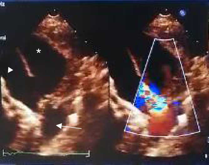
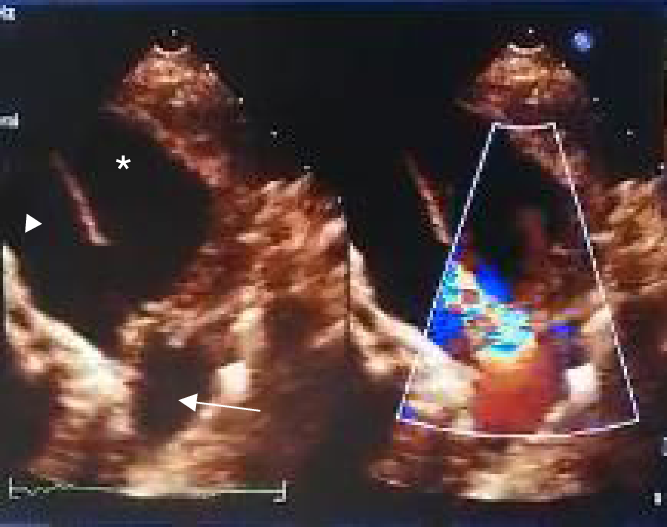
During her evaluation, a new echocardiogram showed a residual defect with left to right shunt with a 74 mmHg pressure gradient. The flow through this structure was turbulent because of a partial obstruction of the clip near the pulmonary insertion where an aneurysmatic lesion was found (Fig. 1).
Echocardiogram. Suprasternal view: We can see a residual ductus arteriosus with turbulent flow and an aneurysm in the distal portion of the pulmonary trunk. (* Aneurysm, >Pulmonary trunk > D. Aorta).
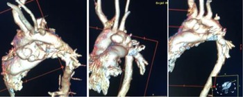
Angiotomography showed a large aneurysm of the ductus arteriosus extending to the distal pulmonary trunk (Fig. 2). The blood culture was positive for S. aureus so she received an antibiotic course for 45 days.
Angiotomography with a large aneurysm of the residual ductus arteriosus with a clip out of its place, extending to the distal pulmonary trunk.
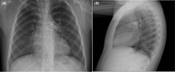
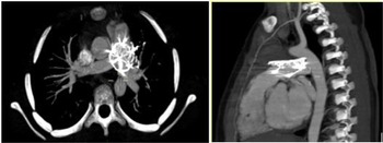
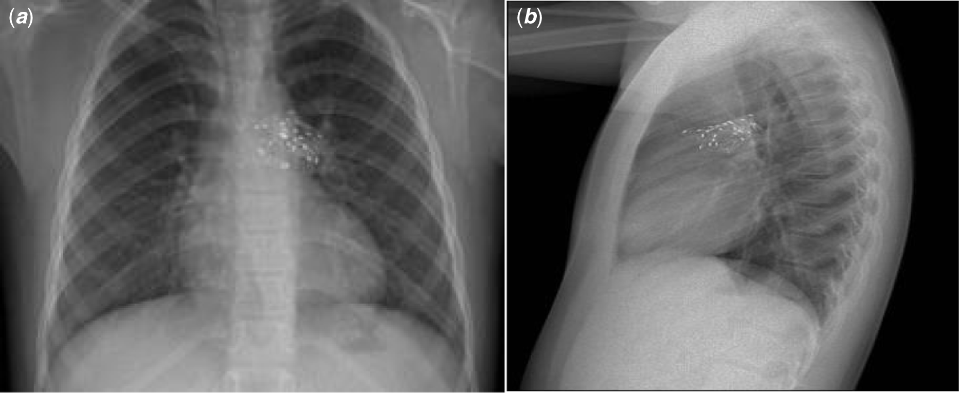
After completing the course of antibiotics, she underwent interventional treatment. The aortic arch angiography showed a 3 mm residual ductus arteriosus with one of the clips occluding 75% of the ductus and an aneurysmatic lesion at its proximal end extending to the distal portion of the pulmonary trunk measuring 20 mm on its origin with a sack of 48 mm. Then, a Cardia ASD occluder of 24 mm (Cardia Inc., Burnsville, MN, USA) was placed in the defect, placing the left disc inside the aneurysm and the right disc in the top portion of the pulmonary trunk. The residual ductus arteriosus was then closed with a Amplatzer Plug vascular II device of 10 mm (AGA Medical Corporation, Golden Valley, MN, USA). Control angiography showed no residual flow through the ductus arteriosus with the aneurysm fully occluded. The total time of procedure was 7 hours. Chest X-ray after the procedure showed an adequate position of both devices (Fig. 3). She stayed stable, and she was discharged 72 hours later.
Chest X-ray A: Posteroanterior protection B: Lateral protection. Showing an adequate position of the device, with normal pulmonary vasculature.
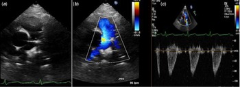
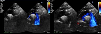
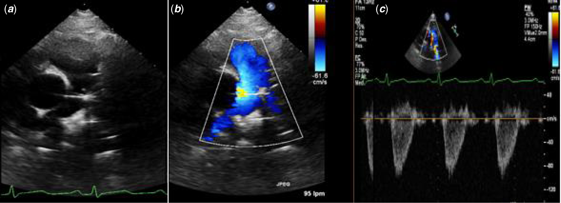
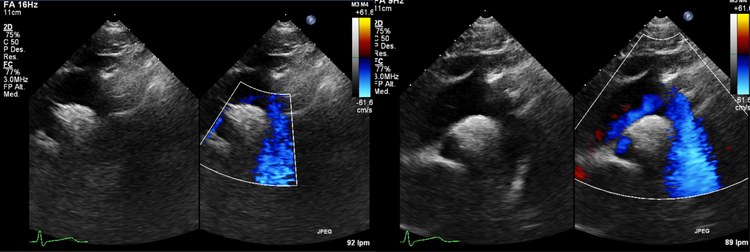
During follow-up 12 months later, cardiovascular physical examination is normal, no murmur is found with normal S2. Chest X-ray shows no cardiomegaly with devices in adequate position. Echocardiogram shows Cardia ASD device fully occluding the aneurysmatic lesion with no residual blood flow. The left side of the device inside the aneurysm, which is collapsed, and the right side of the device in the pulmonary trunk near the left pulmonary branch origin with no obstruction registered (Fig. 4). Also, the ductus arteriosus was evaluated corroborating no residual defect (Fig. 5).
Echocardiogram. A) PSAX: We can see the device in the pulmonary trunk near the left branch origin, B) With colour Doppler with laminar flow. C) Pulsed Doppler with a peak velocity of 1 m/s. No obstruction gradient.
Echocardiogram. Suprasternal view: We can see the aortic arch and pulmonary branch with no residual lesion with the aneurysm fully occluded. There is no flow through the ductus arteriosus.
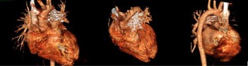
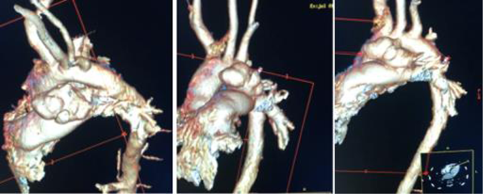
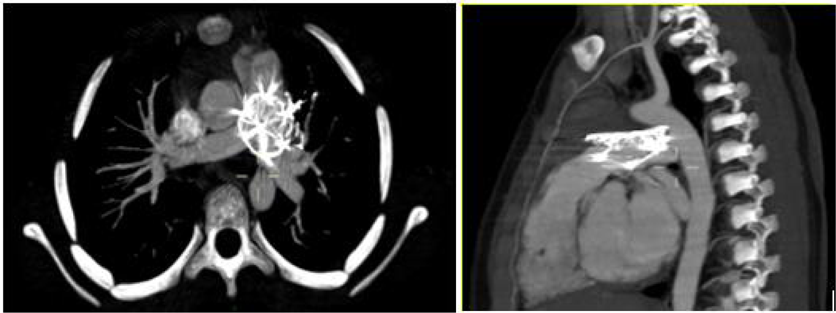
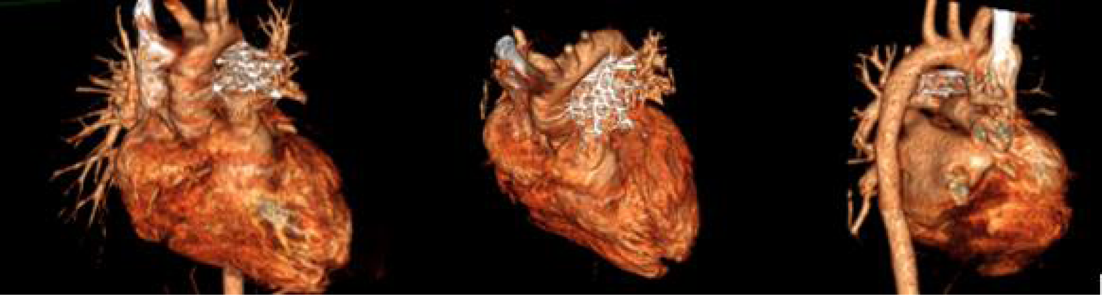
Angiotomography a year after the procedure, we can see the Cardia ASD occluder with good positioning inside the aneurysm, with the ductus arteriosus fully closed (Figs. 6 and 7).
Angiotomography with Cardia device occluding the aneurysm.
3D reconstruction. Both devices are in good position with no residual defects.
Discussion
The ductus arteriosus closure by surgery has been used for many years which has made it a safe procedure with a low morbidity and mortality rate; however, it can still present some complications such as rib deformity, scoliosis, in addition to the time of in-hospital stay and post-surgical pain among others. Video-assisted thoracoscopy has been presented as an option to minimise them, reporting recurrent laryngeal nerve injury, pneumothorax, chylothorax, rupture of the ductal wall, atelectasis, and in some cases the need to convert to thoracotomy or re-intervention as its complications. The development of aneurysm after video-assisted repair is apparently very uncommon. This may occur when one or more of the clips that are placed do not occlude the ductus arteriosus a 100%, and the hydrostatic force exerted by the residual flow displaces the clip partially or totally, when it does partially then the adventitia and some of the muscular layers of the duct remain fixated with the clip and the intima and some layers of the muscle prolapse creating a sac. This pulsatile pressure gradually increases the size of the aneurysm and can cause compression of adjacent structures, be prone to infection or even rupture.
An intentional search for this complication was carried out without finding reported cases; therefore, we consider the report of this case to be of great value for the cardiovascular physicians’ community as well as its resolution to help explore therapeutic options to minimise further complications.
Conclusions
In the case described, we present an apparently unreported complication as well as its resolution through interventionism by placing an ASD occluder device in the aneurysm and a vascular plug in the residual ductus arteriosus, with a satisfactory development, remaining asymptomatic and without any other complications in the follow-up through 12 months.
Acknowledgements
None.
Financial support
This research received no specific grant from any funding agency, commercial, or not-for-profit sectors.
Conflicts of interest
None.