INTRODUCTION
The Leishmania parasite is characterized by its capacity to survive and replicate in the macrophage. After phagocytosis of the stationary-phase promastigotes, a phagosome is formed around the ingested parasite with gradual fusion with lysosomes and endosomes. This results in an acidic (pH 4·7–5·2) parasitophorous vacuole, the phagolysosome, with hydrolytic and proteolytic properties (Burchmore and Barrett, Reference Burchmore and Barrett2001; Vray, Reference Vray, Burke and Lewis2002). One of the major functions of the macrophage is to destruct invading microorganisms. In normal circumstances, macrophages kill the organism with oxygen- and non-oxygen-dependent mechanisms.
Oxygen-dependent mechanisms include the production and intracellular release of reactive oxygen species (ROS), such as superoxide (O2●−), hydrogen peroxide (H2O2) and hydroxyl radical (HO●) derived from the respiratory burst. Leishmania parasites show some sensitivity towards these ROS (Murray, Reference Murray1981; Haidaris and Bonventre, Reference Haidaris and Bonventre1982; Wilson et al. Reference Wilson, Andersen and Britigan1994) and macrophages are able to produce free radicals upon infection. However, Leishmania survives this due to inhibition of the oxidative burst or increase in antioxidant defence (reviewed by Van Assche et al. (Reference Lodge and Descoteaux2011)).
Next to oxidative stress, induction or inhibition of apoptosis in the host cell can play a role in the survival of the parasite. Apoptosis can either be initiated or be down regulated, thereby supporting intracellular survival, modulating the host immune response or facilitating the egress from the host cell to infect neighbouring cells (Luder et al. Reference Luder, Gross and Lopes2001; Heussler et al. Reference Heussler, Kuenzi and Rottenberg2001). On the one hand, induction of apoptosis in host cells can be useful for the dissemination of the parasite and the spreading of the infection without activating the host inflammatory defence system (Getti et al. Reference Getti, Cheke and Humber2008). Indeed, parasite-containing apoptotic bodies with intact membranes could be released and phagocytosed by uninfected macrophages (Getti et al. Reference Getti, Cheke and Humber2008). On the other hand, inhibition of apoptosis of infected cells can decrease the elimination of the parasite through induced phagocytosis (Moore and Matlashewski, Reference Moore and Matlashewski1994; Heussler et al. Reference Heussler, Kuenzi and Rottenberg2001; Luder et al. Reference Luder, Gross and Lopes2001; Akarid et al. Reference Akarid, Arnoult, Micic-Polianski, Sif, Estaquier and Ameisen2004; Lisi et al. Reference Lisi, Sisto, Acquafredda, Spinelli, Schiavone, Mitolo, Brandonisio and Panaro2005; Getti et al. Reference Getti, Cheke and Humber2008).
In the present study, we investigated the production of O2●− and NO in the macrophage after infection with Leishmania using electron paramagnetic resonance (EPR), enabling the measurement of specific free radicals with great sensitivity. It was also studied whether different species (L. donovani vs. L. infantum) and drug-susceptibilities (antimony-resistant vs -susceptible) affect free radical production. In addition, different techniques were used to explore whether infection with L. donovani or L. infantum induces apoptosis in primary mouse macrophages (PMM) and RAW 264.7 macrophages.
MATERIALS AND METHODS
Oxidative stress measurements
Diethyldithiocarbamate sodium salt (DETC) was purchased from Alexis Inc. The O2●− spin probe 1-hydroxy-3-methoxycarbonyl-2,2,5,5-tetramethylpyrrolidine (CM-H) and EPR Krebs Hepes buffer (KHB, 99 mM NaCl, 4·69 mM KCl, CaCl2·2H2O 2·5 mM; MgSO4·7H2O 1·2 mM, NaHCO3 25 mM, KH2PO4 1·03 mM, D(+)glucose 5·6 mM and Na-HEPES 20 mM, the impurity of chemicals with Fe2+ and Cu is ⩽ than 0·005 ppm) were obtained from Noxygen, Germany. D-MEM, RPMI-1640 medium, L-glutamine and inactivated fetal calf serum (iFCS) were purchased from Invitrogen, Belgium. Swiss mice were supplied by Janvier, France. Animal experiments were approved by the ethical committee of the University of Antwerp.
Leishmania strains
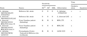
Spleen-derived amastigotes of the drug-sensitive laboratory strains L. donovani (MHOM/ET/67/L82 and L. infantum MHOM/MA/67/ITMAP263 were allowed to transform to promastigotes in M199 medium at 25 °C. Promastigotes of L. infantum MHOM/FR/96/LEM3323 were obtained from Groupe Hospital-Universitaire Caremeau and Centre National de Référence des Leishmania (Dr L. Lachaud/Professor Dedet), France. L. donovani MHOM/NP/03/BPK275/0 clone 18 (SbIII/SbV resistant, clinical outcome patient = non-responder) and MHOM/NP/02/BPK282/0 clone 4 (SbIII/SbV sensitive, clinical outcome patient = definitive cure) are spleen-derived samples from Nepalese visceral Leishmania patients obtained from the Kaladrug consortium. To make the text and figures more clear, the following abbreviations were used: L. infantum ITMAP263, L. donovani L82, L. donovani BPK 275 Cl18, L. donovani BPK 282 Cl 4 and L. infantum LEM 3323 (Table 1). Infection of macrophages is microscopically assessed on Giemsa-stained preparations.
Overview of the Leishmania strains used, the source, drug sensitivity and abbreviation

Macrophages
The murine (BALB/c mouse) macrophage-like cell line RAW 264·7 was grown at 37 °C and 5% CO2 in DMEM supplemented with 10% iFCS. Primary peritoneal mouse macrophages (PMM) were recruited by intraperitoneal administration of 2% (m/v) aqueous starch dispersion to Swiss CD-1 mice (Elevage Janvier, France). Peritoneal macrophages were harvested 48 h later and grown in RPMI-1640 supplemented with 2% penicillin/streptomycin and 10% iFCS. Cells are maintained in tissue culture flasks or multiwell plates at 37 °C and 5% CO2.
Detection of free radicals with EPR
The EPR method for the detection of the overall O2●− and NO production in infected and non-infected macrophages was used as described earlier (Deschacht et al. Reference Deschacht, Horemans, Martinet, Bult, Maes and Cos2010). PMM were grown in 24-well plates (Greiner) at 1 × 10⁶ cells/well 2 days prior to the test. After 48 h, the cells were infected with stationary phase promastigotes at a 10/1 parasites/macrophage ratio. This ratio was always used except when stated explicitly. All EPR measurements were performed in Krebs HEPES buffer (pH 7·4). After an infection of 5 min up to 48 h and 50 min incubation with the O2●− spin probe CM-H (37 °C, 5% CO2), 50 μl of supernatant was sampled into a capillary tube for measurement at 37 °C. In the stimulation experiments, 10 μM PMA was added 5 min before adding the spin probe. For NO experiments, RAW 264.7 cells were seeded in 6-well plates (Greiner) at 0·5 × 106 cells/well and infected 2 days later with stationary-phase promastigotes at a 10/1 parasite/macrophage ratio. At 6, 24 and 48 h post-infection, cells were incubated with [Fe(DETC)2] for 1 h before harvesting. After removal of the supernatant, re-suspended cells were brought in a liquid nitrogen Dewar (Magnettech, Germany). Instrument settings were 10 mW of microwave power, 5 G of amplitude modulation, 100 kHz of modulation frequency and 80 G sweep width. In the stimulation experiments, 100 ng/ml lipopolysaccharide (LPS) and 5 ng/ml interferon-gamma (INFγ) were added simultaneously with infection of the macrophages. Data are expressed as Delta Y values, which represent the peak height of the EPR signal.
Detection of nitrite with the Griess reaction
A Griess reagent kit (Invitrogen G-7921) was used for the determination of extracellular nitrite in macrophages. After infection, the supernatant was transferred to a 96-well plate (150 μl/well). After addition of 20 μl of Griess reagent and 130 μl of demineralized water, samples were incubated for 30 min and absorbance was measured at 550 nm (Labsystems Multiskan MCC/340). A standard calibration curve was set up by diluting the nitrite standard of the kit.
Detection of apoptosis with Annexin V-FITC labelling
PMM or RAW 264.7 cells were seeded in Lab-Tek chamber slides (Nunc, 178599) at a concentration of 50 000 cells/well for PMMs and 10 000 cells/well for RAW 264.7 macrophages. After 24 or 48 h, cells were infected with stationary-phase promastigotes (10/1 parasite/macrophage ratio) of the different Leishmania strains (Table 1). FITC-linked Annexin V (Becton Dickinson 556420) was used to detected apoptotic cells. Propidium iodide (PI) (maximal final concentration of 0·4 μM) was included in the protocol to discriminate between cells in early apoptosis and cells undergoing late apoptosis or necrosis. Cycloheximide (CHX) at 30 μg/ml was included as positive control. Cells were evaluated using fluorescence microscopy (Zeiss Observer ZI) with a 488 nm filter for FITC detection and a 640 nm filter for PI detection. For each parameter, at least 50 cells were counted. Results were represented as the mean±s.e.m. of 3 independent experiments.
Western blotting
Sample preparation
After infection, 1 × 10⁶ cells were lysed in an appropriate volume of 1/20 β-mercaptoethanol/Laemmli buffer (Bio-Rad Laboratories) solution. Samples were heated for 5 min at 100 °C and stored at −80 °C. Procaspase-3 and the largest fragment of cleaved procaspase-3 were detected by a standard Western blot procedure. Samples (30 μl) were loaded on a 4–20% Mini-PROTEAN® TGX™ Precast Gel (Bio-Rad). After gel electrophoresis, proteins were transferred to an Immobilon-P Transfer membrane (Millipore Corporation) according to standard procedures.
Caspase-3 detection
Samples were blocked with 5% non-fat dry milk (Bio-Rad) and then incubated overnight at 4 °C with primary antibody (1:500) (monoclonal rabbit caspase-3 antibody, Cell Signaling 9662) in antibody dilution buffer (TBS-T containing 1% nonfat dry milk). Membranes were washed and incubated with the peroxidase-conjugated secondary antibody (1:1000) (polyclonal swine anti-rabbit immunoglobulins/HRP, Dako P0399) for 1 h at room temperature. Antibody detection was accomplished with SuperSignal West Femto chemiluminescent substrate (Thermo Scientific 34096) using a Lumi-imager (VWR GenoPlex).
Stripping for β-actin detection
Membranes were stripped by incubation in an appropriate strip buffer for 30 min. The buffer contains 1 g/l SDS, 15 g/l glycine, 10 ml of Tween 20 and ultrapure water to make 1 litre. After a washing and a blocking step, membranes were incubated for 1 h with a monoclonal mouse anti-β-actin antibody (1:10 000) (clone AC-15, Sigma Aldrich A5441). Membranes were then incubated with polyclonal rabbit anti-mouse immunoglobulins/HRP (1:10 000) (Dako P0260) for 1 h. Antibody detection was accomplished with SuperSignal West Pico chemiluminescent substrate (Thermo Scientific 34096) using a Lumi-imager (VWR GenoPlex).
STATISTICAL ANALYSIS
All results were expressed as the mean±s.e.m. of at least 3 independent experiments. Statistical analyses were carried out with SPSS PASW Statistics 18 software. The statistical tests used in the present study are noted in the figure legends. P < 0·05 was considered statistically significant. For data that were not evenly distributed, statistic analyses were done on logarithmic transformed data.
RESULTS
Superoxide response of PMMs after Leishmania infection
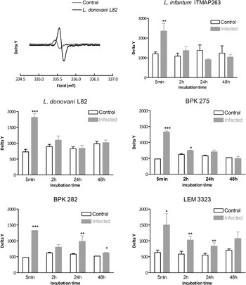
Reaction of the spin probe CM-H with O2●− leads to the formation of the nitroxide radical CM●, which can easily be measured with EPR. To investigate the effect of Leishmania infection on the production of O2●− in macrophages, nitroxide radical (CM●) levels were measured in the supernatants of infected PMMs at 5 min, 2 h, 24 h and 48 h compared to non-infected cells. For all strains, our results demonstrated that the macrophage O2●− levels were increased 5 min after infection and then gradually decreased to basal levels at 48 h post-infection (Fig. 1). Despite some marginal differences between the various species and strains, no biologically relevant differences between the different species and strains with different drug susceptibility were discerned. Subsequently, the effect of Leishmania infection on O2●− levels was investigated in PMA-stimulated macrophages (10 μM, added 5 min post-infection). Comparable to non-stimulated macrophages, all Leishmania strains evoked a similar O2●− production response in PMA-stimulated macrophages: O2●− levels significantly increased 5 min after infection and then gradually decreased to PMA-stimulated control levels at 48 h post-infection (data not shown).
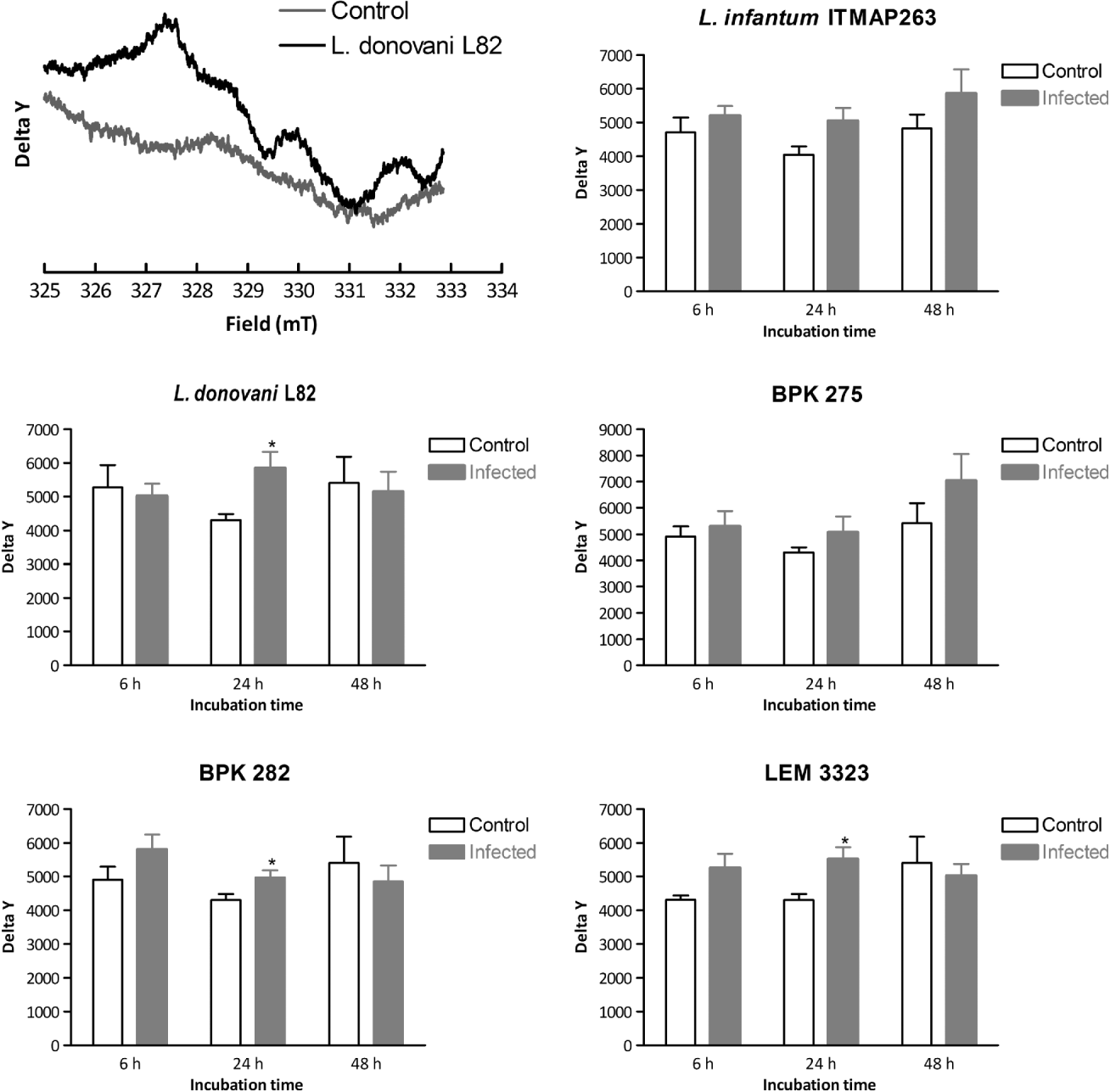
Effect of infection with different Leishmania strains on O2●− production in PMM. Delta Y values (a representative example of the middle peak of the 3-peak CM● signal is given in the first figure and was used to calculate Delta Y values) represent the peak height of nitroxide radical CM● which is formed after the reaction of O2●− with the spin probe CM-H. At each time-point, infected macrophages were compared with non-infected macrophages. For all tested strains, infection led to a significant increase 5 min after infection (independent t-test, n ⩾ 3, ***P <0·001, **P <0·01, *P <0·05).

Nitric oxide response of RAW 264.7 after Leishmania infection
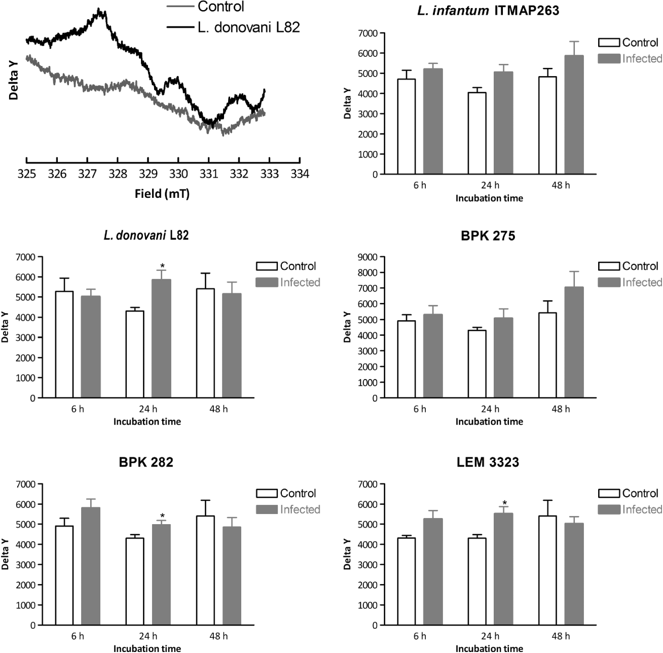
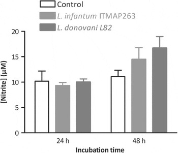
NO was measured as the spin adduct NO-[Fe(DETC)2]. Because a higher number of cells was required compared to the O2●− experiments, a macrophage cell line was selected instead of PMMs. RAW 264.7 cells were infected with stationary phase promastigotes and measurements took place in liquid nitrogen at 6 h, 24 h and 48 h post-infection. At 24 h, a significant increase in NO production was observed for all strains except L. infantum ITMAP263 and L. donovani BPK 275 (Fig. 2). At 48 h, NO production returned to the control level. When nitrite instead of NO was measured, no significant increase could be detected (Fig. 3). Like for O2●−, the effect of infection on NO production was investigated in stimulated macrophages. RAW 264.7 cells were stimulated with 100 ng/ml LPS and 5 ng/ml INFγ and 6 h, 24 h and 48 h post-infection with 2 different Leishmania strains (Table 1); NO was measured as NO-[Fe(DETC)2] complex. The NO response was similar to that of stimulated control cells at 6 h post-infection. The infection led to a small non-significant increased NO production after 24 h, which persisted until 48 h (data not shown).
Effect of infection with different Leishmania strains on NO production in RAW 264.7 macrophages. Delta Y values represent the peak height of the NO-[Fe(DETC)2] complex which is formed after the reaction of NO with the spin trap Fe(DETC)2 (example of Delta Y is given in the first figure, independent t-test, n ⩾ 3, *P <0·05). At each time-point, infected macrophages were compared with non-infected macrophages.
Nitrite levels measured with the Griess reaction after infection of RAW 264.7 macrophages. There was no significant difference between infected and control cells (n = 3, one-way ANOVA with Dunnett's post-hoc test).

Apoptotic cell death in macrophages after Leishmania infection
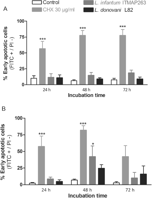
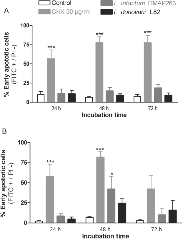
PMMs or RAW 264.7 cells were infected with L. infantum ITMAP 263 or L. donovani L82. After 24 h, 48 h or 72 h infection, the occurrence of apoptosis was checked with the Annexin V-FITC protocol (Fig. 4). Only L. infantum ITMAP263 in RAW 264.7 macrophages caused a significant increase in Annexin V-FITC positive cells at 48 h (Fig. 4). There was also a small increase in FITC and PI (=late apoptotic) positive RAW 264.7 cells 72 h after infection with L. infantum ITMAP263 or L. donovani L82.
Percentage of Annexin V-FITC positive cells in control, CHX treated (30 μg/ml), Leishmania donovani L82 or L. infantum ITMAP263 infected (A) PMMs or (B) RAW 264.7 (10:1 parasite:macrophage ratio). At least 100 cells were counted and results are given as the average±s.e.m. of at least 3 independent experiments. There was no significant difference between control and infected cells, except for RAW 264.7 cells 48 h post-infection with L. infantum 67 (one-way ANOVA with Dunnett's multiple comparison post hoc).
Caspase-3 detection in infected cells
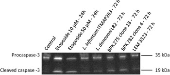
RAW 264.7 cells were infected with different Leishmania strains for 24 h, 48 h or 72 h. After 72 h, a marginal cleavage of procaspase-3 was observed for all tested strains compared to the etoposide control (Fig. 5).
Cleavage of procaspase-3 after 72 h infection of RAW 264.7 with different Leishmania strains. Etoposide was used as positive control (10 or 50 μM, 24 h incubation). Cleavage of procaspase-3 was analysed using Western blotting and β-actin was used as loading control.

DISCUSSION AND CONCLUSION
O2●− is a free radical that plays a major role during phagocytosis (Channon et al. Reference Channon, Roberts and Blackwell1984; Gantt et al. Reference Gantt, Goldman, McCormick, Miller, Jeronimo, Nascimento, Britigan and Wilson2001). In the literature, inhibition of O2●− production by the amastigote stage is described for L. mexicana and L. major (Kantengwa et al. Reference Kantengwa, Muller, Louis and Polla1995; Pham et al. Reference Pham, Mouriz and Kima2005). In these experiments, extracellular O2●− production was determined by reduction of cytochrome c and intracellular O2●− by the nitroblue tetrazolium (NBT) assay. In the present study, O2●− production was measured using EPR. The use of spin traps and probes with EPR allows the sensitive quantification of the actual free radical and not its metabolite (such as the Griess reaction which measures nitrite, a metabolite of NO) or secondary product (such as the measurement of reduced cytochrome C by O2●−). Moreover, with the EPR technique O2●− production can be measured at different time-points. Our measurements showed that phagocytosis of Leishmania promastigotes elicits a general burst of O2●− in macrophages, while during amastigote multiplication later in the infection O2●− production is brought back to the basal level. This is in accordance with the above-mentioned reports for L. mexicana and L. major (Kantengwa et al. Reference Kantengwa, Muller, Louis and Polla1995; Pham et al. Reference Pham, Mouriz and Kima2005) and can be explained by the interference with NADPH oxidase assembly in the phagosomal membrane (Lodge et al. Reference Lodge, Diallo and Descoteaux2006; Lodge and Descoteaux, Reference Lodge and Descoteaux2006). After phagocytosis, promastigotes rapidly transform into amastigotes triggered by the acidic environment in the phagolysosome. Whereas the macrophage predominantly produces O2●− in an attempt to kill the parasite during phagocytosis, NO becomes increasingly important as a defence mechanism during the intracellular amastigote stage. The induction of iNOS explains why NO does not play a role during phagocytosis (Wang et al. Reference Wang, Zhao, Matta, Meng, Liu, Liu, Nelin and Liu2009). Our results showed that infection leads to an increase in NO production 24 h post-infection, after which the NO levels decreased to basal levels. Conversely, the nitrite response as detected with the Griess reaction reached a maximum 48 h post-infection. This can be explained by the fact that the Griess reaction measures an accumulation of the NO metabolites nitrite and nitrate and not the actual NO production at the measured time-point. This nicely demonstrates the added value of the EPR technique to monitor the NO production during Leishmania infection compared to the Griess reaction.
According to the literature, NO production seems to depend on the type of host cell. An increase in NO production was observed in macrophages (PMMs and RAW 264.7) and confirms previously published results for other macrophages and Leishmania strains, such as J774 and PMMs upon infection with L. major (Green et al. Reference Green, Crawford, Hockmeyer, Meltzer and Nacy1990; Cunha et al. Reference Cunha, Assreuy, Moncada and Liew1993). In contrast, this effect was not observed in dog monocytes infected with L. infantum (Panaro et al. Reference Panaro, Lisi, Mitolo, Acquafredda, Fasanella, Carelli and Brandonisio1998), which endorses the importance of the host cell on ROS production. For both O2●− and NO, the response of PMMs after infection was very similar between the different Leishmania species (L. infantum vs L. donovani) and strains (drug-sensitive vs -resistant). Based on literature, it can be hypothesized that SbIII -resistant strains show less ROS production by host macrophages in comparison with SbIII -sensitive strains. Indeed, different Sb-resistant strains have been reported to preserve elevated trypanothione [T(SH)2] levels (Mukhopadhyay et al. Reference Mukhopadhyay, Dey, Xu, Gage, Lightbody, Ouellette and Rosen1996; Haimeur et al. Reference Haimeur, Brochu, Genest, Papadopoulou and Ouellette2000). Thiol-depletion of these strains re-established their susceptibility to SbIII (Mandal et al. Reference Mandal, Wyllie, Singh, Sundar, Fairlamb and Chatterjee2007), indicating that the increased T(SH)2 levels are causing resistance. Other antioxidant enzymes were also overexpressed in Sb-resistant strains, such as tryparedoxin and tryparedoxin peroxidase (Wyllie et al. Reference Wyllie, Vickers and Fairlamb2008, Reference Wyllie, Mandal, Singh, Sundar, Fairlamb and Chatterjee2010). Consequently, it could be expected that the increased antioxidant capacity of the Sb-resistant strain would affect the free radical levels in the macrophages. However, our results did not show a significant difference in NO or O2●− levels between SbIII/SbV sensitive (L. donovani BPK 282 Cl4) and resistant (L. donovani BPK 275 Cl18) strains, which suggests that the effects on thiol levels do not lead to a decrease in NO and O2●− availability during infection. However, it is still possible that the NO and O2●− levels are only locally decreased in the phagolysosomes, containing the Sb-resistant amastigotes and not elsewhere in the macrophage.
A lot of research has been performed on the interaction of Leishmania with NADPH oxidase (Van Assche et al. Reference Van Assche, Deschacht, Inocêncio da Luz, Maes and Cos2011). Amastigotes, but also promastigotes inhibit NADPH oxidase (Kumar et al. Reference Kumar, Pai and Sundar2001, Reference Kumar, Pai, Pandey and Sundar2002; Lodge et al. Reference Lodge, Diallo and Descoteaux2006). It was also demonstrated that O2●− and H2O2 concentrations in monocytes from patients with visceral leishmaniasis were significantly lowered (Kumar et al. Reference Kumar, Pai and Sundar2001). To investigate whether this NADPH oxidase inhibition also affects the O2●− response, PMA-stimulated macrophages were infected with different Leishmania strains. Compared to the controls, the O2●− response was decreased after 2 h. There was a difference in O2●− response dependent on the strain: L. infantum ITMAP263 infection resulted in the highest decrease in O2●− production in PMA-stimulated macrophages showing that Leishmania infection can inhibit the O2●− production in activated PMMs. This is one possible reason why the parasite is able to survive inside macrophages. Whether this inhibition effectively leads to an increase in parasite survival needs further investigation.
Compared to NADPH oxidase activity and O2●− inhibition, there is less known about the effects of infection on NO production. Phagosomal amastigotes are known for their capacity to decrease NO production in infected macrophages: the NO production in LPS-stimulated cells significantly decreased after infection with Leishmania (Wilkins-Rodriguez et al. Reference Wilkins-Rodriguez, Escalona-Montano, Aguirre-Garcia, Becker and Gutierrez-Kobeh2010). As for the inhibition of NADPH oxidase, down-regulation of NO production after Leishmania infection can be explained by lipophosphoglycan (LPG) activity. Treatment of macrophages with LPG inhibits NO synthesis in a time- and dose-dependent manner. These data clearly demonstrate that LPG is able to regulate the iNOS expression in macrophages (Proudfoot et al. Reference Proudfoot, Schneider, Ferguson and McConville1995, Reference Proudfoot, Nikolaev, Feng, Wei, Ferguson, Brimacombe and Liew1996; Bogdan and Rollinghoff, Reference Bogdan and Rollinghoff1998). The leishmanicidal activity in LPG-treated J774 macrophages was reduced compared to untreated control cells (Proudfoot et al. Reference Proudfoot, Nikolaev, Feng, Wei, Ferguson, Brimacombe and Liew1996). When bone marrow-derived dendritic cells (BMDCs) were stimulated with LPS and IFNγ, infection with L. mexicana amastigotes led to a down regulated NO production (Wilkins-Rodriguez et al. Reference Wilkins-Rodriguez, Escalona-Montano, Aguirre-Garcia, Becker and Gutierrez-Kobeh2010). The interference with this cytotoxic mechanism was not sufficient to permit the survival of L. mexicana after 48 h of infection. In accordance to previous studies (Cunha et al. Reference Cunha, Assreuy, Moncada and Liew1993; Panaro et al. Reference Panaro, Brandonisio, Sisto, Acquafredda, Leogrande, Fumarola and Mitolo2001; Shweash et al. Reference Shweash, Adrienne, Schroeder, Neamatallah, Bryant, Millington, Mottram, Alexander and Plevin2011; Coelho-Finamore et al. Reference Coelho-Finamore, Freitas, Assis, Melo, Novozhilova, Secundino, Pimenta, Turco and Soares2011), our results demonstrate that infection with Leishmania in macrophages did not significantly decrease the NO production in stimulated cells.
Our results show some signs of apoptosis at 48 h (PS externalization) and 72 h (PS externalization and cleavage of procaspase-3) post-infection. However, these effects were marginal and the consequence on the infection itself is uncertain. Induction of apoptosis in human monocyte-derived macrophages (THP-1 and U937) and peripheral blood mononuclear cells (PBMC) after Leishmania infection has already been described (Getti et al. Reference Getti, Cheke and Humber2008). Our results did not confirm these observations although the same techniques, incubation times and infection rates were used, suggesting that the effect of infection on host cell apoptosis may be dependent on host cell type and/or Leishmania strain.
In contrast to our experiments, many investigators studied the effects of infection after chemically induced apoptosis in host cells. In these experiments, the effect seems to be parasite stage-dependent. Early stages of infection (within 24 h) showed an inhibitory effect on induced apoptosis, while intracellular amastigotes showed induction of apoptosis at late stages of infection (up to 72 h post-infection) (Moore and Matlashewski, G. Reference Moore and Matlashewski1994; Akarid et al. Reference Akarid, Arnoult, Micic-Polianski, Sif, Estaquier and Ameisen2004; Lisi, S. et al. Reference Lisi, Sisto, Acquafredda, Spinelli, Schiavone, Mitolo, Brandonisio and Panaro2005; Ruhland, A. et al. Reference Ruhland, Leal and Kima2007; Getti, G. T. et al. Reference Getti, Cheke and Humber2008). Although our results show a small increase in early apoptotic cells (Annexin V-FITC test) and caspase-3 cleavage in PMMs and RAW 264.7 cells after infection, none of the effects was statistically significant.
In conclusion, it can be stated that, for the first time, two important cellular macrophage defence systems were tested on the intracellular amastigote stage of a range of Leishmania species and antimony-resistant strains. Our results clearly demonstrate that O2●− is important during phagocytosis of the promastigotes while NO becomes important later during infection. Our tests only showed some marginal effect of infection on apoptosis, questioning the role of apoptosis in infection rate or virulence of Leishmania.
FINANCIAL SUPPORT
This work was supported by a grant from the University of Antwerp (GOA no. 2407) and Kaladrug-R FP7-HEALTH-2007-B (grant number 222 895). P.C. was a Post-Doctoral researcher awarded a grant by the Fund for Scientific Research (FWO) – Flanders (Belgium).