


Highlights
-
• Other than chorea and dystonia, which are common motor manifestations of chorea-acanthocytosis (ChA), oculomotor impairment and parkinsonism are also observed.
-
• Olfactory function is significantly impaired in patients with ChA.
-
• In addition to mood disorders and OCD, approximately 20% of the patients reported suicidal thoughts.
Introduction
Chorea-acanthocytosis (ChA) is an ultra-rare, progressive, autosomal recessive neurodegenerative disorder with an estimated prevalence of one per million individuals. The disease results from mutations in VPS13A, the gene on chromosome 9q21 that encodes chorein. Reference Walker, Peikert, Jung, Hermann and Danek1 Although the precise molecular structure and cellular function of VPS13A remain incompletely understood, chorein deficiency leads to selective putamen and caudate nucleus degeneration, two critical regions of the basal ganglia that play a crucial role in motor regulation. In addition, ChA involves extensive neuronal damage, including medium spiny neurons, striatal interneurons, dopaminergic neurons and motor neurons. Reference García-García, Chaparro-Cabanillas and Coll-Manzano2
Clinically, ChA presents with a combination of motor and non-motor manifestations. Motor symptoms commonly include chorea, orofacial dyskinesia, dystonia and occasionally parkinsonism. Seizures, cognitive impairment and psychiatric disturbances are among the non-motor features. Reference Lin, Meng and Shafeng3 Nevertheless, detailed investigation into certain aspects of the disease remains limited.
The Unified Huntington’s Disease Rating Scale (UHDRS) is a comprehensive clinical tool designed to assess motor function, cognition, behavioral abnormalities and functional capacity in Huntington’s disease (HD). 4 However, due to the clinical similarity between HD and ChA Reference Schneider and Bird5,Reference Suzuki, Sato and Ota6 and the absence of a disease-specific rating scale for ChA, the UHDRS has also been employed in ChA. Reference Liu, Liu and Wan7,Reference Miquel, Spampinato and Latxague8 The UHDRS cognitive component includes only three tests, focusing on executive and psychomotor functions; to perform a broader evaluation of cognition, the Mini-Mental State Examination (MMSE) has been used in a number of previous studies. Reference Chen, Zhang, Wang and Zhang9,Reference Huang, Zhang, Tao, Lv, Xu and Liang10
Olfactory dysfunction has emerged as a potential early biomarker in several neurodegenerative disorders, such as Parkinson’s disease (PD), Alzheimer’s disease (AD), HD and Neurodegeneration with Brain Iron Accumulation (NBIA) syndromes. Reference Amini, Rohani and Habibi11–Reference Chowdhury, Das, Mishra, Jayashree and Bodakhe14 Given this, and the prominent basal ganglia and frontostriatal involvement in ChA, Reference García-García, Chaparro-Cabanillas and Coll-Manzano2 we hypothesized that olfactory impairment may also be present in ChA. To date, no data on olfactory function in ChA patients are available.
The present study aimed to conduct a multidimensional evaluation of ChA patients, with a specific emphasis on underexplored domains, including ocular motor abnormalities, psychiatric symptoms, cognitive deficits and olfactory dysfunction. Through a systematic assessment of these domains, we aimed to broaden the understanding of the clinical spectrum of ChA and clarify how its motor and non-motor features may interact.
Methods
Study design and participants
This study employed a cross-sectional observational design with a case–control component for olfactory evaluation. Twenty-eight patients diagnosed with ChA who attended our movement disorders clinic in a tertiary hospital from January 2022 to December 2024 were asked to participate in the study. Those who completed all intended assessments during the study period were enrolled. Demographic and clinical data were obtained from medical records and standardized examinations (described in the following sections) performed at each patient’s most recent visit. Olfactory function was then compared between these patients and an age-and gender-matched control group (1:1) recruited by convenience sampling from hospital staff without a history of neurological, psychiatric and olfactory disorders.
Inclusion criteria for patients included a confirmed diagnosis of ChA by whole-exome sequencing, no other identified pathogenic or likely pathogenic mutations except in the VPS13A gene, aged 18 years or older and the ability to complete the study assessments independently or with the assistance of a caregiver. Patients were required to have no history of concurrent neurodegenerative disorders or head injury with frontal damage. To minimize cognitive confounding, patients with cognitive impairment severe enough to preclude valid testing were excluded from olfactory evaluation.
All participants or their legally authorized representatives provided informed consent. The study protocol complied with the Helsinki Declaration (1975/2008) and was reviewed and approved by the institutional ethics committee (IR.IUMS.FMD.REC.1399.350).
Motor assessment (UHDRS Part 1)
Motor function was evaluated using all 31 items of the UHDRS motor subscale, with each item scored on a standardized 0–4 scale (0 = normal/no impairment; 4 = severe impairment). The total motor score (TMS) was calculated by summing all items (range: 0–124), with higher scores indicating greater motor impairment. 4 Specifically, the clinical features assessed included choreic movements of the mouth, face, trunk and upper and lower limbs, dystonic movements of the trunk and extremities and ocular motor functions encompassing saccade initiation, saccade velocity and smooth pursuit in both horizontal and vertical directions. Parkinsonism was defined as the presence of bradykinesia, rigidity, impaired finger tapping and/or slowness during hand pronation-supination; to increase specificity, scores of 1 (indicating mild impairment) were not considered. Vocal and motor tics were also documented.
Behavioral assessment (UHDRS Part 3)
The behavioral subscale assessed 10 neuropsychiatric domains through a structured interview, including anxiety, irritability, aggression, obsessive-compulsive disorders (OCD), psychotic symptoms (delusions and hallucinations), sad mood, low self-esteem and suicidal thoughts. Each domain was rated for frequency and severity on a scale of 0–4. The score of each domain was calculated by multiplying the frequency by the severity (range per domain: 0–16). The total behavioral score (TBS) was the cumulative sum of all domain scores (0–160), with higher scores indicating worse behavioral impairment. 4
Functional assessment (UHDRS Part 4)
The UHDRS functional assessment consists of three sections: the functional assessment scale (FAS), the independence scale (IS) and the total functional capacity (TFC). The FAS consists of 25 yes/no questions that provide a detailed picture of the patient’s daily living abilities. Each “yes” response earns one point, yielding a total FAS score of 0 to 25. The IS, a single-item rating, is scored from 10 to 100 in 10-point intervals based on the clinician’s overall judgment of the patient’s level of independence in daily life. The TFC comprises five domains: three (activities of daily living, finances and occupation) are rated on a scale of 0–3, while the remaining two (level of care and household chores) are rated on a scale of 0–2. The scores from these domains are totaled to create a score from 0 to 13. Higher FAS, IS and TFC scores indicate a better functional capacity. 4
Cognition assessment
Two neurologists independently administered the linguistically adapted MMSE to assess patients’ cognitive function. Reference Ansari, Naghdi, Hasson, Valizadeh and Jalaie15 The MMSE consists of 11 items evaluating several cognitive domains, including language, memory, attention/calculation, orientation and, to a lesser extent, executive function. Reference Folstein, Folstein and McHugh16 The test has a maximum score of 30, and scores below 24 are considered indicative of cognitive impairment. Reference Ansari, Naghdi, Hasson, Valizadeh and Jalaie15
Olfactory assessment
Olfactory function was assessed using the culturally adapted Sniffin’ Sticks test, following the protocol described in previous studies. Reference Amini, Rohani and Jalessi13,Reference Kamrava, Hosseini and Farhadi17 This test comprises three subtests: odor threshold (OT), odor discrimination (OD) and odor identification (OI), along with a total score derived from the sum of these subtests (TDI). Reference Hummel, Sekinger, Wolf, Pauli and Kobal18 TDI scores of less than 30.75 were classified as hyposmia. Furthermore, each olfactory subtest was compared to the normative data that specifically matched for gender and age to assess olfaction (scores above the 10th percentile for each subtest were categorized as normosmia). Reference Oleszkiewicz, Schriever, Croy, Hähner and Hummel19
Statistical analysis
Data are presented as means and standard deviations for quantitative variables and as frequencies and percentages for categorical variables. The statistical analyses were performed using both parametric and nonparametric tests as appropriate. Pearson’s correlation coefficient was used to assess relationships between normally distributed continuous variables. To estimate the precision of the correlations, 95% confidence intervals (CI) were obtained using bias-corrected and accelerated (BCa) bootstrap resampling with 5,000 samples. Multiple testing in correlation analyses was controlled using the Benjamini–Hochberg false discovery rate (FDR) procedure (q = 0.05). Categorical variables were compared between groups using the Chi-square or Fisher’s exact test, as appropriate. Continuous variables were evaluated using either the independent samples t-test for normally distributed data or the Mann–Whitney U test for non-normally distributed data.
To address potential non-independence due to family clustering, we conducted a cluster-level resampling sensitivity analysis. One participant per family (16 families in total) was randomly selected, and correlations were recomputed across 1000 iterations. Full-sample correlations (N = 23) were compared with the empirical distribution of the resampled estimates. Robustness was defined as full-sample estimates falling within the 95% percentile interval of the resampled distribution and being comparable in magnitude and direction to the mean resampled correlation coefficients.
The sensitivity analysis was performed in R using the packages dplyr, purrr and tidyr. Correlation patterns are also visualized as a heatmap of the correlation matrix, created with the ggplot2 package in R version 4.5.2. All other analyses were performed using IBM SPSS Statistics version 26, with p-values ≤ 0.05 considered statistically significant.
Results
Demographic and clinical characteristics of participants with ChA
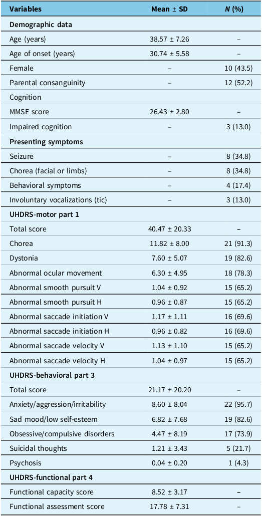
A total of 23 genetically confirmed ChA patients from 16 families were enrolled. Detailed genetic variants are presented in Supplementary Table 1. Parental consanguinity was observed in over half of the cohort, and 14 patients (60.9%) had at least one sibling involved. The male-to-female ratio was 1.3, and the mean disease duration was 7.86 ± 6.04 years. Approximately one-third of the patients initially presented with chorea or seizure, which increased to 91.3% and 60.9% on their last visit, respectively. Vocal tics were initially reported in 13% of the patients, increasing to 65.2% as the disease progressed. Motor tics presented as eye blinking in only three patients. Demographic data and disease features are presented in Table 1. The pharmacological history, as demonstrated in Supplementary Table 2, indicated that 56.5% (n = 13) of cases were receiving neuroleptics, while 82.6% (n = 19) were on anticonvulsant regimens, including five patients (21.7%) who were taking them at mood-stabilizing doses at the time of assessment. Fifteen (65.2%) patients were receiving sodium valproate, and nine (39.1%) were receiving tetrabenazine.
Demographic and clinical characteristics of patients with chorea-acanthocytosis

Table 1. Long description
This is a three-column table with rows grouped into thematic sections, reporting descriptive statistics for 23 patients with chorea-acanthocytosis. The columns are labeled Variables, Mean ± SD, and N (%). Rows are organized into five sections: demographic data, cognition, presenting symptoms, UHDRS-Motor part 1, UHDRS-Behavioral part 3, and UHDRS-Functional part 4.
-
• Demographic variables include age, age at disease onset, sex, and parental consanguinity. Mean age was 38.57 years and mean age at onset was 30.74 years. Ten patients (43.5%) were female, and 12 (52.2%) had parental consanguinity.
-
• Cognitive assessment includes MMSE score and impaired cognition. Mean MMSE score was 26.43, with impaired cognition in 3 patients (13.0%).
-
• Presenting symptoms included seizure and chorea in 8 patients each (34.8%), behavioral symptoms in 4 (17.4%), and involuntary vocalizations in 3 (13.0%).
-
• Motor assessment showed a mean UHDRS total motor score of 40.47. Chorea was present in 21 patients (91.3%), dystonia in 19 (82.6%), and abnormal ocular movements in 18 (78.3%). Smooth pursuit and saccadic abnormalities were frequent, affecting approximately 65-70% of patients.
-
• Behavioral assessment showed a mean UHDRS total behavioral score of 21.17. Anxiety, aggression, or irritability were most common, affecting 22 patients (95.7%), followed by sad mood or low self-esteem in 19 (82.6%), obsessive-compulsive symptoms in 17 (73.9%), suicidal thoughts in 5 (21.7%), and psychosis in 1 (4.3%).
-
• Functional assessment showed mean functional capacity and assessment scores of 8.52 and 17.78, respectively.Overall, the table shows that patients commonly exhibit prominent motor abnormalities, frequent psychiatric or behavioral symptoms, and mild cognitive impairment.
SD = standard deviation; N = number; MMSE = Mini-Mental State Examination; UHDRS = Unified Huntington’s Disease Rating Scale; V = vertical; H = horizontal.
Two patients exhibited atypical phenotypic trajectories. One patient primarily presented with chorea, but after 16 years, chorea improved spontaneously, and the patient gradually developed parkinsonism ((VPS13A (NM_033305.3):c.644A>G (p.Tyr215Cys)) and (VPS13A (NM_033305.3):c.8341del (p.Ser2781Hisfs*2))). The other case, who had had seizures for 13 years, showed parkinsonian features as her first motor symptoms (VPS13A (NM_033305.3):c.3499_3503del (p.Ser1167Ilefs*6)) (Patients 9 and 16, respectively, as indicated in Supplementary Table 1).
Motor and functional profiles
The most prevalent motor symptom in the UHDRS questionnaire was chorea (including limb, truncal, facial and orobuccolingual). Dystonia and eye movement abnormalities were also common. Impairment of saccadic initiation and velocity was more prominent than disturbed smooth pursuit eye movement, and both were more affected in the vertical direction than the horizontal direction. However, five patients exhibited normal eye movements. Eating dystonia and lip and tongue biting were seen in 15 (65.2%) patients. According to the study definition (scoring >1 on at least one parkinsonian feature), two patients (8.7%) met criteria for parkinsonism, with bradykinesia scores of 3 and 4. In contrast, nine patients exhibited only mild signs (score = 1) and were not classified as having parkinsonism. Among these nine patients, six were receiving tetrabenazine. Supplementary Table 2 summarizes medication use in this subgroup.
Among functional measures, the mean IS score was 73.48 ± 21.02 (range: 40–100); 3 patients scored 100, 8 cases scored 90, and the remaining 12 cases (52.2%) had lower scores. The prominent findings of the UHDRS, Parts 1, 3 and 4, are summarized in Table 1. Domain-specific UHDRS results, including subscale scores and individual items, are detailed in Supplementary Table 3.
Behavioral profile and cognitive performance
The median TBS score was 16 (interquartile range = 8–32), with 95.7% and 87.0% of patients scoring less than 80 and 40, respectively. Behavioral assessments revealed that all the patients exhibited at least one behavioral disorder, with anxiety being the most common symptom (95.7%). The second most common symptom was mood disorder (including sad mood and low self-esteem), followed by OCD. Suicidal thoughts were present in 21.7% of the patients. According to the MMSE, 20 patients (87%) had normal cognition. Independent samples t-test showed that mean MMSE, IS and TBS scores were significantly higher in patients with suicidal ideation than those without (MMSE: p = 0.004, Hedges’ g = 1.059; IS: p = 0.020, Hedges’ g = 0.929; and TBS: p = 0.001, Hedges’ g = 2.047). Thorough behavioral evaluation results are presented in Supplementary Table 3.
Olfactory function and case–control comparison
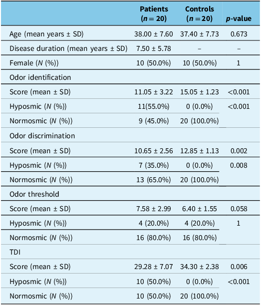
The olfactory assessment was conducted in 20 patients (3 were excluded due to profound cognitive impairment). There were no active nasal/sinus pathologies, history of recent respiratory tract infections (within the month prior to the assessment), chronic rhinosinusitis, previous nasal or paranasal sinus surgeries, COVID-19-related hyposmia/anosmia or self-reported olfactory dysfunction in these patients. The mean TDI score was 29.28 ± 7.07, and 50% of patients had hyposmia, as indicated by a TDI score < 30.75. The TDI (Glass’ ∆ = 2.098), OI (Cohen’s d = 1.626) and OD (Cohen’s d = 1.111) scores showed statistically significant differences in ChA patients compared to the gender-and age-matched controls. However, no significant difference in OT scores was observed between the two groups (Cohen’s d = 0.623). The details of the eligible patients and their olfactory assessment are reported in Table 2.
Olfactory performance in patients with chorea-acanthocytosis compared to healthy controls

Table 2. Long description
This is a four-column comparative table. Columns are labeled Patients (n = 20), Controls (n = 20), and P-value, with the first column listing variables. Rows are grouped into participant characteristics and olfactory test outcomes.
Participant characteristics include age, disease duration, and sex. Groups had similar mean age (38.0 vs. 37.4 years) and identical sex distribution (50% female). Mean disease duration in patients was 7.5 years.
Olfactory outcomes are grouped into odor identification, odor discrimination, odor threshold, and TDI. Each domain includes mean score plus classification as hyposmic or normosmic.
Patients had significantly lower odor identification scores than controls (11.05 vs. 15.05, p < 0.001), with hyposmia present in 55.0% of patients and none of the controls.
Odor discrimination scores were also lower in patients (10.65 vs. 12.85, p = 0.002), with hyposmia in 35.0% of patients and none of the controls.
Odor threshold scores did not differ significantly between groups (7.58 vs. 6.40, p = 0.058), and hyposmia prevalence was identical at 20.0% in both groups.
TDI scores were significantly reduced in patients (29.28 vs. 34.30, p = 0.006). Overall hyposmia based on TDI was present in 50.0% of patients and absent in controls.
Overall, the table demonstrates impaired odor identification, odor discrimination, and TDI scores in chorea-acanthocytosis, while odor threshold is relatively preserved.
SD = standard deviation; N = number; TDI = sum of odor identification, discrimination and threshold scores.
Correlations between motor and non-motor domains
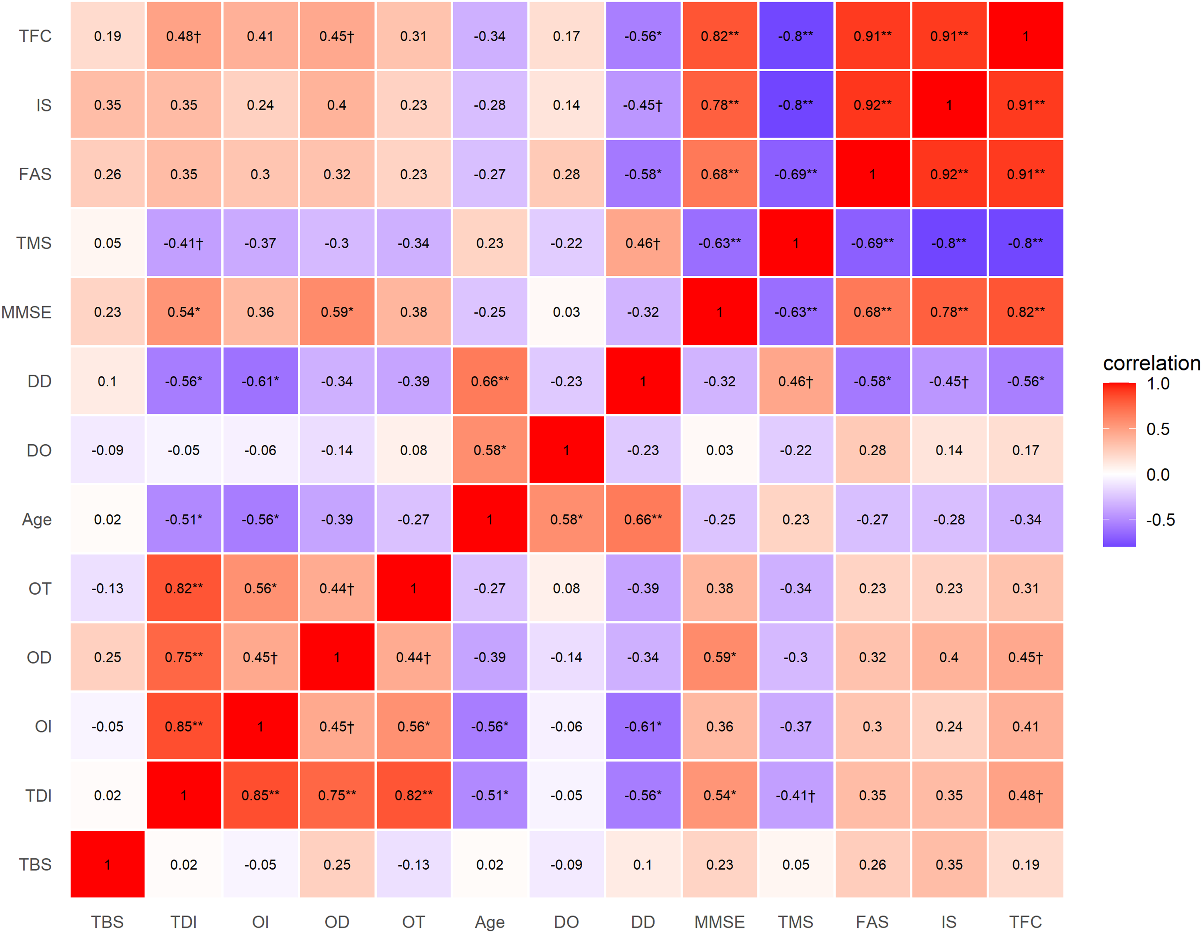
TMS was negatively correlated with TDI (r = −0.415, 95% BCa CI [−0.671, −0.075], p = 0.049), MMSE (r = −0.627, 95% BCa CI [−0.828, −0.383], p = 0.001), FAS (r = −0.693, 95% BCa CI [−0.816, −0.565], p < 0.001), IS (r = −0.800, 95% BCa CI [−0.913, −0.628], p < 0.001) and TFC (r = −0.795, 95% BCa CI [−0.892, −0.644], p < 0.001) scores. It was also correlated with disease duration (r = 0.460, 95% BCa CI [0.056, 0.753], p = 0.027).
The TFC correlated positively with TDI (r = 0.481, 95% BCa CI [0.128, 0.750], p = 0.020) and OD (r = 0.449, 95% BCa CI [0.105, 0.744], p = 0.032) scores, while negatively with disease duration (r = −0.555, 95% BCa CI [−0.798, −0.253], p = 0.006).
TBS did not show any significant correlations with cognition, motor function or olfaction components.
The analysis showed positive linear associations between MMSE scores and FAS (r = 0.675, 95% BCa CI [0.430, 0.898], p < 0.001), IS (r = 0.781, 95% BCa CI [0.579, 0.945], p < 0.001), TFC (r = 0.824, 95% BCa CI [0.671, 0.944], p < 0.001), OD (r = 0.593, 95% BCa CI [0.240, 0.838], p = 0.003) and TDI (r = 0.542, 95% BCa CI [0.231, 0.750], p = 0.008) scores.
The disease duration was negatively correlated with the scores of TDI (r = −0.562, 95% BCa CI [−0.752, −0.316], p = 0.005), OI (r = −0.610, 95% BCa CI [−0.814, −0.346], p = 0.002), FAS (r = −0.578, 95% BCa CI [−0.806, −0.297], p = 0.004) and IS (r = −0.454, 95% BCa CI [−0.717, −0.144], p = 0.030). Age was also negatively correlated with OI (r = −0.561, 95% BCa CI [−0.768, −0.270], p = 0.005) and TDI (r = −0.510, 95% BCa CI [−0.725, −0.283], p = 0.013) scores.
After FDR correction, the associations between TMS and TDI, TMS and disease duration, TFC and TDI, TFC and OD and disease duration and IS did not remain statistically significant.
Figure 1 illustrates a heatmap of the correlation pattern between motor and non-motor symptoms.
Correlation heatmap of the motor and non-motor symptoms in patients with chorea-acanthocytosis. * False discovery rate (FDR) p ≤ 0.05; ** FDR p ≤ 0.01; † p ≤ 0.05 but not significant after FDR correction. TFC = total functional capacity; IS = independence scale; FAS = functional assessment scale; TMS = total motor score; MMSE = Mini-Mental State Examination; DD = disease duration; DO = disease onset; OT = odor threshold; OD = odor discrimination; OI = odor identification; TDI = the sum of OT, OD and OI; TBS = total behavioral score.

Figure 1. Long description
This figure is a square correlation heatmap arranged as a matrix, with the same variables listed on both the horizontal and vertical axes. Each cell represents the correlation between two variables. Variables include total behavioral score (TBS), olfactory scores (TDI, odor identification, odor discrimination, and odor threshold), age, disease onset, disease duration, cognitive score (MMSE), total motor score (TMS), and functional scales (FAS, IS, and TFC). Cells are color-coded by correlation direction and strength. Red indicates positive correlation and blue indicates negative correlation, with color intensity proportional to correlation strength. Statistical significance is marked with symbols indicating corrected and uncorrected p-values. Overall, the heatmap highlights interconnected relationships between olfactory dysfunction, cognition, motor severity, and functional disability in chorea-acanthocytosis.
Sensitivity analysis considering family clustering
The sensitivity analysis demonstrated that significant full-sample correlations were robust to family clustering, except for the TMS–IS correlation. Specifically, these correlations fell within the corresponding resampled 95% percentile intervals and were comparable in both magnitude and direction to the mean resampled coefficients. In contrast, nonsignificant correlations showed greater variability; three estimates fell outside the resampled 95% percentile intervals, and several differed in magnitude from the resampled mean coefficients.
Discussion
This study aimed to present a multidimensional assessment of 23 genetically confirmed ChA patients, with particular attention to underexplored domains, including oculomotor impairment, psychiatric features and olfactory dysfunction. While chorea is commonly observed in ChA, it is a shared feature across several neurodegenerative disorders. Reference Walker20 Our findings suggest that ChA may exhibit more distinctive motor features; approximately 75% of the patients demonstrated oculomotor disturbances, affecting saccadic initiation and velocity as well as smooth pursuit, especially in the vertical plane. Similarly, Hardie et al. described 19 cases of neuroacanthocytosis (NA) syndrome, among which five patients had identified saccade and smooth pursuit abnormalities, upward gaze limitation and poor convergence; however, their study lacked detailed reporting and did not include genetic confirmation of ChA. Reference Hardie, Pullon and Harding21 Another study using magnetic search coil techniques in three ChA cases described frequent hypometric and fragmented vertical saccades, smooth pursuit impairment and fixation instability. Reference Gradstein, Danek, Grafman and Fitzgibbon22 In 2016, a review incorporating these studies revealed some similarities in eye movement abnormalities between NA and two other choreatic disorders. Reference Attoni, Beato, Pinto and Cardoso23 In Sydenham’s chorea, hypometric saccades were observed in 80% of patients. Reference Attoni, Beato, Pinto and Cardoso23 HD has shown a high prevalence of saccadic velocity impairment (96%), initiation latency (91%) and smooth pursuit involvement. Reference Attoni, Beato, Pinto and Cardoso23 These findings reinforce the possibility of midbrain involvement or its connecting pathways with the basal ganglia in ChA. Reference Gradstein, Danek, Grafman and Fitzgibbon22
All patients in our study exhibited at least one neuropsychiatric symptom. These symptoms can precede motor manifestations by over a decade, Reference Rodrigues, Walker, Bader, Danek, Marques and Tumas24,Reference Walterfang, Yucel and Walker25 as four of our patients primarily presented with psychiatric features. Orbitofrontal and anterior cingulate dysfunction may contribute to the presence of OCD, depression and psychosis in ChA. Reference Huang, Rong and Chen26 Although behavioral disturbances were common in this study group, approximately 40% of our participants exhibited mild to no impairment on each psychiatric measure; this, together with the limited sample size, may account for the weak and insignificant correlations observed between TBS and other clinical variables. Nearly 20% of the cases reported suicidal ideas; notably, these patients had markedly better cognitive and functional status and worse behavioral condition compared to those without suicidal thoughts. This could reflect that patients with more intact cognition may have greater insight into their illness, which is not treatable and has a devastating outcome, leading to emotional distress and suicidal thoughts. Similar findings occur in HD and dementia, where suicide risk is highest when insight is still preserved. Reference Paulsen, Hoth, Nehl and Stierman27–Reference Choi, Lee and Han29 The presence of suicidal ideation in one-fifth of our patients emphasizes the need for multidisciplinary care by neurologists, psychiatrists and supporting systems.
Olfactory dysfunction, which is common in PD, AD, HD and NBIA disorders, Reference Amini, Rohani and Habibi11–Reference Chowdhury, Das, Mishra, Jayashree and Bodakhe14 can predate motor or cognitive symptoms. Reference Chen and Kostka30 To our knowledge, there is currently no research regarding olfaction in ChA patients. We found that 50% of our ChA patients had hyposmia, which is lower than the reported rates in PD, AD and HD. Reference Amini, Rohani and Habibi11,Reference Son, Jahanshahi and Yoo12,Reference Chowdhury, Das, Mishra, Jayashree and Bodakhe14 The etiology of olfactory loss in neurodegenerative disorders remains unclear. Reference Amini, Rohani and Jalessi13,Reference Gu, Zhang and Zhao31 In PD, alpha-synuclein (Lewy bodies/neurofilaments) and tau pathology contribute to olfactory deficits. Reference Guo and R-d32–Reference Beauchamp, Chan and Hung34 In HD, huntingtin deposits in the olfactory bulb may play a role in olfactory dysfunction. Reference Highet, Dieriks, Murray, Faull and Curtis35 A postmortem study of seven ChA patients revealed tau pathology in the hippocampus, midbrain and pons. Reference Ditzel, Walker and Nirenberg36 Mild Lewy pathology and glial tau inclusions were also observed, though typical Lewy bodies were absent. Reference Ditzel, Walker and Nirenberg36
In the present study, OI was the most impaired olfactory subtest (55%), followed by OD, whereas OT did not differ significantly between patients and matched controls. A prior investigation of olfactory impairment profiles conducted by Whitcroft et al. has shown that deficits in OI and OD are more pronounced in PD than in sinonasal disorders, which primarily affect OT. In the absence of specific pathological alterations in the olfactory epithelium in PD, they concluded that OI and OD reflect central olfactory pathway function, whereas OT is more sensitive to peripheral pathology. Reference Whitcroft, Cuevas, Haehner and Hummel37 Subsequent work has further supported this distinction, attributing OT impairment to peripheral nervous system dysfunction and OD/OI deficits, which rely on higher-order cognitive processes, to central mechanisms. Reference Marin, Vilas and Langdon38 Moreover, associations between OT – but not OI – and the nasal microbiome Reference Thangaleela, Sivamaruthi, Kesika, Bharathi and Chaiyasut39 may further support the peripheral basis of OT dysfunction.
Accordingly, our findings suggest predominant involvement of central olfactory pathways rather than peripheral sensory dysfunction. Central olfactory system involvement has also been documented in PD, Reference Whitcroft, Cuevas, Haehner and Hummel37,Reference Marin, Vilas and Langdon38,Reference Iannilli, Stephan, Hummel, Reichmann and Haehner40 with impairments typically observed across all subtests, Reference De Cleene, Schwarzová and Labrecque41 but more marked in OI than in OT. Reference Franco, Garrigós and Lillo42 In contrast, a recent study in HD identified OT as the most affected domain, suggesting a greater contribution of peripheral mechanisms. Reference Amini, Rohani and Habibi11 Although reported olfactory deficits in HD vary across studies, OT impairment appears prominent in manifest – but not in premanifest – HD compared with controls. Reference De Cleene, Schwarzová and Labrecque41 These differences in olfactory profiles may aid in the differential diagnosis of ChA. However, the small sample size and limited adjustment for confounding factors require cautious interpretation. Validation in larger, well-controlled studies is necessary.
The primary correlation patterns identified in this study remained largely unaffected by family clustering. Significant associations were robust to relatedness, whereas weaker, nonsignificant associations showed reduced stability in this small sample. Among the observed associations, moderate-to-strong correlations between the TFC score and both the TDI and OD scores suggest that olfactory performance may be relatively preserved in patients with better functional status. As the TFC score can reflect the severity of disease, this relationship may imply that olfactory impairment is another sign of the underlying neurodegeneration. Although this correlation did not survive FDR correction, the magnitude of the effect suggests a potentially meaningful relationship warranting further investigation. In addition, functional measures were strongly correlated with MMSE score, supporting the concept of ChA as a multisystem neurodegenerative condition.
Parkinsonism may occur in either the early or late stages of ChA, Reference Reilmann43 while the Westphal variant of HD is primarily characterized by parkinsonism and dystonia and typically presents before the age of 20 years. Reference Martino, Stamelou and Bhatia44 A recent study described a case of ChA with early-onset parkinsonism accompanied by dystonia but without chorea. Reference Monfrini, Di Fonzo and Morgante45 Another case involved coexisting VPS13A and PARK2 mutations, suggesting digenic inheritance. Reference Mitchell, Albin, Dauer, Goudreau and Sidiropoulos46 A case report of two ChA patients demonstrated transitioning of symptoms from chorea to parkinsonism, potentially reflecting progressive presynaptic nigrostriatal degeneration, as shown by dopamine transporter single-photon emission CT (SPECT) imaging. Reference Nagy, Noyce, Velayos-Baeza, Lees, Warner and Ling47 Nigral degeneration has been described in some, Reference Hardie, Pullon and Harding21,Reference Rinne, Daniel, Scaravilli, Harding and Marsden48 but not in all, Reference Connolly, Hazrati and Lang49 studies; a study on two ChA cases found no significant nigral neuronal loss but noted striatal substance P-positive neuron degeneration, implying involvement of the striato-pallido-thalamo-cortical pathway in phenotype variation. Reference Connolly, Hazrati and Lang49
In this study, parkinsonism was observed in two patients. One individual developed parkinsonian features 16 years after the initial onset of chorea, with symptoms persisting despite discontinuation of tetrabenazine, a drug known to cause parkinsonian side effects. The second patient, with a history of epilepsy and long-term treatment with sodium valproate (1000 mg/day for 13 years), presented parkinsonism as the first motor manifestation of ChA at age 37 (confirmed by genetic testing). Although both tetrabenazine and sodium valproate have been associated with drug-induced parkinsonism (DIP), the persistence of parkinsonian symptoms six years after tetrabenazine discontinuation and the emergence of marked parkinsonism after long-term sodium valproate use raise the possibility of an underlying neurodegenerative mechanism rather than a purely pharmacological effect. This pattern may represent a phenotypic shift from hyperkinetic to hypokinetic features, with progression from prominent chorea to bradykinesia and rigidity in later stages of the disease, as reported in late-stage adult-onset HD. Reference Novak and Tabrizi50
Neuroleptics, tetrabenazine and, to a lesser extent, anticonvulsants are known to induce drug-related movement disorders, including parkinsonism. Symptoms of DIP typically develop within several weeks after treatment initiation. Reference Shin and Chung51,Reference De Faria Sousa and Espinosa52 Also, clinical improvement usually occurs within weeks to months following drug discontinuation or dose reduction, though symptoms may persist or worsen in a subset of patients. Reference Shin and Chung51,Reference De Faria Sousa and Espinosa52 Owing to the study’s cross-sectional design, distinguishing DIP from neurodegenerative parkinsonism was challenging. Although dopamine transporter SPECT imaging may assist in this distinction, Reference Shin and Chung51,Reference De Faria Sousa and Espinosa52 it was not among the primary objectives of the present study.
Beyond the two patients with overt parkinsonism, nine individuals (39.1%) exhibited minimal parkinsonian signs (score of 1 across the predefined motor items). However, due to prolonged disease duration in this subgroup (8.66 ± 3.93 years) and chronic exposure to anticonvulsant and/or neuroleptic medications (Supplementary Table 2), these patients were not classified as parkinsonism patients.
We acknowledge several important limitations. The extreme rarity of ChA resulted in a small sample size, limiting statistical power and generalizability. Given the exploratory design and the number of correlation analyses performed, we controlled for type I error using FDR correction; however, replication in larger cohorts is warranted. Furthermore, some participants were from the same families, which may have inflated correlation estimates due to clustering. Nevertheless, the one-per-family sensitivity analysis yielded results comparable to the primary findings. As our clinic is located within a tertiary referral center, cases may have been more complex or severe than those in the general population, introducing potential referral bias. Five of the 28 invited patients did not complete the assessments; their exclusion may have resulted in bias if they differed systematically from the participants. Additionally, recruiting controls from hospital staff may have introduced selection bias, potentially overestimating case–control differences, as controls were likely to have higher educational or socioeconomic status – factors that may influence olfactory performance and the distribution of risk factors for olfactory dysfunction, such as smoking. Although patients with cognitive impairment were excluded from olfactory testing, residual cognitive influences on OD and OI performance cannot be entirely ruled out and should be considered when interpreting the olfactory findings. Moreover, the MMSE may underestimate cognitive impairment in ChA because of its limited sensitivity to subcortical and executive dysfunction. Finally, a substantial proportion of participants were treated with neuroleptic agents, anticonvulsants and/or tetrabenazine, which may have confounded the interpretation of motor, behavioral and cognitive findings. The cross-sectional design precluded causal inference and assessment of disease progression. These limitations underscore the need for future multicenter studies with larger cohorts and prespecified subgroup analyses to better characterize disease-related manifestations and longitudinal trajectories.
Conclusion
In conclusion, this study presents the multifaceted clinical profile of ChA and highlights the critical contribution of non-motor symptoms, such as psychiatric disturbances and olfactory impairment. To the best of our knowledge, this is the first study that investigated olfaction in ChA patients. The observed correlations between olfaction, cognition and clinical severity provide additional insight into the broader clinical spectrum of the disease and may help inform future diagnostic and management approaches. Future studies should focus on uncovering the underlying mechanisms of non-motor symptoms and on developing targeted interventions that address both motor and non-motor aspects of ChA.
Supplementary material
The supplementary material for this article can be found at https://doi.org/10.1017/cjn.2026.10624.
Acknowledgments
The authors gratefully acknowledge the participants and their families, as well as the clinical and research staff who contributed to data collection. Editorial assistance for English grammar and academic style was provided using ChatGPT (OpenAI); this support did not involve the generation of scientific content, data analysis, interpretation, study design or conclusions.
Author contributions
EA: Conceptualization, data curation, formal analysis, investigation, methodology, project administration, writing – original draft, review, editing. MR: Conceptualization, methodology, supervision, writing – review, editing. AJA: Data curation, formal analysis, investigation, writing – original draft. EC: Supervision, critical review. AL: Supervision, critical review. AF: Data curation, formal analysis, investigation, writing – original draft. AA: Genetic data curation and validation, formal analysis, writing – review, editing. MJ: Supervision, writing – review, editing.
Funding statement
This study did not receive any funding.
Competing interests
The authors declare they have no conflicts of interest.
Ethical standards
This study was approved by the ethical committee of Iran University of Medical Sciences (IR.IUMS.FMD.REC.1399.350) and conducted in accordance with the Declaration of Helsinki.
Patient consent statement
Written informed consent was obtained from patients or their legally authorized guardians.




Target article
Chorea-Acanthocytosis: From Motor to Behavioral and Olfactory Profiles
Related commentaries (1)
Reviewer Comment on Amini et al. “Chorea-Acanthocytosis: From Motor to Behavioral and Olfactory Profiles”