Introduction
Bovine respiratory syncytial virus (BRSV) is a pathogen of cattle that can cause disease by itself and also as a member of the bovine respiratory disease complex (BRDC). As such it is an extremely important bovine pathogen. The impact of BRSV on the economy of the cattle industry is considerable. Some studies estimate that more than 60% of epizootic respiratory disease in dairy herds is caused by BRSV infection (Meyer et al., Reference Meyer, Deplanche and Schelcher2007). In addition, this figure may be as high as 70% in beef herds. With mortality varying from 2–3% to as high as 20% in some outbreaks, the disease can exert a strong negative impact on the dairy and beef industries.
BRSV is closely related to a human virus, respiratory syncytial virus (RSV), which is the most important cause of infectious lung disease in children less than two years of age, as well as the elderly and immune compromised human population (Falsey, Reference Falsey2007; van Drunen Littel-van den Hurk et al., Reference van Drunen Littel-van den Hurk, Mapletoft, Arsic and Kovacs-Nolan2007). The pathogenesis of disease in both the bovine and human hosts is closely related to the host response to virus infection. Immune modulation by vaccination has been shown to induce disease exacerbation in humans and in cattle (reviewed in Meyer et al., Reference Meyer, Deplanche and Schelcher2007). Much has been learned about BRSV during the 30 plus years since its original discovery. However, there are still unanswered questions regarding the variable pathogenesis, mechanisms of immune protection and modes of transmission within the cattle population. Several excellent reviews have recently dealt with molecular aspects of BRSV (Valarcher and Taylor, Reference Valarcher and Taylor2007) and with recent vaccine research (Meyer et al., Reference Meyer, Deplanche and Schelcher2007). Thus, this review will concentrate on the immunopathogenesis resulting from the interaction of BRSV with the host's immune system.
Characteristics of the virus
BRSV is an enveloped single negative strand RNA virus in the genus Pnuemovirus within the family Paramyxoviridae. Examination of the virus using electron microscopy shows the virus to be pleomorphic or filamentous. The size ranges from 35 to 150 nm in diameter; in addition, it can attain a filamentous shape with a diameter between 60 and 100 nm (reviewed in Trudel et al., Reference Trudel, Nadon, Simard, Bélanger, Alain, Séguin and Lussier1989; Valarcher and Taylor, Reference Valarcher and Taylor2007). The envelope is composed of host cell lipid and contains three transmembrane glycoproteins. There are 11 proteins encoded by the genome of BRSV. The G protein is the largest [approximately 260 amino acids (AAs)]; the fusion (F) protein is synthesized as a 547 AA precursor that is cleaved into F1 and F2 subunits; and the smallest of the three membrane proteins is the small hydrophobic (SH) protein (Samal and Zamora, Reference Samal and Zamora1991). The role of the G protein in cell attachment prior to infection has been established. An immune response to the heavily glycosylated G protein has been associated with protection (Taylor et al., Reference Taylor, Thomas, Furze, Cook, Wyld, Lerch, Hardy and Wertz1997). In addition to the membrane bound form of G protein a soluble form has been described (Hendricks et al., Reference Hendricks, Baradaran, McIntosh and Patterson1987). The F protein binds the virus to the cell thereby facilitating infection. The F protein causes fusion of infected cells to form syncytia with uninfected cells thereby facilitating virus spread. A 27 AA peptide released from the F protein after proteolytic cleavage into F1 and F2 may have a role in pathogenesis as a virokinin with smooth muscle constriction activity (Zimmer et al., Reference Zimmer, Rohn, McGregor, Schemann, Conzelmann and Herrler2003).
The viral proteins forming the nucleocapsid include the nucleocapsid protein (N), the phosphoprotein (P) and the RNA-dependent RNA polymerase (L). In addition to these proteins, there is a matrix protein composed of three forms (M, M2.1 and M2.2) and there are two non-structural proteins NS1 and NS2. These non-structural proteins are important in resistance to interferons (IFNs) α and β (reviewed by Valarcher and Taylor, Reference Valarcher and Taylor2007).
BRSV has been considered to be relatively stable genetically. However, recent studies by Deplanche et al. (Reference Deplanche, Lemaire, Mirandette, Bonnet, Schelcher and Meyer2007) have shown that although there is great genetic stability of the G protein there is genetic heterogeneity resulting from frequent mutations in the G-coding region, resulting in quasispecies. Earlier studies on field isolates from Europe and North America showed that there has been a continuous evolution of sequences of the N, G, and F proteins in BRSV isolates since 1967 – possibly the result of selective pressure from vaccination (Valarcher et al., Reference Valarcher, Schelcher and Bourhy2000).
BRSV infection
Much like human RSV infection, which is most severe in small children, BRSV infection of calves is generally more severe than infection in older animals (Van der Poel et al., Reference Van der Poel, Brand, Kramps and Van Oirschot1994). Adult cattle can become infected, but generally the disease is less severe than in the younger animals. Infection is characterized by pyrexia, anorexia, depression, cough, increased respiratory rate, and in the most severe cases dyspnea with open-mouth breathing and wheezes. Pathological evaluation of lungs of animals that die shows large areas of consolidation, occasionally with emphysematous bulla in those most severely affected. Histologically, there is evidence of bronchiolitis and interstitial pneumonia. Staining sections for BRSV antigen using immunoperoxidase reveals the presence of virus in bronchial epithelial cells and sometimes in type 2 alveolar cells. In experimental models in which the day of infection is known, viral shedding usually begins around day 3 or 4, and rarely lasts past day 10. Virus can usually be isolated at necropsy on day 7 but will usually not be detectable in the lung by day 10 of infection (personal observation).
In BRSV infection, the extent of lung damage does not correlate with the relatively short duration of the viral infection in the host and the limited distribution of the virus in the body. The host response to the virus accounts for the majority of the lung pathology. In fact, the ability of the virus to modulate the host immune response has far ranging implications, which impact on the response to vaccination, to secondary bacterial invaders and to inhaled environmental antigens.
Experimental BRSV infection
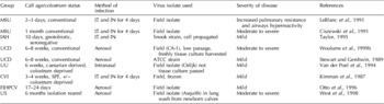
The lack of a successful infection protocol for inducing severe BRSV disease hampered early attempts to study pathogenesis of the infection. Thus, a variety of different protocols have been developed to induce experimental infection with BRSV. These are summarized in Table 1. Gnotobiotic calves taken by caesarian section and kept in isolation units were used by three groups (Kimman et al., Reference Kimman, Westenbrink, Schreuder and Straver1987; Van der Poel et al., Reference Van der Poel, Brand, Kramps and Van Oirschot1994; McInnes et al., Reference McInnes, Sopp, Howard and Taylor1999). The ages of the calves varied from 2 days (LeBlanc et al., Reference LeBlanc, Baker, Gray, Robinson and Derksen1991) to 6 months (West et al., Reference West, Bogdan, Hamel, Nayar, Morley, Haines and Ellis1998). Two groups administered the virus by a combination of intratracheal and intranasal routes for four consecutive days (Kimman et al., Reference Kimman, Westenbrink, Schreuder and Straver1987; Ciszewski et al., Reference Ciszewski, Baker, Slocombe, Reindel, Haines and Clark1991). Three groups (Otto et al., Reference Otto, Elschner, Reinhold, Köhler, Streckert, Philippou, Werchau and Morgenroth1996; West et al., Reference West, Bogdan, Hamel, Nayar, Morley, Haines and Ellis1998; Woolums et al., Reference Woolums, Singer, Boyle and Gershwin1999a, Reference Woolums, Anderson, Gunther, Schelegle, LaRochelle, Singer, Boyle, Friebertshauser and Gershwinb) administered the virus by aerosol. Minimal tissue culture passage appears to be important for induction of moderate to severe clinical signs of disease. The presence of low titers of maternal antibodies specific for BRSV does not appear to prevent infection. These various infection protocols have been used to establish details on BRSV pathogenesis as well as potential efficacy of vaccine formulations and vaccination protocols.
Models of experimental BRSV infection

MSU=Michigan State University, UCD=University of California, Davis, UU=University of Utrecht, The Netherlands, CVI=Central Veterinary Institute, Lelystad, The Netherlands, University of Saskatoon, Canada, FIHPCV=Federal Institute for Health Protection of Consumers and Veterinary Medicine, Germany, IAH=Institute of Animal Health, Compton, England. IT=intratracheal and IN=intranasal.
Immunopathogenesis of BRSV infection: host–viral interaction
Early observations of children infected with RSV showed that some developed severe disease, characterized by wheezing, while others developed only mild upper respiratory tract symptoms. Further examination of these children showed that many of the severely affected patients had detectable RSV-specific IgE antibodies in the serum and nasopharyngeal secretions (Welliver et al., Reference Welliver, Wong, Sun, Middleton, Vaughan and Ogra1981). Furthermore, many children had increased levels of histamine in nasal secretions. Later studies demonstrated a T-helper cell type skew in RSV-infected children with severe disease. Demonstration of interleukin-4 (IL-4) production by lymphocytes from these patients was supportive of a Th2 modulation of the immune response (Román et al., Reference Román, Calhoun, Hinton, Avendaño, Simon, Escobar, Gaggero and Díaz1997). Early studies by Stewart and Gershwin (Reference Stewart and Gershwin1989) demonstrated the presence of BRSV-specific IgE in the serum of calves experimentally infected with BRSV. Subsequent studies by this group using a posterior mediastinal lymph cannulation procedure to obtain efferent lymph from the lung demonstrated that in experimentally-infected calves there was a correlation between severity of clinical signs and the presence of BRSV-specific IgE in serum and lymph. Moreover, the presence of RNA coding for IL-4 in lymphocytes draining the lung was temporally related to BRSV infection (Gershwin et al., Reference Gershwin, Gunther, Anderson, Woolums, McArthur-Vaughan, Randel, Boyle, Friebertshauser and McInturff2000).
The lack of a commercially available vaccine for RSV to protect the human population is a result of a serious vaccine failure in the 1960s (Kim et al., Reference Kim, Canchola, Brandt, Pyles, Chanock, Jensen and Parrott1969). At that time, a formalin inactivated alum adjuvanted vaccine in clinical trials caused disease exacerbation in vaccinated children subsequently infected naturally with field virus; there were two deaths and 80% of those infected were hospitalized (Openshaw et al., Reference Openshaw, Culley and Olszewska2001). The cattle industry has not been hampered with concerns that BRSV killed vaccine might enhance disease and has had several products in heavy use for many years. However, there have been numerous reports of vaccine-induced BRSV exacerbation (some published observations, many unpublished observations). One report documented a severe outbreak among cattle vaccinated with a modified live vaccine in the Netherlands (Kimman et al., Reference Kimman, Sol, Westenbrink and Straver1989). IgM levels indicated the presence of the virus in the herd at the time of vaccination; yet vaccinated cattle were more severely affected than unvaccinated cattle. A report from Belgium documented severe fatal BRSV in cattle vaccinated with a killed BRSV vaccine (Schreiber et al., Reference Schreiber, Matheise, Dessy, Heimann, Letesson, Coppe and Collard2000). The reported lung lesions were reminiscent of those seen in the children who had vaccine-exacerbated disease, including eosinophilia, severe proliferative alveolitis, bronchiolitis obliterans and areas of emphysema. The vaccine administered to the calves described by Schreiber et al. (Reference Schreiber, Matheise, Dessy, Heimann, Letesson, Coppe and Collard2000) was killed with β-propiolactone and was incorporated in a saponin/aluminum hydroxide adjuvant.
Experimental reproduction of vaccine exacerbated BRSV disease has been accomplished by several groups. Gershwin et al. (Reference Gershwin, Schelegle, Gunther, Anderson, Woolums, Larochelle, Boyle, Friebertshauser and Singer1998) used a protocol fashioned after that described in the ill-fated human trial (Kim et al., Reference Kim, Canchola, Brandt, Pyles, Chanock, Jensen and Parrott1969) to produce a formalin killed alum adjuvanted vaccine, and a sham vaccine lacking virus for control animals. In this study, conventionally raised calves were immunized twice at a 2-week interval followed by challenge with virulent BRSV by aerosol exposure 42 days after the initial vaccination. Clinical signs, pulmonary function, virus shedding and antibody responses were monitored during the 10 days after infection. Two calves in the vaccinated/infected group had to be humanely euthanized on the seventh day post infection due to severe dyspnea and a moribund state. Overall the vaccinated group was significantly more severely affected than the control sham vaccinated/infected group. Moreover, pulmonary function testing revealed significantly greater pulmonary resistance and decreased lung compliance in the vaccinated calves. Additionally, lung pathology scoring revealed lesions characteristic of the vaccine exacerbated disease seen in the children (severe alveolitis, bronchiolitis obliterans, etc.) (Gershwin et al., Reference Gershwin, Schelegle, Gunther, Anderson, Woolums, Larochelle, Boyle, Friebertshauser and Singer1998).
Further studies using sera from the calves in the vaccine enhancement experiment demonstrated a correlation between severity of clinical signs and serum BRSV-specific IgE levels. Using Western blot analysis on sequential serum samples it was shown that vaccination induced a strong IgE response to several BRSV proteins in the most severely affected calves. In comparison, calves in another experiment in which vaccine enhanced disease did not occur showed little or no IgE, but IgG antibodies were induced by vaccination (Kalina et al., Reference Kalina, Woolums, Berghaus and Gershwin2004).
The induction of a Th2 response by vaccination with formalin killed BRSV vaccine was further confirmed by demonstration of significantly decreased levels of IFN-γ produced by peripheral blood lymphocytes from the vaccinated/infected calves compared with the sham vaccinated/infected calves in the above described study (Woolums et al., Reference Woolums, Singer, Boyle and Gershwin1999a). These results support the contention that immunomodulation towards the Th2 response is involved in pathogenesis of vaccine-induced disease exacerbation.
Disease exacerbation following vaccination with formalin inactivated alum adjuvanted BRSV vaccine has also been demonstrated by Antonis et al. (Reference Antonis, Schrijver, Daus, Steverink, Stockhofe, Hensen, Langedijk and van der Most2003) in the Netherlands. This group used colostrum deprived specific pathogen free calves obtained by caesarean section and housed in isolators, unlike the conventionally raised calves used in the previously described study. The duration of time between the final vaccination and experimental BRSV challenge was also different (5 months in the Antonis study versus 3 weeks in the Gershwin study). Despite these differences, both experiments demonstrated more severe disease in vaccinated calves and the presence of IgE antibodies in the serum of vaccinated/infected calves.
Recognition of the disease enhancing effects of killed adjuvanted RSV vaccines prompted Mapletoft et al. (Reference Mapletoft, Oumouna, Townsend, Gomis, Babiuk and van Drunen Littel-van den Hurk2006) to formulate an inactivated BRSV vaccine with CpG containing deoxyoligonucleotides (ODN) intended to switch the immune response towards a protective Th1 response. Numerous studies using CpG ODN in murine and human cells have shown that these sequences bind to Toll-like receptors and stimulate secretion of IL-1, IL-6, IL-12 by antigen presenting cells, and IL-6, IL-10, and IFN-γ by lymphocytes (Bharadwaj et al., Reference Bharadwaj, Bewtra and Agrawal2007). In this trial, which included calves vaccinated with formalin-inactivated BRSV with and without the CpG ODN, the observations included decreased clinical signs and virus titers in lung as well as increases in IgG2 levels and IFN-γ production by peripheral blood mononuclear cells. The presence of IgG2 and IFN-γ in cattle are hallmarks of a Th1 type response (Estes and Brown, Reference Estes and Brown2002).
A presumed association between RSV and allergic asthma in children has been substantiated by epidemiological studies in humans (Juntti et al., Reference Juntti, Kokkonen, Dunder, Renko, Niinimäki and Uhari2003; Becker, Reference Becker2006). Experimentally, mouse models have been used to demonstrate that RSV infection can predispose for development of an allergic response to allergen by its immunomodulatory effect (Barends et al., Reference Barends, Van Oosten, De Rond, Dormans, Osterhaus, Neijens and Kimman2004). Moreover, studies with mouse models have shown that the RSV G protein contains a domain that likely functions to skew the Th1/Th2 cytokine balance (Johnson and Graham, Reference Johnson and Graham1999; reviewed in Becker, Reference Becker2006). Although cattle do not develop asthma as a clinical syndrome, the development of IgE antibodies to inhaled pollens and molds has been documented (Olchowy et al., Reference Olchowy, Gershwin and Dean1995). Studies by our group examined whether BRSV infection could facilitate development of IgE antibodies to Micropolyspora faeni (renamed Saccharopolyspora rectivirgula), the causal agent of Farmer's lung and Bovine Farmer's lung disease (Gershwin et al., Reference Gershwin, Himes, Dungworth, Giri, Friebertshauser and Camacho1994), and/or cause exacerbation of IgE responses of calves already sensitized to the mold allergen prior to BRSV infection. In one experiment, calves were either exposed to virus only, exposed to aerosolized M. faeni over a 24-day period and then challenged with M. faeni during BRSV infection, similarly M. faeni aerosol exposed and challenged but sham infected, or exposed to aerosolized M. faeni, infected with BRSV without M. faeni challenge. BRSV-specific IgE was associated with increased lung pathology and clinical disease and M. faeni aerosolization enhanced development of BRSV-specific IgE. Significantly increased concentrations of prostaglandin I2 were associated with M. faeni aerosol exposure, particularly when combined with BRSV infection. After M. faeni challenge exposure, thromboxane B2 concentrations were significantly greater in the BRSV and M. faeni challenge-exposed calves. M. faeni-specific IgE concentrations had a highly significant direct correlation with prostaglandin E2 concentrations in samples taken 30 min after the second M. faeni challenge exposure. Histamine concentrations were significantly greater in calves infected with BRSV than in uninfected controls regardless of M. faeni exposure (Gershwin and Giri, Reference Gershwin and Giri1992). These studies confirmed the synergistic effect of allergen exposure and BRSV infection in modulating the lung environment for a type 1 hypersensitivity response.
More recently, we used technetium labeled ovalbumin (OA) to examine the clearance of allergen from the lungs of calves during a BRSV infection. In this study, after baseline clearance of labeled OA was performed, each calf was infected by aerosol with BRSV; an aerosol of technetium labeled OA was administered on several days throughout infection. Gamma camera imaging was performed to determine the half time for clearance of the OA from the lungs. BRSV infection caused an increase in the time it took for the allergen to be cleared from the lungs (Gershwin et al., Reference Meyer, Deplanche and Schelcherin press). Increased exposure to allergen coupled with the Th2 environment suggests that BRSV may facilitate enhanced sensitization to inhalants. Potentially, sensitization to allergens present in dust may play a role in the development of chronic respiratory disease in some cattle.
Kalina et al. (Reference Kalina, Anderson and Gershwin2006) extended these observations to examine the effect of fungal allergen Alternaria alternata aerosol exposure (prior to and during BRSV infection) on both the primary immune response and the clinical outcome of a secondary BRSV infection. Experimental groups included BRSV infected calves aerosol exposed to Alternaria and to sham Alternaria; exposures were initiated 6 days before BRSV infection and continued through the sixth day of infection. The second set of aerosols/infection began just over 3 months from the primary BRSV infection and continued as described for the primary infection. Alternaria aerosols did not influence the clinical course of the primary infection; however, the Alternaria exposed/BRSV infected animals had a significantly lower mean clinical score for the secondary infection than those exposed to sham Alternaria and BRSV infection. Exposure to Alternaria facilitated IgE antibody production in BRSV infected calves during the primary and secondary infections. At necropsy the Alternaria exposed BRSV infected calves had greater numbers of lung eosinophils and IL-4 secreting peripheral blood mononuclear cells, as determined by flow cytometry. Paradoxically, BRSV infection enhanced Th2 responses against inhaled Alternaria. Yet animals with higher IgE responses to inhaled Alternaria did not have more severe disease when infected a second time with BRSV.
BRSV infection and polymicrobial infections
BRSV has been associated with several bacterial pathogens in the BRDC. Like bovine herpes virus, BRSV can predispose to infection with Mannheimia haemolytica, Pasteurella multocida and Histophilus somni. However, the immunomodulatory capability of BRSV adds another dimension to the pathogen interaction. Our group examined the effect of BRSV infection on subsequent H. somni infection, using well-developed model systems (Gogolewski et al., Reference Gogolewski, Schaefer, Wasson, Corbeil and Corbeil1989; Woolums et al., Reference Woolums, Singer, Boyle and Gershwin1999a, Reference Woolums, Anderson, Gunther, Schelegle, LaRochelle, Singer, Boyle, Friebertshauser and Gershwinb; Gershwin et al., Reference Gershwin, Berghaus, Arnold, Anderson and Corbeil2005). The working hypothesis was that H. somni infection would be enhanced because of the need for bacteria-specific IgG2, in cattle the product of a Th1 response (Estes and Brown, Reference Estes and Brown2002). The BRSV infection was well established by day 6 after virus aerosol thereby inducing a Th2 environment when the bacteria were administered by intratracheal inoculation. Indeed infection was more severe and bacteria persisted longer in the lungs in dually infected calves as compared with those infected with single pathogens. Moreover, a H. somni-specific IgE response was detected in dually infected cattle. Protein specificity of the IgE has been examined (Corbeil et al., Reference Berghaus, Corbeil, Berghaus, Kalina, Kimball and Gershwin2006). Further details on this work are presented in the review on H. somni.
The effect of BRSV infection on a previously established immune response to H. somni appears to be less effective in modulation. In a subsequent experiment, we hypothesized that combining BRSV vaccination with H. somni vaccination would induce cytokine shifts resulting in greater IgE production to both pathogens and enhanced disease. Concurrent vaccination followed by concurrent infection with single agents or both BRSV and H. somni showed increased BRSV-specific serum IgE levels for the calves with the most severe disease, while no difference was detected in the IgE levels to H. somni among groups. Moreover, those calves with the least evidence of disease showed increased levels of IFN-γ message and H. somni-specific IgG2 antibodies. The BRSV vaccine induced the expected Th2 response, but the H. somni bacterin induced a strong IgG2 response, which proved to be protective (Berghaus et al., Reference Berghaus, Corbeil, Berghaus, Kalina, Kimball and Gershwin2006). Taken together with the experiments on unvaccinated calves described above, it is suggestive that viral replication may be an important factor for modulation by BRSV of the immune response to H. somni towards a Th2 response. Additionally, a well established Th2 environment induced by the BRSV replication prior to introduction of H. somni antigens may be required for BRSV modulatory effects on the immune response to H. somni.
Summary
It is well documented that much of the clinical and pathological observations in BRSV infection result from the host response to the virus. There are unique aspects of this virus that facilitate development of pro-inflammatory responses that fail to induce protection and long-lived immunity. When examined, most observations made on human RSV have proven to be shared with BRSV. Thus, it is likely that continued efforts to develop new vaccination strategies would have reciprocal benefits for both species.