Implications
Tenderness of meat is a major quality attribute affecting consumers purchasing decision. Variability in tenderness of meat produced from young and old buffaloes results in huge economic loss to producers. Meat produced from young and old buffaloes beset with their own sensory and technological advantages and disadvantages and therefore, requires different processing strategies for their effective utilization. Outcomes of this research may provide control tools of meat quality improvement from young and old buffaloes to engineer innovative processing strategies to minimize tenderness variability induced revenue loss to the meat industries.
Introduction
Buffalo (Bubalus bubalis) meat is emerging as an important red meat source in several Southeast Asian nations and few European countries to augment nutritional security. India has 57.8% of world’s buffaloes and Indian buffalo meat accounts for 20% of world’s beef trade. India is a largest producer and exporter of buffalo meat in the world with an annual turnover of US$ 3.29 billion. Majority of buffalo meat is produced in India mainly from old and culled animals (>10 years age) after completion of their productive/reproductive life. Meat from such animals is tough and less juicy, characterized by high amounts of connective tissue (Naveena et al., Reference Naveena, Kiran, Sudhakar Reddy, Ramakrishna, Vaithiyanathan and Devatkal2011). On the other hand meat produced from young buffalo calves (<2 years age) is considered tender, juicy and fetches premium price in the export market.
Meat produced from young and old animals have distinctly different biochemical characteristics that will affect the meat quality traits especially tenderness. Meat from young and old buffaloes were studied with respect to their physical and chemical aspects and showed that meat from young buffalo was more tender than old buffalo (Syed Ziauddin et al., Reference Syed Ziauddin, Mahendrakar, Rao, Ramesh and Amla1994). Buffalo meat was considered tough with coarse texture and tenderization of tough buffalo meat has been achieved using different enzymatic (Naveena et al., Reference Naveena, Mendiratta and Anjaneyulu2004) and chemical (Naveena et al., Reference Naveena, Kiran, Sudhakar Reddy, Ramakrishna, Vaithiyanathan and Devatkal2011) methods. On the contrary, Neath et al. (Reference Neath, Del Barrio, Lapitan, Herrera, Cruz, Fujihara, Muroya, Chikuni, Hirabayashi and Kanai2007) compared the tenderness and some characteristics of water buffalo meat and beef during postmortem ageing and demonstrated superior tenderness in water buffalo meat relative to Brahman beef. Thus, meat produced from young and old buffaloes offer a valuable model to investigate tenderization process with reference to biochemistry, ultrastructure and proteome characteristics.
In the post-genomic era, proteomic tools such as two-dimensional gel electrophoresis (2DE) coupled with mass spectrometry (MS) have been extensively utilized to elucidate the biochemical mechanisms influencing muscle to meat conversion and meat tenderness in beef and pork (Lametsch et al., Reference Lametsch, Karlsson, Rosenvold, Andersen, Roepstorff and Benedixen2003; Laville et al., Reference Laville, Sayd, Morzel, Blinet, Chambon, Lepetit, Renand and Hocquette2009). Most of the studies on tenderness prediction revealed the heat shock proteins (HSPs) as potential biomarkers (Kim et al., Reference Kim, Cho, Lee, Park, Lee, Cho, Choy, Yoon, Im and Park2008; Polati et al., Reference Polati, Menini, Robotti, Millioni, Marengo, Novelli, Balzan and Cecconi2012). Effect of ageing on sensorial and textural quality parameters of bovine meat is well documented (Monson et al., Reference Monson, Sanudo and Sierra2005; Vieira et al., Reference Vieira, Cerdeño, Seffano, Lavín and Mantecón2007) with few studies on proteolysis (Jia, et al., Reference Jia, Hollung, Therkildsen, Hildrum and Bendixen2006; Bjarnadottir et al., Reference Bjarnadottir, Hollung, Hoy, Bendixen, Codrea and Veiseth-Kent2012). However, there are no published literatures comparing the proteome characteristics of meat from young and old animals specific to tenderness during postmortem ageing. Further, to our knowledge 2DE of buffalo meat has not been reported so far. Meat scientists around the world are looking for biological markers of tenderness which would help classification based on their potential tenderness and to optimize genetic selection of meat animals. The protein biomarkers of tenderness identified so far have been sorted and grouped according to their common biological functions belonging to different biological pathways like glycolytic and oxidative energy production, cell detoxification, protease inhibition and production of heat shock proteins, along with some unusual biomarkers (Ouali et al., Reference Ouali, Gagaoua, Boudida, Becila, Boudjellal, Herrera-Mendez and Sentandreu2013). Biochemical basis, proteome profile and ultrastructure of meat influencing tenderness may vary significantly between young and old animals. Better understanding of the proteome characters from animals of different age group (young and old) and proteomic changes that occur during postmortem ageing is essential to develop strategies for improving fresh meat quality. However, proteome characterization of meat from young and old buffaloes has not been reported. Therefore, the objective of this study was to define the underlying biochemical, proteomic and ultrastructural differences that govern tenderness and palatability between young and old buffalo meat.
Material and methods
Procurement of buffalo meat
The longissimus thoracis et lumborum or longissimus dorsi (LD) muscles were harvested from hot boned carcass from 18 young (<2 years) and 18 old (>10 years) water buffaloes (Bubalus bubalis) immediately after exsanguination from local municipal slaughterhouse of Hyderabad, India. Buffaloes were slaughtered according to traditional halal method followed in India and weighed ~100 kg for young and 400 kg for old animals, respectively. Each muscle was cut into four portions, packed in low density polyethylene (2325 to 6510 cm3 O2/m3 per 24 h at 3°C) bags under atmospheric conditions and randomly assigned to four different ageing periods (0, 2, 4 and 6 days) at 4±1°C in domestic refrigerator. At each designated ageing period 6 randomly assigned portions of LD were evaluated for proteome changes. Fresh meat samples were analysed for pH, water-holding capacity, protein extractability, muscle fibre diameter, myofibrillar fragmentation index (MFI), myoglobin content and %metmyoglobin. Cooked meat steaks were also evaluated for Warner-Bratzler shear force (WBSF) values and cooking yield.
Sample analysis
Proximate composition
Moisture, fat, protein and ash content of raw buffalo meat was determined according to AOAC (2005).
pH and water-holding capacity
Muscle sample of 10 g was homogenized with 50 ml chilled distilled water in a tissue homogenizer (WiseMix, HG-15D; Daihan Scientifics, Gangwon-do, Korea) and the pH value was measured with a digital pH meter (Model 420A+, Thermo Orion, Waltham, MA, USA). For determining water-holding capacity, 20 g of minced meat sample was stirred with 30 ml, NaCl (0.6 M) and centrifuged at 3000× g using refrigerated centrifuge (Sorvall, Biofuge Stratos; Thermo electron LED GmbH, Osterode, Germany) for 15 min. The supernatant was measured and amount of water retained by samples was expressed in percentage (Wardlaw et al., Reference Wardlaw, Maccaskill and Acton1973).
Protein extractability
Protein extractability was determined according to procedure of Joo et al. (Reference Joo, Kauffman, Kim and Park1999). Sarcoplasmic and total proteins (sarcoplasmic+myofibrillar) were extracted separately by homogenizing 2 g sample with 20 ml of ice-cold 0.025 M potassium phosphate buffer (pH 7.2) or 20 ml of ice-cold 1.1 M potassium iodide in 0.1 M phosphate buffer (pH 7.2), respectively, and kept overnight at 4°C with frequent shaking. All Samples were centrifuged at 5000× g for 20 min and concentration of protein in the supernatant was determined by the Biuret method. Myofibrillar protein extractability was calculated by obtaining difference between total and sarcoplasmic protein extractability.
MFI
MFI was determined as per the procedure outlined by Hawkins et al. (Reference Hawkins, Davis, Cable and Ramsey1987). Minced meat of 10 g was added to 50 ml of cold sucrose (0.24 M) and Potassium chloride (0.02 M) solution in a 150 ml homogenization tube and blended for 40 s at high speed using tissue homogenizer (WiseMix, HG-15D). The resulting homogenate was filtered through a pre-weighed cheese cloth and the MFI was reported as the weight of the residue in grams times one hundred.
Myoglobin content and %metmyoglobin
Myoglobin (Mb) was extracted from raw meat using a modified procedure of Warris (Reference Warris1979). Samples were blended with five volumes of cold 0.04 M phosphate buffer at pH 6.8 for 10 s in a homogenizer. After keeping at 1°C for 1 h, the mixtures were centrifuged at 3500× g (Sorvall, BiofugeStratos) and 4°C for 30 min. The supernatant was further clarified by filtration through Whatman No. 1 filter paper. The absorbance of filtrate was measured at 525, 572 and 700 nm using a UV-VIS spectrophotometer (Model: UV-1700, PharmaSpec; Shimadzu, Tokyo, Japan). The myoglobin concentration, %metmyoglobin (Met Mb) were calculated according to Trout (Reference Trout1989).
 $$\eqalignno{ & {\rm Mb}\,\left( {{\rm mg}/{\rm g}} \right)\,{\equals}\,\left( {{\rm A}_{{{\rm 525}}} {\minus}{\rm A}_{{{\rm 7}00}} } \right){\rm }{\times}{\rm 2}.{\rm 3}0{\rm 3 }{\times}{\rm dilution}\,{\rm factor}\,\,\cr &\%\,\,{\rm Met}\,{\rm Mb}\,{\equals}\,{\rm 1}.{\rm 395}{\minus}\left[ {\left( {{\rm A}_{{{\rm 572}}} {\minus}{\rm A}_{{{\rm 7}00}} } \right)/\left( {{\rm A}_{{{\rm 525}}} {\minus}{\rm A}_{{{\rm 7}00}} } \right)} \right]{\rm }{\times}{\rm 1}00$$
WBSF
Control and treated muscle samples were sealed in low-density polyethylene bags and placed in water bath maintained at 100°C for 30 min, followed by overnight chilling at 4±1°C. Chilled samples were equilibrated to room temperature and 1.25 cm cores were taken using tissue borer with muscle fibres parallel to the direction of the borer. The WBSF of the cores were measured using texturometer (Model H1KF; Tinius Olsen, Redhill, UK) with V-shaped stainless steel blade (60° angle). The cores were sheared perpendicular to the muscle fibre orientation with 75 N load range and a crosshead speed set at 200 mm/min. The force required to shear the samples were recorded in Newtons.
Ultrastructural study
Muscle fibre diameter
Muscle fibre diameter was determined as described by Tuma et al. (Reference Tuma, Venable, Wuthier and Henrickson1962). A small core (1.00 cm) of muscle tissue was fixed in formal saline for 24 h and was blended in a homogenizer at low speed for 30 s. A drop of the homogenate was placed over a glass slide, covered with cover slip and observed under a microscope with 10× eyepiece containing a calibrated micrometer.
Histological study
Tissues were fixed in 10% neutral buffered formalin for histological studies. The fixed tissues were processed and stained with haematoxylin and eosin stain as described by Singh and Sulochana (Reference Singh and Sulochana1977).
Scanning electron microscopy (SEM)
Samples were fixed in 2.5% glutaraldehyde in 0.1 M phosphate buffer (pH 7.2) for 24 h at 4°C and post fixed in 2% aqueous osmium tetroxide for 4 h. The processed samples were mounted over the stubs with double-sided carbon conductivity tape, and a thin layer of gold coat over the samples was applied by using an automated sputter coater (Model: JEOL JFC-1600) for 3 min and scanned under SEM (Model: JOEL-JSM 5600) at 1000×, 600× and 150× magnifications as per the standard procedures (Bozzola and Russell, Reference Bozzola and Russell1998).
Transmission electron microscopy (TEM)
Samples were fixed in 2.5% to 3% glutaraldehyde in 0.1 M phosphate buffer (pH 7.2) for 24 h at 4°C and post fixed in 2% aqueous osmium tetroxide in the same buffer for 2 h. Dehydrated in series of graded alcohols, infiltrated and embedded in araldite 6005 resin or spur resin (Spurr, Reference Spurr1969). Ultrathin (50 to 70 nm) sections were made with a glass knife on ultra microtome (Leica Ultra cut UCT-GA-D/E-1/00), mounted on copper grids and stained with saturated aqueous uranyl acetate and counter stained with Reynolds lead citrate. Viewed under TEM (Model: Hitachi, H-7500, Japan) at required magnification as per the standard procedures.
Proteome analysis
2DE
Muscle tissue (100 mg) was washed and extracted in lysis buffer (8 M urea, 2 M thiourea, 1% DTT, 2% CHAPS, 2% Protease inhibitor and 2% IPG buffer pH 3 to 10). Protein quantification of the supernatant was performed using 2-D Quant Kit (GE Healthcare, Pittsburgh, USA). The immobilized pH gradient (IPG) strips (Immobiline®drystrip, 11 cm, pH range 3 to 10) were passively rehydrated in 200 µl of protein solution containing 750 to 800 µg proteins for 12 h. Rehydrated IPG strips were subjected to iso electric focusing (IEF) in a Ettan IPGPhor-3 (GE Healthcare, Uppsala, Sweden) gel apparatus at 18°C for a total of 10 800 Vh. Focused IPG strips were equilibrated in 6 M urea, 20% glycerol, 2% SDS, 0.375 M Tris (pH 8.8), for 15 min in 2% DTT and 5% iodoacetamide, respectively. After equilibration, proteins were separated in the second dimension, with the SE 600 Ruby apparatus at 100 V with 60 mA/gel until tracking dye reached lower end of gel. The gel was removed and stained with Colloidal Coomassie blue for 3 h followed by overnight destaining. Protein samples from LD muscles of young and old buffaloes were run under the same conditions and the five gels for each young and old meat samples were produced.
2DE gel image analysis
Stained gels were scanned on an Image Scanner III using labscan 6.0 software. Spot detection and quantification were performed with Image Master Platinum7.0 software (GE Healthcare). The images were grouped into different groups according to required study and the intensity and area of individual spots were analysed for comparative image analysis. For each spot in young or old buffalo meat sample, spot quantity values in triplicate gels were averaged for statistical analysis. A spot was considered to be significant in differential abundance when it was associated with P<0.05.
In-gel digestion and protein identification by MALDI-TOF/TOF MS
In-gel digestion of the proteins separated by 2DE was performed as described by Shevchenko et al. (Reference Shevchenko, Wilm, Vorm and Mann1996) with slight modifications. Subsequent to enrichment and purification through the Zip-Tip pipette tips, peptide mixtures were dissolved in 0.5 µl of CHCA matrix solution (5 mg/ml CHCA in 50% ACN/0.1% TFA) and spotted onto a freshly cleaned MALDI target plate. Spots were allowed to dry for 30 min at room temperature. After air drying, the crystallized spots were analysed using a 4800 MALDI-TOF/TOF mass spectrometer (AB Sciex, Farmingham, MA, USA) linked to 4000 series explorer software (version 3.5.3). All mass spectra were recorded in a reflector mode within a mass range from 800 to 4000 Da, using a Nd:YAG 355 nm laser. The acceleration voltage and extraction voltage were kept at 20 and 18 kV, respectively. All the MS spectra were obtained from accumulation of 900 shots. MS/MS spectra were acquired for the 15 most abundant precursor ions, with a total accumulation of 1500 laser shots and collision energy of 1 kV. The combined MS and MS/MS peak lists were searched using the GPSTM Explorer software version 3.6 (AB Sciex). Protein identification was performed by MS/MS ion search using MASCOT version 2.1 (http://www.matrixscience.com) search engine against the Swiss-Prot database.
Statistical analysis
The experimental design was a completely randomized design with repeated measures. Fixed effects included age of the animal (young and old) and four different ageing period. Statistical analysis was performed with the ANOVA using SPSS (SPSS version 13.0 for windows; SPSS, Chicago, IL, USA). Least square means for F-tests were calculated by using Duncan’s multiple range tests and were considered significant at P<0.05.
Results and discussion
Proximate composition
Proximate composition (data not shown) varies considerably between young and old buffalo meat. Mean moisture content was lower (P>0.05) in old buffalo meat (75.67%) compared with young buffalo meat (77.33%). Decrease in moisture content with increase in the age of beef due to increase in fat content was reported by Lawrie (Reference Lawrie1998). Higher (P>0.05) protein content was observed in old buffalo meat (21.87%) relative to meat from young buffaloes (20.81%). The total fat content was found to be 0.41 and 0.37% for young and old buffaloes, respectively. Relatively low fat content in buffalo meat is attributed to poor marbling (Lapitan et al., Reference Lapitan, Delbarrio, Katsube, Ban-Tokuda, Orden, Robles, Fujihara, Cruz, Homma and Kannai2007). Relatively higher ash content was observed in old buffalo meat (1.63%) compared with young buffalo meat (1.39%). Proximate composition of buffalo meat observed in the present study are in agreement with earlier reports (Keshava Rao et al., Reference Keshava Rao, Anjaneyulu, Lakshmanan, Sharma and Kondaiah1985).
pH and water-holding capacity
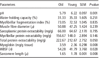
Ultimate pH and water-holding capacity of meat influence protein extractability/solubility which is an important parameter showing protein functionality. Mean pH value (Table 1) was found to be significantly greater in young (6.22) compared with old (5.79) buffaloes. This might be due to the variation in muscle glycogen reserve and the difference in postmortem aerobic and anaerobic metabolism between young and old buffalo meat. A pH of 5.64 and 5.69 was also reported in ground buffalo meat (Sahoo and Anjaneyulu, Reference Sahoo and Anjaneyulu2000) and buffalo meat cubes (Naveena et al., Reference Naveena, Mendiratta and Anjaneyulu2004), respectively. Significantly lower pH in old buffalo meat might be due to increased sensitivity of older animals to stress during slaughter which results in rapid breakdown of muscle glycogen. No difference was observed in water-holding capacity between old and young buffalo meat, eventhough ultimate pH differed between them.
Biochemical, histological and textural qualities of longissimus dorsi muscle from young (<2 years) and old buffaloes (>10 years)

n=3.
Protein extractability and MFI
When the sarcoplasmic and myofibrillar proteins were extracted individually from young and old buffalo meat, no difference (P>0.05) was observed between them. However, total protein extractability of buffalo meat as influenced by age showed significant (P<0.05) difference between old (222.67 mg/g protein) and young (212.67 mg/g protein) buffalo meat. Sarcoplasmic and myofibrillar protein concentration of 5.12% and 7.19% were recorded in male buffalo calf meat (Anjaneyulu et al., Reference Anjaneyulu, Sengar, Lakshmanan and Joshi1985). Whereas, in old female buffalo meat a salt soluble protein content of 8.2% was reported (Anjaneyulu et al., Reference Anjaneyulu, Sharma and Kondaiah1989). Old male buffalo meat had significantly (P<0.05) higher salt soluble protein compared with the meat from young male and old female buffaloes (Kandeepan et al., Reference Kandeepan, Anjaneyulu, Kondaiah, Mendiratta and Lakshmanan2009). These findings indicate the higher sarcoplasmic and myofibrillar protein content in meat from old buffaloes relative to young buffaloes resulting in greater total protein extractability in old buffalo meat which influences protein functionality. MFI did not differ (P>0.05) between young (72.53) and old (73.05) buffalo meat. In contrast to our study, significantly higher MFI was observed in young buffalo meat compared with meat from old buffaloes (Kandeepan et al., Reference Kandeepan, Anjaneyulu, Kondaiah, Mendiratta and Lakshmanan2009).
Myoglobin and % metmyoglobin content
Myoglobin concentration found to vary significantly (P<0.01) between old and young buffalo meat with 3.59 and 2.36 mg myoglobin/g tissue, respectively. Myoglobin content was reported to vary from 2.7 to 9.4 mg/g depending upon the type of muscle and age and meat becomes darker with increasing age (Valin et al., Reference Valin, Pinkas, Drahnev, Boikovski and Polikronov1984). Higher total meat pigment concentration was also reported in old male buffalo meat relative to young males due to higher content of heme pigment and myoglobin. The %metmyoglobin was significantly (P<0.01) higher in young (31.60%) compared with old (23.94%) buffalo meat indicating better colour stability in old buffalo meat.
WBSF
WBSF value, which is an objective measure of tenderness is higher (P<0.05) in old buffalo meat (54.28 N) relative to young buffalo meat (41.79 N). Shear force values observed in the current study are consistent with findings of Syed Ziauddin et al. (Reference Syed Ziauddin, Mahendrakar, Rao, Ramesh and Amla1994) and Kandeepan et al. (Reference Kandeepan, Anjaneyulu, Kondaiah, Mendiratta and Lakshmanan2009). The shear force value was reported to have positive correlation with fibre diameter. Higher muscle fibre diameter, smaller sarcomere length and relatively lower MFI in old buffalo meat correlates with increased WBSF values indicating toughness.
Ultrastructural study
Muscle fibre diameter of 47.25 and 50.88 µ was observed for young and old buffalo meat, respectively (P>0.05). Kandeepan et al. (Reference Kandeepan, Anjaneyulu, Kondaiah, Mendiratta and Lakshmanan2009) reported a significantly (P<0.05) higher muscle fibre diameter in old buffaloes compared with young buffalo meat. A muscle fibre diameter of 60.76 μ was recorded in meat cubes from old female buffaloes (Naveena et al., Reference Naveena, Mendiratta and Anjaneyulu2004). It was agreed that, fibre diameter increases and tenderness decreases with animal age (Tuma et al., Reference Tuma, Venable, Wuthier and Henrickson1962). However, these authors further reported that, the correlation between these two variables differed greatly with age of animals and hence muscle fibre diameter is a poor indicator of tenderness. Sarcomere length was significantly (P<0.01) lower in old buffalo (1.65 µ) compared with young buffalo (1.78 µ) meat. Similar results were also reported by several authors (Kandeepan et al., Reference Kandeepan, Anjaneyulu, Kondaiah, Mendiratta and Lakshmanan2009) indicating higher sarcomere length in young buffalo compared with old buffalo meat.
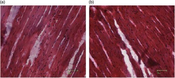
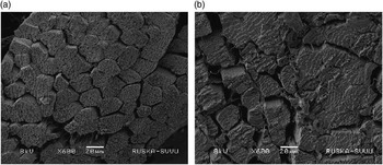
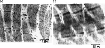
The structural details as observed under different magnification during light microscopy, SEM and TEM are shown in Figures 1, 2 and 3, respectively. The light microscopy photographs revealed the variation in muscle fibre diameter between young and old buffalo meat. Inter-myofibrillar space as evidenced by white or light grey areas between muscle fibres appeared to be higher in young buffaloes. The muscle fibres appeared more-dark and densely packed in old buffalo meat samples. These initial results prompted us to further characterize the structure using scanning and TEM. The SEM gives images with great depth-of-field yielding a characteristic 3D display that provides greater insight into the surface structure of a biological sample. The SEM images revealed coarse muscle fibres in old buffalo meat samples with large muscle fibre bundles compared with young buffalo muscles. Large gap between muscle fibre bundles in old buffalo meat samples indicates thick perimysium layer which will significantly contribute to toughness of meat with increase in age of the animal. The sarcomere is a structural unit of muscle fibre. The sarcomere length of young buffalo meat was higher compared with old buffalo meat. The TEM has been used to observe changes in Z-line during tenderization of culled cow meat by proteolytic enzymes and by calcium chloride following osmotic dehydration (Gerelt et al., Reference Gerelt, Ikeuchi, Nishiumi and Suzuki2002). The TEM images were utilized for calculating sarcomere length which is the distance between two Z-disks. In the present study, TEM images depicted the longer sarcomeres in young buffalo meat samples relative to old buffalo meat. The photographs from old buffalo meat revealed the orderly arranged structure of Z-disks with clear view. However, in young buffalo meat samples, the Z-disks are degraded and more fragmentation can be observed due to higher proteolysis and MFI in younger buffaloes.
Light microscopy images showing the ultrastructure of longissimus dorsi muscles from (a) young and (b) old buffalo meat.

Scanning electron microscopy (SEM) images showing the ultrastructure of longissimus dorsi muscles from (a) young and (b) old buffalo meat.

Transmission electron microscopy (TEM) images showing the ultrastructure of longissimus dorsi muscles from (a) young and (b) old buffalo meat.

Proteome characterization
The 2DE gel analysis of total skeletal muscle proteins from LD muscles revealed separation of 415 and 558 spots, respectively, in old and young buffalo meat. The class analysis table by ANOVA of old and young buffalo meat gels indicated 93 spots which had protein spot expression of 1.5-fold or more between them. Around 93 spots were differentially expressed between young and old buffaloes. Three significantly differentially expressed spots were subjected to tandem MS (MALDI-TOF/TOF MS). Similar to our results, 41 spots were reported to be differentially expressed with age in pork (Hollung et al., Reference Hollung, Grove, Faergestad, Sidhu and Berg2009). The greater number of differentially expressed spots in the current study may be due to extraction of total meat proteins (both water and salt soluble) in contrast to other studies wherein only sarcoplasmic proteins were studied. The protein DJ (PARK7) protecting cells from oxidative stress and death is frequently over expressed in tender meat (Laville et al., Reference Laville, Sayd, Morzel, Blinet, Chambon, Lepetit, Renand and Hocquette2009). The glycolytic enzymes or proteins involved in the structure and the contraction like, troponin T, α actin or heavy (MYH1) and light (MYL1, MYL2 and MYL6B) myosin II chains have been reported to have altered abundance with tenderness (Polati et al., Reference Polati, Menini, Robotti, Millioni, Marengo, Novelli, Balzan and Cecconi2012).
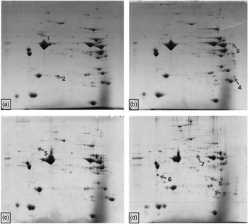
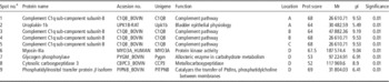
During ageing, the 2DE gel analysis showed significant difference in protein separation and expression pattern between old and young buffaloes (Figure 4). In old buffaloes 560 (Figure 4a) and 718 (Figure 4b) spots were separated with 108 spots being differentially expressed among them. Whereas, In young buffaloes 516 (Figure 4c) and 772 (Figure 4d) spots were separated, respectively, on 0 and 6th day of ageing with 124 spots being differentially expressed between both ageing period. Differentially expressed protein spots were selected from old and young buffalo meat during 6th day of ageing as indicated in Figure 4. The selected protein spots were digested and subjected to tandem MS (MALDI-TOF/TOF MS). The proteins were identified by database searching in National Centre for Biotechnology Information (NCBI). The proteins identified by MS/MS are presented in Table 2 with their name, NCBI accession number, molecular weight, isoelectric pH (pI), sequence coverage, functional roles, gene coding for respective proteins, spot ratio and significance level.
Total skeletal muscle proteins from longissimus dorsi muscles of young and old buffalo meat during ageing separated on two-dimensional gel electrophoresis (2DE). (a) Old 0’day; (b) Old 6’day; (c) Young 0’ day; (d) Young 6’day. Nine differentially expressed protein spots are numbered and indicated on the gel photographs.

Functional roles, molecular weight, isoelectric point, accession number of differentially abundant proteins separated by 2DE from young and old buffalo meat during ageing

2DE=two-dimensional gel electrophoresis; A=Old 0’day; B=Old 6’day ageing; C=Young 0’ day; D=Young 6’day ageing; Mr=molecular weight; pI=isoelectric point.
a Nine differentially expressed protein spots are numbered and indicated on the gel photographs.
Marino et al. (Reference Marino, Albenzio, della Malva, Santillo, Loizzo and Sevi2013) reported increase in number of spots during postmortem ageing of beef, ascribed to the presence of sarcoplasmic proteins and fragments deriving from MHC (fMHC). Other studies have also reported the increase in the quantity of fragments of glycolytic enzymes in the myofibrillar fraction are processes that characterize meat ageing (Bjarnadottìr et al., 2010). Similarly Lametsch et al. (Reference Lametsch, Karlsson, Rosenvold, Andersen, Roepstorff and Benedixen2003) observed that 103 spots changed significantly over 72 h ageing in pig longissimus muscle. This increased number of spots with ageing confirms proteolytic degradation with ageing. The change in spot intensity with ageing has been proposed to be from three different causes viz, proteolysis, post-translational changes and release from protein structures or complexes. In addition, other cytoskeletal proteins including troponin-T and desmin were related to meat quality traits, as shown by earlier studies (Wheeler and Koomaraie, Reference Wheeler and Koohmaraie1999). Using proteomic analysis Laville et al. (2009) showed that during beef muscle ageing the solubility of some cytoplasmic proteins (HSPs and glycolytic enzymes) decreased, some proteins were fragmented (glycolytic enzymes, structural proteins) and some full length proteins were released from cellular structures (actin, troponin). A study suggests a relationship between ageing-related levels of proteolysis and meat quality traits by using proteomics and surface enhanced laser desorption-time-of-flight (SELDI-TOF) technology in longissimus muscle of Yorkshire and Duroc (Tepas et al., Reference Tepas, Jansen, Konrad, Broekman, Henny and Heuven2008). The differentially expressed proteins between young and old buffaloes was found to be Uroplakin-1b, where as other protein Complement C1q subcomponent subunit B spots was identified as the same protein although present at different positions on the 2DE gel. This may reflect different isoforms of the proteins caused by modifications that influence the charge of the protein (Lametsch et al., Reference Lametsch, Karlsson, Rosenvold, Andersen, Roepstorff and Benedixen2003). There are major differences in the abundance of proteins involved in energy metabolism, stress response and structural proteins between different age groups during ageing. Increased number of spots with progress in ageing indicates proteolytic degradation with ageing. In old animal ageing, same protein Complement C1q subcomponent subunit B was expressed at different location which may reflect the fragmentation of protein and appearance of the same as isoforms in different location (Lametsch et al., Reference Lametsch, Karlsson, Rosenvold, Andersen, Roepstorff and Benedixen2003). In young animals, during ageing new proteins were expressed like myosin-IIIa, glycogen phosphorylase, cytosolic carboxypeptidase 3, and phosphatidylinositol transfer protein β isoform. Their appearance indicate higher rate of proteolysis in young animals in comparison to old animals. Similarly higher rate of myofibrillar fragmentaion has been reported in young buffaloes in comparison to old buffalo meat (Kandeepan et al., Reference Kandeepan, Anjaneyulu, Kondaiah, Mendiratta and Lakshmanan2009). Similar to our study glycogen-phosphorylase has been reported as biomarker of meat quality in pig during ageing (Lametsch et al., Reference Lametsch, Karlsson, Rosenvold, Andersen, Roepstorff and Benedixen2003; Laville et al., Reference Laville, Sayd, Morzel, Blinet, Chambon, Lepetit, Renand and Hocquette2009), where in determined the most susceptible proteins to endogenous enzyme action in the first 48 h postmortem. The glycogen phosphorylase has also been reported to be associated with PSE condition in pig meat (Laville et al., Reference Laville, Sayd, Morzel, Blinet, Chambon, Lepetit, Renand and Hocquette2009). Peptidases has also been reported to be involved in postmortem proteolysis in beef and role of different endopeptidase on tenderness has been reviewed in detail by Sentandreu et al. (Reference Sentandreu, Coulis and Ouali2002). The biomarkers so far identified have been sorted and grouped according to their common biological functions. All of them refer to a series of biological pathways including glycolytic (Bouley et al., Reference Bouley, Chambon and Picard2004; Laville et al., Reference Laville, Sayd, Morzel, Blinet, Chambon, Lepetit, Renand and Hocquette2009) and oxidative energy production (Zapata et al., 2012), cell detoxification (Zapata et al., Reference Zapata, Zerby and Wick2009; Polati et al., Reference Polati, Menini, Robotti, Millioni, Marengo, Novelli, Balzan and Cecconi2012), protease inhibitors (Gagaoua et al., Reference Gagaoua, Boudida, Becila, Picard, Boudjellal, Sentandreu and Ouali2012), and production of Heat Shock Proteins (Flower et al., Reference Flower, Chesnokova, Froelich, Dixon and Witt2005; Bjarnadottir et al., Reference Bjarnadottir, Hollung, Hoy, Bendixen, Codrea and Veiseth-Kent2012). Some unusual biomarkers are also presented: annexins, galectins and peroxiredoxins (Zapata et al., Reference Zapata, Zerby and Wick2009; Bjarnadottir et al., Reference Bjarnadottir, Hollung, Hoy, Bendixen, Codrea and Veiseth-Kent2012; Polati et al., Reference Polati, Menini, Robotti, Millioni, Marengo, Novelli, Balzan and Cecconi2012).
Conclusions
Present study revealed that, meat from young and old buffaloes differ significantly in their quality and each beset with their own advantages and disadvantages and therefore, requires different processing strategies for their effective utilization. Meat from old buffaloes had higher myoglobin redox stability and total protein extractability whereas, young buffalo meat had lower WBSF and sarcomere length. Present study has demonstrated that variation in tenderness between young and old buffalo meat can be reduced through ageing process. Using 2DE and MALDI-TOF/TOF MS we have successfully identified few important proteins whose expression levels are associated with meat tenderness. The proteins identified in the present study includes: complement C1q subcomponent subunit B, uroplakin-1b, aspartate aminotransferase, myosin-IIIa, glycogen phosphorylase, cytosolic carboxypeptidase 3 and phosphatidylinositol transfer protein β isoform. Positive correlation of these proteins with other objective measures of tenderness indicates their potential to influence meat quality especially tenderness. Hence, these proteins may be useful as biomarkers for meat quality in buffalo meat.
Acknowledgements
Financial assistance from Indian Council of Agricultural Research (ICAR) is acknowledged. The authors acknowledge the support of Dr Laxman, Ruska Lab, SVVU, Tirupati and Dr C. Ramakrishna, NRC on Meat, Hyderabad for their assistance with ultrastructure studies. This research was a part of doctoral work of first author (Kiran, M.) submitted to Sri Venkateswara Veterinary University, Tirupati (India). First author would like to thank University Grants Commission, Govt. of India for providing fellowship for his doctoral programme.