Implications
Excessive intake of high-fat/high-cholesterol food is typical of Western countries nutrition. Surprisingly, only a few data are available on the lipid metabolism modulation in response to different nutrition styles. Our results highlight the potentially beneficial effects of periodic withdrawals from an atherogenic diet compared with a regular administration. Considering the major impact of nutrition on current health epidemics such as atherosclerosis, hypercholesterolemia and obesity, this study should be considered as an initial step to further investigate this issue, which bears considerable social and economic repercussions.
Introduction
Obesity related to a Western ‘fast-food’ diet is rapidly reaching epidemic proportions and is becoming a major health problem (Lobstein and Rigby, Reference Lobstein, Rigby and Leach2005). In theory, the increase in lipid metabolism following dietary fat supplementation might be modulated by different nutrition styles, such as regular compared with sporadic or periodic fat consumption even though no information on this issue has been hitherto provided by clinical or experimental studies. Nevertheless, much effort has been made to understand the mechanisms regulating lipid homeostasis. In particular, many studies have focused on defining the biological roles and mechanisms of action of the nuclear receptors liver X receptor α (LXRα) and peroxisome proliferator-activated receptor α (PPARα), which serve as lipid sensors (Lefebvre et al., Reference Lefebvre, Chinetti, Fruchart and Staels2006; Baranowski et al., Reference Baranowski2008). Endogenous agonists of LXRα include a variety of oxidized cholesterol derivatives referred to as oxysterols (Baranowski et al., Reference Baranowski2008). The activation of LXRα results in the regulation of many genes involved in reverse cholesterol transfer and conversion of cholesterol to bile acids, protecting cells from lipid overload. However, activation of LXRα also induces expression of the sterol regulatory element-binding protein 1 (SREBP1) and the consequent activation of genes involved in fatty acid and triglyceride biosynthesis, resulting in hepatic lipid accumulation and hypertriglyceridemia. Multiple effects exerted by LXRα activation also include the control of cholesterol absorption through the regulation of the expression of various membrane transporters belonging to the ATP-binding cassette family, such as ABCA1, ABCG5 and ABCG8 (Ji et al., Reference Ji and Gong2008). Moreover, LXRα is known to be able to downregulate SREBP2 expression – a transcriptional factor that constitutes the main regulator of cholesterol biosynthesis – although the molecular mechanism underlying this modulation remains unclear (Millatt et al., Reference Millatt, Bocher, Fruchart and Staels2003).
PPARα is a nuclear receptor involved in inflammation and lipid homeostasis that can be activated by natural ligands such as free fatty acids or eicosanoids (Luci et al., Reference Luci, König, Giemsa, Huber, Hause, Kluge, Stangl and Eder2007). Synthetic agonists of this nuclear receptor – such as fibrates – are commonly used as hypolipidemic agents due to their effectiveness in reducing low-density lipoproteins (LDL) cholesterol and triglyceride levels and improving high-density lipoproteins (HDL) cholesterol levels (Abourbih et al., Reference Abourbih, Filion, Joseph, Schiffrin, Rinfret, Poirier, Pilote, Genest and Eisenberg2009). Extensive cross-talk between LXRα and PPARα has been shown (Millatt et al., Reference Millatt, Bocher, Fruchart and Staels2003). For instance, PPARα activation can induce transcription of the LXR α gene in rat hepatocytes and in rats in vivo (Tobin et al., Reference Tobin, Steineger, Alberti, Spydevold, Auwerx, Gustafsson and Nebb2000) and can influence the activity of SREBP1 affecting cholesterol and triglyceride synthesis (Luci et al., Reference Luci, König, Giemsa, Huber, Hause, Kluge, Stangl and Eder2007; Hebbachi et al., Reference Hebbachi, Knight, Wiggins, Patel and Gibbons2008).
Rodents are the most common model for hyperlipidemia studies. There are few reports on the modulation of lipid homeostasis by diet in pigs despite the fact that pigs and minipigs are widely used as animal models for important human pathologies such as atherosclerosis. A hypercholesterolemic diet is a validated model for inducing experimental atherosclerosis (Dixon et al., Reference Dixon, Stoops, Parker, Laughlin, Weisman and Sturek1999; Geeraert et al., Reference Geeraert, De Keyzer, Davey, Crombé, Benhabilès and Holvoet2007; Chatzizisis et al., Reference Chatzizisis, Jonas, Coskun, Beigel, Stone, Maynard, Gerrity, Daley, Rogers, Edelman, Feldman and Stone2008). Pigs can develop hypercholesterolemia and atherosclerotic lesions after the administration of high-cholesterol diets, reaching plasma cholesterol levels similar to those in humans. After a 50-day period with a standard hypercholesterolemic diet they develop early atherosclerotic lesions (fatty streaks) localized in the abdominal aorta and to a lesser extent in the coronary arteries. In such cases, lesion composition is similar to that of early-stage human atherosclerosis (Casani et al., Reference Casani, Sanchez-Gomez, Vilahur and Badimon2005). Plasma lipid common markers in pigs after a high-fat/high-cholesterol diet have been reported by many authors (Dixon et al., Reference Dixon, Stoops, Parker, Laughlin, Weisman and Sturek1999; Geeraert et al., Reference Geeraert, De Keyzer, Davey, Crombé, Benhabilès and Holvoet2007). However, only two papers determined the mRNA levels of PPARα, SREBP1 and some of their target genes in liver and extra-hepatic tissues of pigs subjected to different fat-enriched diets in absence of cholesterol (Duran-Montgé et al., Reference Duran-Montgé, Theil, Lauridsen and Esteve-Garcia2009a and Reference Duran-Montgé, Theil, Lauridsen and Esteve-Garcia2009b). The aim of this study is to verify whether periodic withdrawals from an atherogenic diet, as often occurs in human nutrition, may elicit a better adaptive response of lipid metabolism and lead to a different plasma lipid profile.
Material and methods
Animals
The protocol was performed in 19 castrated male domestic pigs (4 to 5 months old) weighing about 30 to 40 kg, supplied by a commercial farm (Large White×Landrace hybrid pigs). All the animals received water ad libitum and were maintained on a 12-h light/dark cycle in floored indoor pens. The animals were divided into three groups: control pigs (C, n=7); continuous high-fat diet-treated animals (CHF, n=5); intermittent high-fat diet-treated animals (IHF, n=7). Average body weight in the three groups at baseline was not significantly different. Animal instrumentation and experimental protocols were approved by the Animal Care Committee of the Italian Ministry of Health and was in accordance with Italian law (DL-116, 27 January 1992), which is in compliance with the National Institute of Health publication Guide for the Care and Use of Laboratory Animals.
Animal diet
High-lipid diet (4% cholesterol, 20% lard, 17.5% proteins, 33% carbohydrates, 7% fibers, 15.5% vitamins, minerals and moisture, 1.5% Na cholate) was administered to CHF animals for 10 weeks consecutively every day. In IHF animals, the same diet was administered every other week for 10 weeks. Standard diet (4.2% vegetable fat, 15% proteins, 54% carbohydrates, 4.8% fibers, 22% vitamins, minerals and moisture) was administered for 10 weeks to controls. The fat content was animal in origin, with very low contamination in % of vegetable fats due to content of soybean meal and corn. Fats in the standard diet were: total saturated 0.6% (palmitic 0.5, stearic 0.1), total monounsaturated 0.7% (oleic 0.7), total polyunsaturated 1.6% (linoleic 1.5, α-linolenic 0.1). Fats in the hyperlipidic diet were: cholesterol 4%, total saturated 9.2% (palmitic 5.6%, stearic 3.2%, myristic 0.3%+others), total monounsaturated 9.38% (oleic 8.6%, palmitoleic 0.6%+others), total polyunsaturated 3.2% (linoleic 2.88, α-linolenic 0.16, other n-3 fatty acid 0.16). Total energy from standard diet was 3170 Kcal/kg with fatty energy of 378 Kcal (12% of total energy). Total energy from hypercholesterolemic diet was 4450 Kcal/kg with fatty energy of 2430 Kcal (54.6% of total energy).
Experimental protocol
Blood and tissue samples were collected just before animals were killed, on the last day of the last high-fat diet week. Blood samples were collected at baseline as well. Blood samples were immediately processed, whereas liver tissue samples were immediately frozen in liquid nitrogen and stored at −80°C until use.
Laboratory measurements
Total cholesterol (TC), HDL-C, triglycerides (TG), glucose, aspartate aminotransferase (AST), alanine aminotransferase (ALT) and γ-glutamyltransferase (GGT) were measured by standard enzymatic techniques (Synchron CX9 Pro, Beckman Coulter Inc. Fullerton, CA, USA). LDL-C were calculated according to Friedewald et al. (Reference Friedewald, Levy and Fredrickson1972). TC/HDL-C was calculated as ratio between the values. Hematocrit and white blood cell (WBC) count were measured by standard techniques (ADVIA 2120 White Blood Cell Technology, Siemens Healthcare Diagnostics, Milan, Italy).
Histology
At the end of the in vivo study, the entire heart was excised, washed in isotonic physiologic solution, examined macroscopically and immersed in 5% buffered formalin for tissue fixation (7 to 10 days). Thereafter, 10-mm-thick arterial samples were collected from left main, left anterior descending, left circumflex and right coronary arteries for routine histological processing. After Hematoxylin and Eosin, Mallory trichrome and Weigert staining for fibrous tissue and for elastic laminas, consecutive sections from each coronary segment were examined under light microscopy (Olympus BX43, Tokyo, Japan) from 2× to 40× original magnification and digitized by a video system (Olympus DP20 camera) interfaced with a computer with dedicated software (CellSens Dimension, Olympus) for image acquisition and morphometric analysis.
Purification of RNA and cDNA synthesis
Total RNA was isolated from frozen pig liver by RNeasy Midi Kit (Qiagen, Valencia, CA, USA) following the protocols supplied. Tissue homogenization was performed using Tissue Lyser (Qiagen). Purified RNA was quantified using NanoDrop (Celbio, Milan, Italy). RNA purity and integrity were evaluated by checking the absorbance ratios A260/280 and A260/230 and assessing the sharpness of 18S and 28S ribosomal RNA bands on 1% agarose gel stained with GelRedTM (Biotium, Hayward, CA, USA). RNA samples were stored at −80°C until use. For prolonged preservation, RNA samples were added with 1/10 volume of 3M sodium acetate and 2.5 volumes of cold absolute ethanol. Genomic DNA elimination and reverse transcription were performed using QuantiTect Reverse Transcription Kit (Qiagen) following the supplied protocol. The obtained cDNA samples were stored at −20°C until use.
Real-time PCR
Real-time amplification reactions (45 cycles) were performed using Rotor-Gene 3000 (Corbett, Sidney, Australia) by adding 2 μl of 10-fold diluted cDNA and 400 nM of each primer to a real-time PCR supermix (SsoFast EvaGreen Supermix, Bio-Rad, Hercules, CA, USA). The amplification program used was the one suggested by the supplied manual. Primer pairs specific for LXRα, ABCA1, ABCG5, ABCG8, CYP7A1, Apo (apoprotein) A1, ApoC3, SREBP1, SREBP2, SCD (stearoyl CoA desaturase), ACC (acetyl-CoA carboxylase), LDLr (low density lipoprotein receptor), PPARα, ACO (acyl-CoA oxidase), CPT-1 (carnitine palmitoyl transferase-1), TGFβ1, β-actin and GAPDH are reported in Supplementary Table S1 along with the relative annealing temperatures. All the primer pairs were designed with the Beacon Designer 5.0 software and synthesized by Sigma (St. Louis, MO, USA). GAPDH and β-actin were utilized as endogenous controls to normalize for RNA loading or differences in reverse transcription efficiency. Primer pairs were tested with different temperatures to find the best annealing temperature. Before performing real-time PCR experiments, each primer pair was checked performing a standard RT-PCR reaction. The amplified DNA fragments were separated on 1% agarose gel stained with GelRedTM (Biotium) and visualized under ultraviolet light. PCR products were purified by the Wizard SV Gel and PCR Clean-Up system (Promega, Madison, WI, USA) as indicated by the manufacturer, and sequenced by automated fluorescent cycle sequencing (BMR Genomics, Padova, Italy). Sequencing results were analyzed using various software programs (CLUSTAL X, BLASTN) to verify the identity of PCR products. In order to calculate primer efficiency, each gene was first amplified in five scalar dilutions (1 : 10 v/v) of a control cDNA. C t values for each dilution were plotted against the arbitrary number of copies, and the slope of the resulted linear graph was used to calculate the efficiency. Amplification reaction efficiency of each sample was checked to be similar and higher than 1.6. The relative expression levels were calculated with respect to the normalized expression of the control mean to which was assigned a value of 1.
Statistics
Statistical comparison of the three groups was performed using ANOVA with Bonferroni post-hoc test. Linear regression analysis and rank correlation test (Kendall’s) were also used when appropriate. Biohumoral data and expression analysis data are expressed as means, and pooled s.e.m. is also reported together with P-value from ANOVA. P<0.05 was considered statistically significant.
Results
Clinical observations
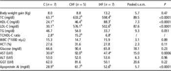
As reported in Table 1, animals of both CHF and IHF groups were fed on a high-fat diet and did not significantly differ in body weight gain after 10 weeks (P=0.065). After 10 weeks, the high-fat diet induced a similar increase in TC in both CHF and IHF groups, showing significantly higher levels of TC (P<0.0001) and LDL cholesterol (P<0.0001) compared with C (Table 1). Both CHF and IHF showed significantly higher values of HDL cholesterol compared to C (P<0.0001) and IHF showed significantly higher values of HDL cholesterol with respect to CHF (P<0.0001). CHF and IHF showed significantly higher values of TC/HDL cholesterol as compared with C (P<0.0001 and P<0.005, respectively). CHF also showed significantly higher values of TC/HDL cholesterol with respect to IHF (P<0.0001). No difference was found between CHF, IHF and C regarding the levels of TG, WBC, hematocrit, serum glucose and serum GGT. AST level was significantly higher in CHF group compared with C and IHF groups (P<0.0001 and P<0.001, respectively). No significant changes were found in ALT levels between groups.
Clinical data measured in pigs administered for 10 weeks with a standard diet (C), a high-fat/high-cholesterol diet continuously (CHF) and a high-fat/high-cholesterol diet intermittently (IHF)

TG=triglycerides; TC=total cholesterol; WBC=white blood cell; HCT=hematocrit; AST=aspartate amino transferase; ALT=alanine amino transferase; GGT=γ-glutamyltransferase; LDL=low-density lipoproteins.
Data are reported as means of 7 (C and IHF) or 5 (CHF) animals. Pooled s.e.m. and P are also reported. LDL cholesterol has been estimated by the Friedewald formula. Statistical analysis: ANOVA followed by Bonferroni post-hoc test. In the same row, significantly different means (P<0.05) are marked with different letters.
Histology
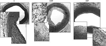
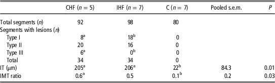
Initial preatheroma coronary lesions (Type I to III according to Stary staging (Stary et al., Reference Stary, Blankenhorn, Chandler, Glagov, Insull, Richardson, Rosenfeld, Schaffer, Schwartz and Wagner1992) or early preatherosclerotic lesions according to the Virmani revision (Virmani et al., Reference Virmani, Kolodgie, Burke, Farb and Schwartz2000) were observed at histology after 10 weeks of diet in both CHF and IHF groups (Figure 1). Total number of lesions, incidence and distribution of each lesion type in all analyzed segments and corresponding morphometric data (mean intimal thickening and intima to media thickness ratio) are summarized in Table 2 and compared with intact coronary segments of standard diet-treated animals (C). Although more advanced lesions were observed in CHF as compared with IHF cases, average morphometric data of all lesions had comparable values in the two groups.
Histopathology of the right coronary artery (proximal 10-mm segment) in a typical example of C, continuous high-fat diet (CHF), and intermittant high-fat diet (IHF) groups. Type II and initial type I fatty streaks are evident in CHF and IHF cases, respectively (H&E stain, 2× to 40× original magnification).

Total number (n) and percentage of preatherosclerotic coronary lesions on total number of analyzed segments in pigs administered for 10 weeks with a high-fat/high-cholesterol diet continuously (CHF), a high-fat/high-cholesterol diet intermittently (IHF) and in standard diet (C)

IT=intimal thickness; IMT=intima/media thickness.
Mean intimal thickness (IT) and intima to media thickness ratio (IMT) of CHF and IHF lesions and of all C segments are also reported.
Data are reported as means of 7 (C and IHF) or 5 (CHF) animals. For IT and IMT ratio, pooled s.e.m. and P are also reported. Statistical analysis of IT and IMT ratio: ANOVA followed by Bonferroni post-hoc test. In the same row, significantly different means (P<0.05) are marked with different letters.
Expression analysis
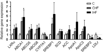
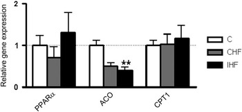
We performed real-time PCR experiments with liver cDNA samples using specific primers for many genes involved in lipid homeostasis in order to investigate the response patterns activated by administration of a high-fat diet. As illustrated in Figure 2, we found an increasing trend in the expression of the nuclear receptor LXRα in the CHF group and especially in the IHF group. As expected, we found a similar increasing trend in the expression of many genes regulated by LXRα, such as the transporters ABCA1, ABCG5 and ABCG8, and the transcriptional factor SREBP1. In particular, we highlighted a significant increase (about a fivefold induction) in the expression of ABCA1. Also, a comparable increasing trend was found for the expression of SCD, ACC, ApoA1 and ApoC3 genes. On the contrary, a significant reduction in the expression of SREBP2 and LDLr genes was found as a consequence of the administration of the high-fat diet both continually and on alternate weeks. No modulation of mRNA level was noticed for cytochrome P450 (CYP) 7A1 (Figure 2). Interestingly, no modulation was observed for PPARα mRNA or its target gene CPT-1 (Figure 3). Instead, a decreasing trend was noticed in hepatic mRNA levels of ACO after following the continuous high-fat diet, whereas a significant reduction was observed when the high-fat diet was administered on alternate weeks.
Relative gene expression of LXRα and its regulated genes. Relative gene expression of LXRα, ABCA1, ABCG5, ABCG8, CYP7A1, SREBP1, SCD, ACC, ApoA1, ApoC3, SREBP2 and LDLr measured by real-time RT-PCR in liver samples from control pigs (C, n=7), pigs subjected to the high-fat diet continually (CHF, n=5) and pigs subjected to the high-fat diet on alternate weeks (IHF, n=7). Results are normalized against two different housekeeping genes (GAPDH and β-actin), expressed as mean±s.e.m., and referred to the controls mean at which was assigned value=1. Statistical significance has been evaluated by one-way ANOVA followed by Bonferroni post-test. *Significantly different from control values, P<0.05; **significantly different from control values, P<0.01.

Relative gene expression of PPARα and its target genes. Relative gene expression of PPARα and its target genes ACO and CPT-1 measured by real-time RT-PCR in liver samples from control pigs (C, n=7), pigs subjected to the high-fat diet continually (CHF, n=5) and pigs subjected to the high-fat diet on alternate weeks (IHF, n=7). Results are normalized against two different housekeeping genes (GAPDH and β-actin), expressed as mean±s.e.m. and referred to the controls mean at which was assigned value=1. Statistical significance has been evaluated by one-way ANOVA followed by Bonferroni post-test. **Significantly different from control values, P<0.01.

Correlation analysis
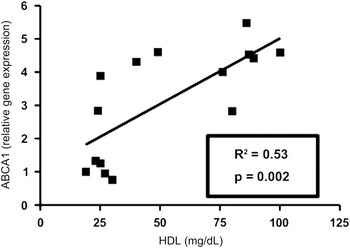
In order to link the systemic status with tissue expression results, we correlated some biohumoral data such as DHDL, LDL and TC with the hepatic expression of the genes, which were modulated by the administration of the high-fat diet (ABCA1, SREBP2 and LDLr). In Figure 4, the positive comparisons we found are illustrated. As expected, we found a significant correlation (R 2=0.53; P=0.002) between hepatic ABCA1 expression levels and HDL-C levels.
Correlation analysis. Significant correlation of ABCA1 expression with circulating HDL-C levels (R 2=0.53; P=0.002).

Discussion
In this study, we investigated the effects of the administration of a high-fat/high-cholesterol diet on lipid homeostasis in a porcine model. We found that the liver of the pigs was able to manage the overload of fatty acids efficiently in a short period of time (10 weeks). In fact, the high-fat diet resulted in a strong modulation of total plasma cholesterol (see Table 1), whereas plasma triglyceride levels were not significantly different, as previously reported by Dixon et al. (Reference Dixon, Stoops, Parker, Laughlin, Weisman and Sturek1999) in minipigs fed an atherogenic diet for 8 weeks. We observed no significant modulation for WBCs, hematocrit or glucose levels between the three experimental groups. The serum AST level was increased in CHF group only and not in the IHF group, whereas no changes were found in serum ALT levels between groups. Since ALT is considered a specific marker of liver damage, whereas AST is also present in many other tissues, we concluded that pig livers are not significantly damaged after the administration of a short-term high-fat diet, as reported in a previous paper (Puccinelli et al., Reference Puccinelli, Gervasi, Pelosi, Puntoni and Longo2012). Obviously, these results need to be further investigated in future studies, increasing the number of animals.
Our results indicate that there is a significant difference in the plasma lipid profile of the IHF group compared with the CHF group. A significant increase in TC and LDL-C levels in both CHF and IHF groups was observed. In addition, IHF pigs showed significantly higher values of HDL cholesterol with respect to CHF pigs. HDL-C levels are known to be inversely correlated with clinical events resulting in atherosclerosis, whereas LDL-C levels are directly related to the rate at which these events occur (Gordon et al., Reference Gordon, Castelli, Hjortland, Kannel and Dawber1977; Lewington et al., Reference Lewington, Whitlock, Clarke, Sherliker, Emberson, Halsey, Qizilbash, Peto and Collins2007). A number of dietary factors, genetic factors, and putatively, the timing of fat food intake, may all alter the amount of cholesterol carried in plasma LDL-C and HDL-C (Schaefer et al., Reference Schaefer, Levy, Ernst, Van Sant and Brewer1981) and may be directly implicated in either atherosclerosis promotion or prevention.
The ratio of total to HDL cholesterol (which is considered more important than the total or lipoprotein cholesterol concentrations in estimating the risk of cardiovascular events) showed a significant difference between the three experimental groups. The CHF group showed a fivefold increased risk of developing cardiovascular disease compared with the IHF group, which has a twofold increased risk compared with the C group. In our experimental model, the higher TC to HDL-C ratio of CHF group was associated to a greater incidence of Stary type II and III preatherosclerotic lesions as compared with IHF group, suggesting a mild atheroprotective effect of HDL-C plasma level, not confirmed by morphometric average values of lesion size. On the other hand, an intra-group direct correlation between individual values of total to HDL-C ratio and corresponding lesion thickness of each case has been observed (data not shown). Altogether these findings support the current hypothesis that a relatively higher HDL-C plasma concentration may be ‘protective’ at endothelial artery level in the context of a diet-induced elevation of circulating TC. To exclude that the fibrotic process was activated by the dietary cholate used in the animals diet to improve absorption of fat, the expression of transforming growth factor β1 (TGFβ1) gene, a fibrosis marker, was examined. The major source of collagen and other extracellular matrix proteins in liver fibrosis is hepatic stellate cells, a population of peri-sinusoidal cells comprising 15% of the resident liver cells (Friedman et al., Reference Friedman2000; Kmiec, Reference Kmiec2001). Hepatic stellate cells typically exist in a quiescent state, serving as the principal storage site for retinoids. In response to stimuli such as bacterial infection or inflammation, the stellate cells become activated and transform into proliferative, fibrogenic cells. The CHF diet slightly increased the expression of TGFβ1 above the control level, whereas the IHF intermittent diet was devoid of this effect (data not shown).
In terms of lipid homeostasis, the hepatic expression of various genes was analyzed in order to investigate patterns activated by high-fat/high-cholesterol diet in pigs. The oxysterols derived from CYP-dependent cholesterol metabolism such as 24-, 25- and 27-hydroxycholesterols are potent LXRα ligands (Janowschki et al., Reference Janowschki, Willy, Devi, Falck and Mangelsdorf1996; Millatt et al., Reference Millatt, Bocher, Fruchart and Staels2003; Baranowski et al., Reference Baranowski2008). LXRα activation and the consequent upregulation of ABCA1 constitute a key step in the process of reverse cholesterol transport, by which HDL particles transport excess cholesterol from tissues back to the liver (Lewis et al., Reference Lewis and Rader2005). In our study, we found in liver an increasing trend in mRNA levels of LXRα and many of its target genes, namely ApoA1, ApoC3, SREBP1 and SCD and ACC (see Figure 2). Hepatic ABCA1 expression was significantly correlated with plasma HDL cholesterol levels (see Figure 4). These findings are in agreement with previous results obtained in rodents and are probably a part of a compensatory response of the liver in order to keep fatty acid/cholesterol balance, thereby preventing the possibility of deleterious changes in membrane structure.
In the present study, we found no modulation of PPARα mRNA in the three experimental groups after the administration of various fat-enriched diets, in agreement with Duran-Montgé et al. (Reference Duran-Montgé, Theil, Lauridsen and Esteve-Garcia2009a and Reference Duran-Montgé, Theil, Lauridsen and Esteve-Garcia2009b). Furthermore, we showed a lack of induction of ACO and CPT-1, two genes involved in fatty acid β-oxidation, which are directly regulated by PPARα. Administration of the high-fat diet resulted in unchanged hepatic levels of CPT-1 mRNA but decreased levels of ACO mRNA. A differential regulation of these two genes was previously reported in literature (Cabrero et al., Reference Cabrero, Jové, Planavila, Merlos, Laguna and Vazquez-Carrera2003). It is worth noting that the pig is a so-called ‘non-proliferating species’ like humans but unlike rodents, due to a different expression and structure of PPARα across the species (Stott et al., Reference Stott, Yano, Williams, Barnard, Hannah, Cieszlak and Herman1995). In fact, expression of PPARα in the porcine liver is much lower than in rat liver (about 10-fold lower), resulting in a much weaker response to PPARα activators when compared with proliferating species (Cheon et al., Reference Cheon, Nara, Band, Beever, Wallig and Nakamura2005; Luci et al., 2007b). It is also possible, as reported by Chakrawarthy et al. (Reference Chakravarthy, Lodhi, Yin, Malapaka, Xu, Turk and Semenkovich2009), that the lipids of a diet originate lipid droplets instead of being ligands for PPARα. Contributing to the lack of PPARα, activation could be the accumulation of a triol derived from cholesterol metabolism (Dussault et al., Reference Dussault, Yoo, Lin, Wang, Fan, Batta, Salen, Erickson and Forman2003) and the consequent activation of PXR (pregnane X receptor), which in turn can inhibit PPARα (Zhou et al., Reference Zhou, Zhai, Mu, Gong, Uppal, Toma, Ren, Evans and Xie2006). We can hypothesize PPARα activation by fatty acids and a concurrent inhibition by PXR, resulting in a lack of effect. Interestingly, we did not find significant CYP7A1 mRNA modulations in the three porcine experimental groups. CYP7A1 is regulated by LXRα in rodents but not in humans, due to a lack of the LXRα-binding response element (LXRE) in the promoter of the human gene (Chiang et al., Reference Chiang, Kimmel and Stroup2001; Chiang, Reference Chiang2004). The possibility that the porcine CYP7A1 gene actually misses the LXRE as in humans should be considered.
Finally, we showed a significant decrease in the expression of SREBP2 – and consequently of its target gene LDLr – as a consequence of cholesterol excess. Indeed, SREBP2 is an important regulator of genes for cholesterol biosynthesis. It is known that the activation of LXRα is able to downregulate SREBP2 expression, although the molecular mechanism underlying this modulation remains unclear (Millatt et al., Reference Millatt, Bocher, Fruchart and Staels2003). Thus, a double action of this response to cholesterol excess was highlighted: the inhibition of cholesterol biosynthesis on one hand and the inhibition of lipid uptake from plasma circulating particles on the other.
Although administration of the high-fat diet resulted in the activation of LXRα and SREBP1, no modulation was noticed for PPARα and its target genes. Interestingly, we found a better adaptive response when a fat-supplemented diet was administered on alternate weeks instead of every week. Sporadic and/or discontinuous high-fat/high-cholesterol food intake is a general characteristic of Western countries’ nutrition styles. The results of our study highlight the potentially beneficial effects of irregular atherogenic food intake, resulting from the combined effect of higher HDL-C plasma concentration and upregulation of several genes involved in hepatic lipid metabolism. Of course, these results should be considered as initial steps for future studies where these data will be investigated more thoroughly. Increasingly often, humans spend periods/days eating in a completely unregulated way, eating excessive amounts of food rich in oil and fat, alternating periods/days when food intake is more or less correct. Periodic withdrawals from a high-fat atherogenic diet, compared with a regular administration, could result in a different adaptive response of lipoprotein metabolism, leading to significantly higher plasma levels of HDL-C and lower TC/HDL-C.
Acknowledgments
The study was funded in part by a grant from the ARTreat project, FP7-ICT-2007 (grant agreement 224297). The authors gratefully acknowledge Dr Claudia Kusmic for her professional assistance in diet formulation, Mr F. Bernini for his help in animal experiments and Mrs Alison Frank for her support in editing the English of the manuscript.
Supplementary material
To view supplementary material for this article, please visit http://dx.doi.org/10.1017/S1751731114003292