Introduction
Minimally invasive surgical techniques are appealing in paediatric heart surgery but offer new challenges to the anaesthesiologist. Dodge-Khatami et al. describe the advantages of the mid-axillary incision for trans-atrial repairable congenital cardiac defects. Reference Dodge-Khatami and Salazar1,Reference Prêtre, Kadner, Dave, Dodge-Khatami, Bettex and Berger2 These include a cosmetically superior scar hidden by the resting arm and an incision far away from breast tissue. Reference Said, Greathouse and McCarthy3 Comparative studies suggest that this approach may improve postoperative quality of life and reduce ICU and hospital stay compared to median sternotomy. Reference Dodge-Khatami and Dodge-Khatami4 This technique allows for extubation in the operating room, minimal complications, and a favourable recovery profile. Reference Said, Greathouse and McCarthy3 While the mid-axillary surgical approach has been previously described, the anaesthetic and perioperative strategies that directly influence recovery, length of stay and overall success have not been reported. The minimally invasive surgical approach is becoming the new norm in many specialised paediatric cardiac centres. Reference Dodge-Khatami, Dodge-Khatami, Nguyen and Rüffer5 This minimally invasive approach does not appear to compromise surgical safety or outcomes, even during early adoption phases. Reference Lo Rito, Brindicci and Moscatiello6 Our manuscript provides an enhanced recovery after surgery template for those centres using the minimally invasive surgical approach.
The analgesic options for surgeries approached via a thoracotomy include neuraxial blocks, chest wall blocks, paracostal interpleural nerve blocks, intravenous narcotics, or any combination thereof. The potential advantages of epidural neuraxial analgesia include reduced morbidity via targeted thoracic sympathectomy, early initiation of feeding, and lower intensity postoperative care in infants recovering from thoracotomy surgeries. Reference Peterson, DeCampli, Pike, Robbins and Reitz7–Reference Tsui, Wagner, Cave and Kearney9 Recent prospective evidence also demonstrates that high thoracic epidural analgesia significantly reduces inflammatory markers, mechanical ventilation duration, and hospital length of stay compared to systemic opioid regimens in paediatric cardiac surgery. Reference Caruso, Lawrence and Tsui10 Additional observational data reinforce the role of thoracic epidurals in attenuating perioperative inflammation and improving renal perfusion, pulmonary function, and glycemic control in this population. Reference Kumar, Ghotra and Dwivedi11 However, continuous epidural analgesia remains an underutilised modality in children and infants due to its technical challenges, variability in results, and rare but catastrophic complications. Reference Tsui, Wagner, Cave and Kearney9,Reference Kumar, Kapoor, Mishra, Joshi, Tiwari and Ramamurthy12 The success of the neuraxial technique is also dependent on the anaesthesiologist’s experience, the method used for its placement, and the limitations to confirmation of the appropriate level and position of the catheter tip. Our enhanced recovery after surgery protocol addresses the variability of the epidural by using transesophageal echocardiography to confirm catheter tip level in patients under general anaesthesia without the need for fluoroscopy.
With these challenges, a multimodal pain management protocol for enhanced recovery after surgery was devised with the input of the surgical, critical care and anaesthetic teams (Supplementary file). The protocol included the placement of a thoracic epidural catheter, under transesophageal echocardiography guidance, at least one hour prior to systemic heparinisation, low-dose intraoperative opioid use, encouraging early extubation, early feeding, scheduled non-narcotic-based analgesic medications in the immediate postoperative period, and early mobilisation. The primary aim of this study was to evaluate the feasibility and clinical outcomes of a standardised enhanced recovery after surgery protocol, including thoracic epidural analgesia, in children undergoing trans-atrial cardiac surgery via the right mid-axillary thoracotomy approach.
Materials and methods
This was an IRB-approved (# HSC-MS-19-0389), single-centre, retrospective study of paediatric patients under 18 years of age who underwent trans-atrial cardiac surgery through the right mini mid-axillary thoracotomy approach. The duration of the study was from July 2018 to May 2020. The start date corresponds to the formalisation of the anaesthetic protocol and the introduction of this approach at the study institution. A detailed institutional protocol for the right mid-axillary approach, including perioperative workflow and anaesthetic steps, is provided in Supplementary File 1. The majority of the data are collected as observational and prospective, being stored electronically as part of a pre-existing anaesthetic and surgical database.
The congenital heart conditions amenable to repair via the right mid-axillary thoracotomy trans-atrial approach are atrial septal defects, ventricular septal defects, partial anomalous pulmonary venous return including the Warden operation, partial atrio-ventricular canal with cleft mitral valve, double-chambered right ventricle, scimitar syndrome, and cor triatriatum, amongst others. Reference Dodge-Khatami, Gil-Jaurena and Hörer13 To ensure feasibility of the thoracotomy approach, the protocol called for performing an early transesophageal echocardiographic assessment of the defect immediately after endotracheal intubation prior to any other anaesthetic intervention. Relative anatomic challenges precluding this approach include outlet-type ventricular septal defects, defects requiring surgical repair of the aortic or pulmonary valve, and the presence of a large patent ductus arteriosus.
Continuous epidural analgesia was chosen as the primary strategy for intraoperative and postoperative pain control once the thoracotomy approach was confirmed. One of the goals of this protocol was to decrease the variability of the epidural catheter tip location and to improve the clinical reliability of the block. Previously, transesophageal sonography has been used to confirm epidural catheter advancement from the caudal approach. Reference Tsui, Wagner, Cave and Kearney9,Reference Caruso, Lawrence and Tsui10 However, the long path of the caudally placed catheter has considerable variability in achieving optimal catheter tip positioning, limiting its utility. Reference Tsui, Wagner, Cave and Kearney9,Reference Caruso, Lawrence and Tsui10,Reference Ueda, Shields and Brennan14,Reference Blanco, Llamazares, Rincón, Ortiz and Vidal15 We minimised this clinical variability in multiple ways. Based on the patient’s spinal anatomy, we accessed the epidural interspaces by loss of resistance technique in the lower thoracic interspace (T6-L1). A stimulating catheter was then advanced under transesophageal echocardiography guidance, and catheter tip location was targeted to be just above the level of the aortic valve in short-axis view. The catheter used in this protocol was a Pajunk stimulating-tip epidural catheter, which is both nerve-stimulating and radio-opaque, allowing for dual-mode confirmation of tip position. The expansion of the epidural space was observed with the injection of saline via the epidural catheter. Figure 1 shows a representative transesophageal echocardiography image captured during real-time catheter placement, with the epidural space visualised as expanding during saline injection. The catheter was then secured in a standard fashion, a bolus of 0.4 mg/kg of ropivacaine 0.2% administered, and a continuous infusion was started. The protocol addressed concerns regarding systemic heparinisation during the procedure and postoperative catheter care (Supplementary file). If the epidural was declined or unsuccessful (more than three attempts) or was deemed unsafe (aspiration of blood during localisation), then a lateral paracostal, interpleural catheter was placed under direct vision at the completion of surgery before closing the thoracotomy by the surgeon. This technique builds upon growing evidence that transesophageal echocardiography can visualise thoracic spinal structures in real time, allowing confirmation of catheter trajectory and expansion. Reference Hanada, Reinking, Kim and Ueda16 Similar success has been reported using fluoroscopy-guided techniques for thoracic epidural placement in children with CHD. Reference Schmehil, Lee, Casella and Millan17
Transesophageal echocardiography (TEE) image demonstrating real-time expansion of the thoracic epidural space during catheter placement.

Cardiopulmonary bypass practice was standardised throughout the study period. Packed red cells and fresh frozen plasma were added to the cardiopulmonary bypass circuit as needed. One dose of steroids (methylprednisolone 15 mg/kg) was given on bypass. Dexmedetomidine infusion (0.5–1 mg/kg/hour) was started after induction of anaesthesia and continued until tracheal extubation. Acetaminophen was administered before tracheal extubation and was continued on a scheduled basis. In the postoperative period, all opioids and non-opioid analgesic medications were administered as per the paediatric pain service order set.
Data analysis and statistics
Data analysis included the prospectively collected congenital anaesthesia database, regional anaesthesia database, surgical database, and electronic medical records until hospital discharge. Clinical outcomes included regional technique outcomes such as the level of epidural space localisation, level of placement, duration of placement, use of opioid and non-opioid medication, duration of mechanical ventilation, and hospital length of stay. As per intensive care assessment protocols, the face, leg, activity, crying, and consolability scale (range 0 to 10) was used to document pain scores. The face, leg, activity, crying, and consolability scores, recorded hourly along with rescue medication log in the intensive care unit, were collated. Scores for the first twelve postoperative hours were used for data analysis. The normality of continuous variables was assessed using the Shapiro-Wilk test and histograms. Variables with satisfactory normal distributions were summarised using the mean and standard deviation. For variables with skewed distributions, the median, first and third quartiles, minimum, and maximum were reported.
Results
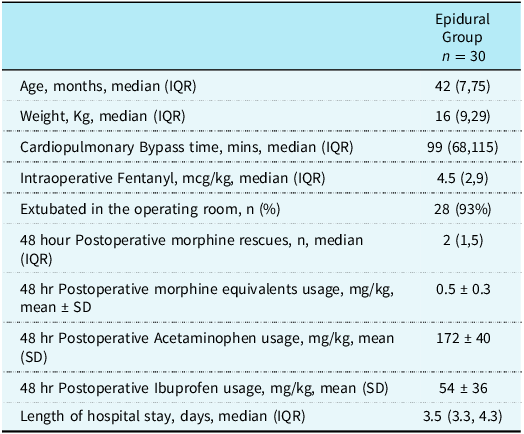
Forty-two paediatric patients underwent a thoracotomy for cardiac surgery during the study period (Table 1). Five patients were transferred to the operating room from the cardiac catheterisation suite when the defect was not amenable to percutaneous device closure. An epidural consent was obtained in 32 patients, and the epidural space was localised by the loss of resistance technique in the T6-L1 interspaces in 25 patients (83%) and L1–L3 interspaces in 5 patients (17%). Once the epidural space was localised, a stimulating catheter was advanced under trans-oesophageal ultrasonography guidance, and the tip placed between the third and fifth thoracic interspace levels in all 30 patients. The location of the catheter tip was later confirmed by radiography. The average time between epidural placement and systemic heparinisation was 106 ± 40 minutes. Epidural infusions were continued for a median of 52 hours postoperatively.
Demographics and outcomes

IQR = interquartile range.
The median age of patients with epidural analgesia was 42 months ( range 3 months to 13 years), and median weight was 15.7 kg ( range 4.9 to 61 kg), Table 1. The intraoperative fentanyl requirement was a median of 4.5 mcg/kg (2–9 mcg/kg) in this cohort. Twenty-eight of the 30 patients (93%) were extubated in the operating room, and two patients were extubated within six hours of ICU admission. The face, leg, activity, crying, and consolability scores for the first 12 hours after extubation were a median of zero (average of 0.8 ± 1.7). The number of intravenous morphine rescues needed after extubation in the intensive care unit was a median of 2, and the 48-hour total opioid requirement was a mean of 0.5 ± 0.3 mg/kg of morphine equivalents. There were no complications related to the surgical approach, and no conversion in approach from a thoracotomy to sternotomy occurred in any patients. There were no re-intubations, neuraxial blockade-related complications, or other anaesthesia-related adverse events. Patients were discharged home in 3.5 days.
Discussion
Paediatric cardiac surgery via the mid-axillary approach is gaining popularity with its superior cosmetic results. However, this approach presents new challenges to the anaesthesiologists and perioperative care team. Without adequate analgesia after a thoracotomy, the clinical goals of rapid extubation, reduced opioid requirements, shortened intensive care stay, and early discharge after cardiac surgery are not feasible. While the optimal choice of a regional anaesthetic block for thoracotomy in children is still debated, the need for such techniques is evident.
Our study demonstrates that a collaborative, multimodal analgesic protocol, centred around a successful thoracic epidural, is both feasible and safe to attain aforesaid clinical goals. Despite the technical challenges of performing thoracic epidurals in infants and small children, catheters were successfully placed at the desired thoracic dermatome in all patients where the epidural space was accessed. This is in part due to the utilisation of transesophageal echocardiography to guide and confirm epidural placement. The utilisation of targeted neuraxial techniques resulted in low intraoperative opioid requirements and enabled a high rate of fast-track tracheal extubation. In the postoperative period, rescue opioid requirements were minimal, likely due to continuous local anaesthetic infusions and scheduled non-narcotic medications. Median hospital length of stay was 3.5 days. In addition, systemic heparinisation was not deemed to be an issue for either placement or removal of epidural catheters, as all procedures adhered to established guidelines. Reference Horlocker, Vandermeuelen and Kopp18
We also acknowledge that a large part of this protocol’s success may be attributed to the collaboration and input of all clinical providers and caregivers. Clear expectations for early extubation, adoption of neuraxial analgesia, minimal opioid utilisation, standardising non-narcotic analgesic medications, early feeding, early ambulation, and neuraxial safe anticoagulation strategies were agreed upon among the care teams, helping to reduce conflicting protocols and smooth transitions of care between services. The role of the paediatric pain team as the sole manager of postoperative analgesia helped reduce confusion among caregivers.
Limitations of this study include lack of a control group not receiving epidural analgesia, acknowledging that our institutional protocol was not designed to look for differences between patients with and without epidural analgesia. Nevertheless, our study serves as a pilot for the safety and feasibility of an enhanced recovery protocol for mid-axillary thoracotomy paediatric cardiac surgeries. The included protocol provides a template for other institutions wanting to adopt this surgical approach. While our emphasis was to maximise benefits of epidural analgesia, we do recognise the need for prospective, randomised studies to further elucidate the best regional modality for post-thoracotomy pain after paediatric minimally invasive cardiac operations.
Supplementary material
The supplementary material for this article can be found at https://doi.org/10.1017/S1047951125109761.
Acknowledgements
We are grateful for the support of Xu Zhang, PhD, Associate Professor in Clinical and Translational Sciences, for assistance with the data analysis carried out in this work.
Financial support
This research received no specific grant from any funding agency, commercial, or not-for-profit sectors.
Competing interests
Nischal Gautam reports a contract with Hemonetics unrelated to this manuscript. All other authors declare no conflicts of interest.