INTRODUCTION
Diarrhoeal diseases are extremely common throughout the world, both in developed and developing countries. In the industrialized countries in particular protozoa are an important factor in the aetiology of diarrhoeal diseases in children. Traditionally, microscopic examination of faecal samples is the diagnostic method that is used for the detection of parasites causing diarrhoea. However, a diversity of additional techniques for each individual organism and multiple sampling is needed to increase the sensitivity and specificity of the detection of the different parasites. In the last 10 years, many diagnostic laboratories have become equipped with the facilities to perform molecular diagnostics. Through the introduction of real-time PCR, problems of contamination with PCR products are greatly reduced as it is a closed system. Using probe-based real-time PCRs it is relatively simple to combine multiple DNA targets in a multiplex assay with the use of different fluorescent labels for each specific detection probe (e.g. Taqman and Minor Groove Binding (MGB) probes, Molecular beacons and fluorescence energy transfer (FRET)-based real-time PCR Logan et al. Reference Logan, Edwards and Saunders2009 – ). Moreover, the implementation of automated DNA isolation methods has made it possible to use DNA-based detection techniques in a high-throughput format in a routine diagnostic setting (van Lieshout and Verweij, Reference van Lieshout and Verweij2010; Stensvold et al. Reference Stensvold, Lebbad and Verweij2011). In this paper, the most relevant parasitic infections causing diarrhoea in children in industrialized countries and the impact of using molecular diagnostics in the routine laboratory setting will be discussed.
INTRODUCTION TO THE MOST RELEVANT INTESTINAL PROTOZOA
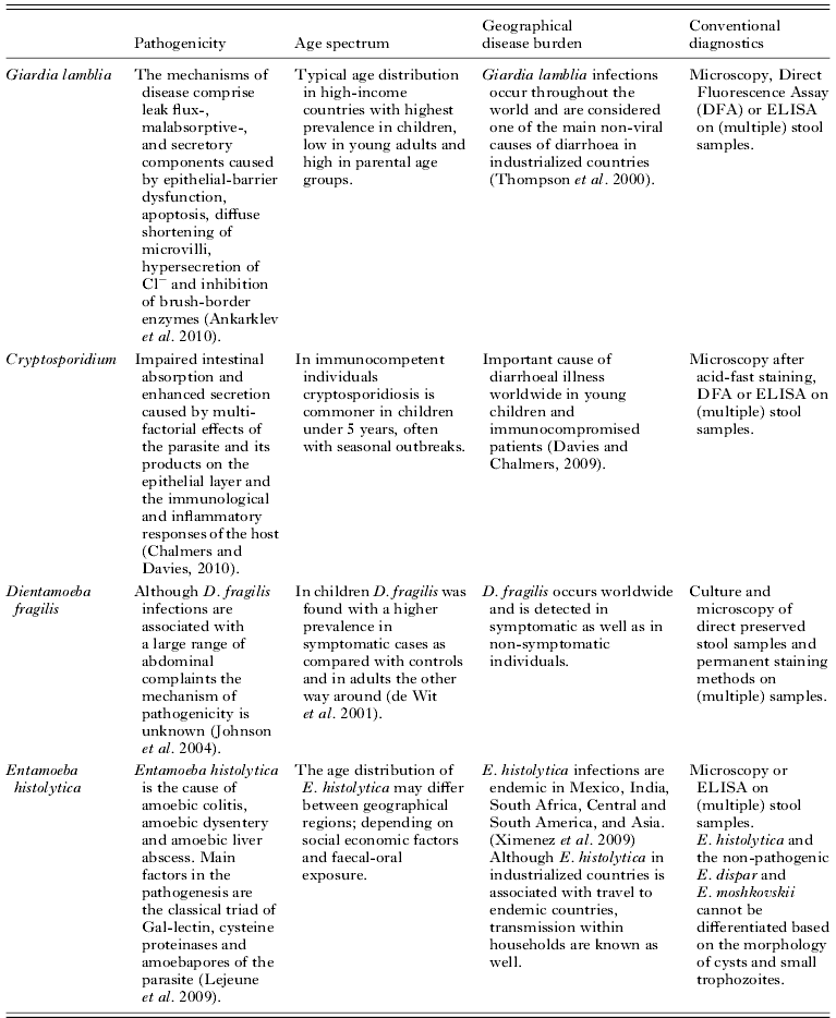
Giardia lamblia infections occur throughout the world and are considered one of the main non-viral causes of diarrhoea in industrialized countries with high prevalence rates in day-care centres and within families (Thompson et al. Reference Thompson, Hopkins and Homan2000). Traditionally, the diagnosis is based on microscopic examination of (multiple) faecal samples using concentration techniques. Direct fluorescent assays (DFA) and the detection of Giardia antigen by ELISA have been accepted as cost-effective alternative diagnostic methods (Garcia and Shimizu, Reference Garcia and Shimizu1997; Mank et al. Reference Mank, Zaat, Deelder, van Eijk and Polderman1997). DNA amplification techniques have shown an excellent sensitivity and a 100% specificity as compared with microscopy and antigen detection (Ghosh et al. Reference Ghosh, Debnath, Sil, De, Chattopadhyay and Das2000; Verweij et al. 2003, Reference Verweij, Blange, Templeton, Schinkel, Brienen, van Rooyen, van Lieshout and Polderman2004; Haque et al. Reference Haque, Roy, Siddique, Mondal, Rahman, Mondal, Houpt and Petri2007; Calderaro et al. Reference Calderaro, Gorrini, Montecchini, Peruzzi, Piccolo, Rossi, Gargiulo, Manca, Dettori and Chezzi2010a).
Cryptosporidium-associated diarrhoea is especially known for its severe symptoms in AIDS patients. Subsequently, improved diagnostic methods were developed and Cryptosporidium is now known as a major cause of water- and food-related outbreaks of gastroenteritis in immunocompetent persons as well (Current and Garcia, Reference Current and Garcia1991; Nichols, Reference Nichols2000). Although probably underestimated, cryptosporidiosis is believed to be one of the major causes of childhood diarrhoea worldwide (Lee and Greig, Reference Lee and Greig2010; Guyot and Derouin, Reference Guyot and Derouin2010; Putignani and Menichella, Reference Putignani and Menichella2010). Modified acid-fast staining techniques are often used for microscopic detection of Cryptosporidium oocysts in stools. The sensitivity and specificity appear to be low as a correct identification is highly dependent on the experience and skills of the microscopist (Weber et al. Reference Weber, Bryan, Bishop, Wahlquist, Sullivan and Juranek1991). Immunological detection methods are available, but due to lack of sensitivity and specificity are not as well accepted as they are in the diagnosis of Giardia infections (Webster et al. Reference Webster, Smith, Giles, Dawson and Robertson1996). PCR has been shown to be a sensitive and specific alternative for the detection of Cryptosporidium in faecal samples (Morgan et al. Reference Morgan, Pallant, Dwyer, Forbes, Rich and Thompson1998; Verweij et al. Reference Verweij, Blange, Templeton, Schinkel, Brienen, van Rooyen, van Lieshout and Polderman2004).
Dientamoeba fragilis was first described in 1918 as a commensal amoeba of the gastrointestinal tract of humans (Jepps and Dobell, Reference Jepps and Dobell1918). In later years classified as a flagellate, through antigen and ultrastructural studies and analysis of the 16S ribosomal RNA, although lacking a flagellum (Camp et al. Reference Camp, Mattern and Honigber1974; Silberman et al. Reference Silberman, Clark and Sogin1996). Since its discovery, the pathogenicity of this organism has remained controversial. In recent years, several authors have published on the clinical importance of D. fragilis as a cause of gastrointestinal problems (Johnson et al. Reference Johnson, Windsor and Clark2004; Stark et al. Reference Stark, Barratt, Roberts, Marriott, Harkness and Ellis2010b). A consensus on the pathogenicity, however, remains unsettled, especially since many D. fragilis infections remain asymptomatic (Okhuysen Reference Okhuysen2001; Gendrel et al. Reference Gendrel, Treluyer and Richard-Lenoble2003; Farthing Reference Farthing2006). In a case-control study conducted in the Netherlands, D. fragilis was indeed found in a higher prevalence in the control group. In children, however, the prevalence of D. fragilis was significantly higher in the cases presenting with abdominal complaints (de Wit et al. Reference de Wit, Koopmans, Kortbeek, van Leeuwen, Bartelds and van Duynhoven2001a, Reference de Wit, Koopmans, Kortbeek, van Leeuwen, Vinje and van Duynhovenb). Because a cyst stage in D. fragilis is missing, microscopic examination should be performed on fresh stools or using fixatives and permanent stains to detect the fragile trophozoites. The sensitivity of examining a single sample is not high because the day-to-day variation of excreted D. fragilis seems even more irregular than that observed in other intestinal protozoan infections such as G. lamblia and E. histolytica (van Gool et al. Reference van Gool, Weijts, Lommerse and Mank2003; Johnson et al. Reference Johnson, Windsor and Clark2004). Immunological techniques for detection of D. fragilis are currently lacking. Real-time PCR is a highly sensitive and specific alternative for the diagnosis of D. fragilis (Verweij et al. Reference ten Hove, Schuurman, Kooistra, Moller, van Lieshout and Verweij2007; Bruijnesteijn van Coppenraet et al. Reference Bruijnesteijn van Coppenraet, Wallinga, Ruijs, Bruins and Verweij2009; Stark et al. Reference Stark, Barratt, Roberts, Marriott, Harkness and Ellis2010a; Calderaro et al. Reference Calderaro, Gorrini, Montecchini, Peruzzi, Piccollo, Rossi, Gargiulo, Manca, Dettori and Chezzi2010b). After at least 8 weeks storage of the faecal samples at 4°, Dientamoeba DNA could still successfully be demonstrated with real-time PCR without loss of sensitivity. Moreover, the influence of the day-to-day variation on the outcome is significantly reduced (Verweij et al. Reference ten Hove, Schuurman, Kooistra, Moller, van Lieshout and Verweij2007).
Entamoeba histolytica is the cause of amoebic colitis, amoebic dysentery and amoebic liver abscess and is responsible for an estimated 100 000 deaths per year (Walsh, Reference Walsh1988). Until 20 years ago, differences in clinical symptoms were explained by the existence of pathogenic and non-pathogenic strains of E. histolytica. Biochemical, immunological and genetic differences between these ‘strains’ have proved sufficient that it is now generally accepted that they represent two different species, E. histolytica and E. dispar, respectively (Diamond and Clark, Reference Diamond and Clark1993; Gonzalez-Ruiz and Wright, Reference Gonzalez-Ruiz and Wright1998). Given that the potentially invasive E. histolytica is morphologically indistinguishable from the non-invasive E. dispar, microscopic examination only is not sufficient for the definitive diagnosis of an E. histolytica infection and additional methods, such as the detection of specific antigens or specific DNA are necessary (Verweij et al. Reference Verweij, Oostvogel, Brienen, Nang-Neifubah, Ziem and Polderman2003a; Qvarnstrom et al. Reference Qvarnstrom, James, Xayavong, Holloway, Visvesvara, Sriram and Da Silva2005; Fotedar et al. Reference Fotedar, Stark, Beebe, Marriott, Ellis and Harkness2007). Antigen detection is highly specific but has a low sensitivity in comparison with PCR (Mirelman et al. Reference Mirelman, Nuchamowitz and Stolarsky1997; Visser et al. Reference Visser, Verweij, Van Esbroeck, Edeling, Clerinx and Polderman2006). Although, amoebiasis might not be the first in line when thinking of parasitic infection in children in the western world, family outbreaks are reported quite frequently and might be underestimated as well (Gatti et al. Reference Gatti, Cevini, Bruno, Novati and Scaglia1995; Vreden et al. Reference Vreden, Visser, Verweij, Blotkamp, Stuiver, Aguirre and Polderman2000; Edeling et al. Reference Edeling, Verweij, Ponsioen and Visser2004).
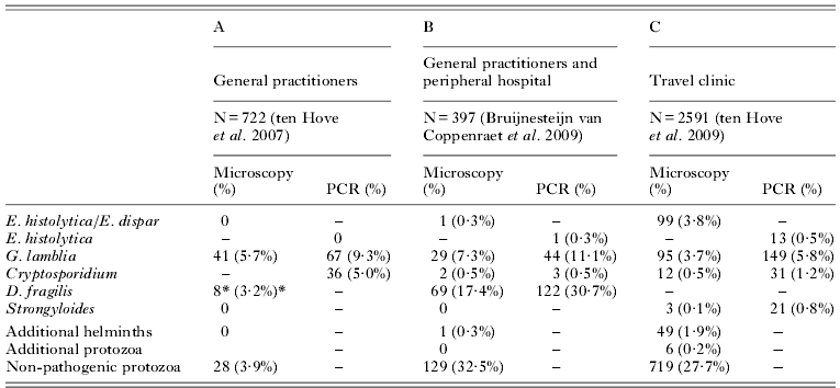
In summary, conventional detection methods of these intestinal protozoa have their limitations in specificity and sensitivity. Particular techniques or adaptations for each organism separately are required to obtain the most optimal diagnosis whereas multiplex real-time PCR offers a sensitive and specific detection of each of these parasites. As an illustration of the application of real-time PCR for the detection of intestinal protozoa three previously published studies in routine settings with a different patient population are summarized and discussed with an emphasis on parasitic infections in children (ten Hove et al. Reference ten Hove, Schuurman, Kooistra, Moller, van Lieshout and Verweij2007, Reference ten Hove, Van Esbroeck, Vervoort, Van den Ende, van Lieshout and Verweij2009; Bruijnesteijn van Coppenraet et al. Reference Bruijnesteijn van Coppenraet, Wallinga, Ruijs, Bruins and Verweij2009). The comparison of microscopic examination and real-time PCR results in these studies are summarized in Table 1.
Comparison of microscopic examination and real-time PCR for the detection of intestinal parasites in faecal samples from subjects attending their general practitioner (A), their general practitioner or medical specialist (B) with gastrointestinal complaints or attending a travel clinic with or without gastrointestinal complaints (C)

– Not performed.
* Only 247 SAF preserved faecal samples examined.
COMPARISON OF MICROSCOPY AND A PCR APPROACH IN DIFFERENT SETTINGS
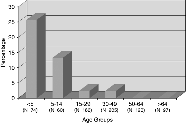
In the first study (Table 1A), 722 faecal DNA samples were available from patients attending their general practitioner (GP) with gastrointestinal symptoms between June and September 2005 (ten Hove et al. Reference ten Hove, Schuurman, Kooistra, Moller, van Lieshout and Verweij2007). A multiplex real-time PCR for the detection of E. histolytica, G. lamblia and Cryptosporidium (HGC-PCR) (Verweij et al. Reference Verweij, Blange, Templeton, Schinkel, Brienen, van Rooyen, van Lieshout and Polderman2004) was performed and compared with routine microscopy using formalin-ether concentration. Giardia was detected with real-time PCR in 67 of 722 samples (9·3%) compared with 5·7% using microscopy. The expected age distribution of G. lamblia infections with higher prevalence rates in children, lower in teenagers and higher rates again from around 30 years of age becomes even more pronounced by the use of PCR. Especially in the children and in the “parents’ age group” of 30–49 years of age, many extra Giardia infections are detected with real-time PCR. In this group of symptomatic cases with apparently low numbers of parasites that are excreted, the infection would have been missed with routine microscopy. In this setting, additional staining for the detection of Cryptosporidium was performed only if requested and because this was not done in any of the cases in this study group Cryptosporidium was not detected. The real-time PCR for Cryptosporidium tested positive in 36 of 722 samples (5·0%) and the detection rate in children under five years of age in this specific study population was more than 25 percent (n=74) (Fig. 1). As a follow-up of this study, additional samples collected from October until January showed that Cryptosporidium reached the highest prevalence rates in August and September. E. histolytica/E. dispar was not detected in any of the samples. Non-pathogenic protozoa were detected in 3·9% of the samples with microscopy and additional parasitic pathogens were not detected in this study population. D. fragilis could be detected only in 8 of 247 cases (3·2%) in which SAF-preserved faecal samples were available. This prevalence seems very low as compared to previous studies conducted in similar population in the Netherlands (de Wit et al. Reference de Wit, Koopmans, Kortbeek, van Leeuwen, Vinje and van Duynhoven2001b). At the time of this study D. fragilis-specific PCR was not yet available but could be used in the second study discussed below conducted by Bruinensteijn van Coppenraet and co-workers. In conclusion, the study of Ten Hove (Reference ten Hove, Schuurman, Kooistra, Moller, van Lieshout and Verweij2007) showed that in a population of patients attending their family doctor with abdominal complaints, the real-time PCR approach revealed significantly higher detection rates of Giardia and unexpected high rates of Cryptosporidium especially in children.
Prevalence of Cryptosporidium parvum ⁄ Cryptosporidium hominis in different age groups, detected with real-time PCR (n=722). The number of cases examined in each category is indicated below each bar.

Key characteristics of the most relevant intestinal protozoa in an industrialized country

The second study (Table 1B), based in a diagnostic laboratory situated in a peripheral hospital, samples from patients (N=397) visiting their family doctor or a specialist in the hospital with gastrointestinal complaints were collected between January 2007 and April 2007 (Bruijnesteijn van Coppenraet et al. Reference Bruijnesteijn van Coppenraet, Wallinga, Ruijs, Bruins and Verweij2009). Although ages were not available for analysis it can be assumed that the age distribution is quite similar as the study in the general practitioners’ population as discussed above. Microscopy for each patient was performed on three consecutive three faecal samples (Triple Faeces Test, TFT) of which the first and third samples were preserved using SAF and the second sample was unpreserved (van Gool et al. Reference van Gool, Weijts, Lommerse and Mank2003). Microscopy was performed using direct wet mounts of both SAF-preserved samples and positive or suspected direct smears were further examined using Chlorazol Black permanent staining. Microscopy for the presence of helminth ova, larvae and cysts was performed using a formalin-ether concentrate of the unpreserved sample. Modified acid-fast staining for detection of Cryptosporidium on the formalin–ether concentrate was performed, on specific request, on liquid stool samples, and for samples from subjects under 16 years of age. Microscopy results were compared with real-time HGC PCR and D. fragilis real-time PCR (Verweij et al. Reference ten Hove, Schuurman, Kooistra, Moller, van Lieshout and Verweij2007) performed on faecal DNA obtained from the unpreserved sample. E. histolytica/E. dispar was detected with microscopy in one case and was detected and specified as E. histolytica with PCR. G. lamblia was detected in one or more faecal samples from the TFT set in 7·3% of the patients as compared to 11·1% detected with PCR performed on one sample only. Cryptosporidium was detected in modified acid-fast stained preparation and could be confirmed with real-time PCR in 2 samples, one additional sample tested positive in the Cryptosporidium PCR. This low positivity rate is probably due to the known low incidence of Cryptosporidium in the winter period. Microscopy revealed D. fragilis in one or two SAF-preserved samples in 69 of 397 cases (17·4%) as compared to 30·7% detected with real-time PCR performed on one sample. Also in an academic hospital setting D. fragilis was detected with real-time PCR in samples from children (n=739) under 15 years of age submitted to the diagnostic laboratory in 38% of the cases (unpublished preliminary data). These high positivity rates, particularly in children, reinforce the need for further study into the role of D. fragilis as a cause of gastroenteritis and its treatment. In the study of Bruinensteijn van Coppenraet, non-pathogenic protozoa were detected with microscopy in 32·5% of the cases and hookworm eggs were detected in one case. Additionally, 2887 requests for parasitological examination from previous months were reviewed to define an algorithm for which cases microscopy is required to detect parasites that are not included as targets in the PCR approach. Helminth ova were found in only 9 of all 2887 reviewed requests. Additional microscopy, performed on the basis of additional anamnestic data such as eosinophilia, elevated IgE and adoption or travel; would have revealed 7 of these 9 cases. This study confirmed again the unsurpassed sensitivity of real-time PCR in the detection diarrhoea-causing pathogens and shows an extreme high detection rate of D. fragilis as a possible cause of abdominal complaints.
In a third study (Table 1C), 2591 stool samples were collected between April 2005 and May 2006 from patients attending the travel clinic of the Institute of Tropical Medicine (ITM) in Antwerp (ten Hove et al. Reference ten Hove, Van Esbroeck, Vervoort, Van den Ende, van Lieshout and Verweij2009). Microscopy was performed daily according to the standard procedures at the ITM and real-time PCR for the detection of E. histolytica, Giardia, Cryptosporidium and Strongyloides stercoralis (Verweij et al. Reference Verweij, Canales, Polman, Ziem, Brienen, Polderman and van Lieshout2009) was performed on a weekly basis. Microscopy and PCRs were performed blind and data were exchanged immediately after testing each week. Using microscopy E. histolytica/E. dispar was detected in 99 of 2591 (3·8%) cases, whereas E. histolytica specific DNA could be detected in 13 (0·5%) cases, only. This ∼1/10 ratio for the number of E. histolytica cases is in agreement with the expected ten percent of E. histolytica of all E. histolytica/E. dispar cases worldwide. Using PCR, the detection rate of G. lamblia, Cryptosporidium and S. stercoralis was increased when compared to microscopy from 3·7% to 5·8%, 0·5% to 1·2%, and 0·1% to 0·8%, respectively. The techniques, such as Baermann and copro-culture, which are used to increase the detection rate of microscopic examination of S. stercoralis, depend on the presence of living larvae in the stool sample. The higher detection rate of S. stercoralis with real-time PCR can be explained through dying of the larvae during transport as most of the samples were sent by regular mail to the laboratory. Microscopy revealed additional helminth eggs and pathogenic protozoa in 2·1% of the cases, only. Non-pathogenic protozoa were found in 27·7% of the cases. Although in this study, only 6·2% of the subjects in this traveller's population were below 15 years of age, it is expected that more and more families with young children will undertake holiday trips to long distance and exotic destinations in the future. Children are especially vulnerable for gastro-intestinal infections because of swimming and walking barefoot, and they are less careful in consumption of potential contaminated drinks and food. With decreasing age of the traveller's population more parasitic infections can be expected.
Sensitivity and specificity of real-time PCR for the detection of these parasitic pathogens has proven to be higher as compared to microscopy, which will probably increase since in many countries the expertise in microscopy is waning. Moreover, the labour time is greatly reduced by using the same DNA isolation and PCR protocols for these intestinal parasites in comparison with the variety of organism- dependent concentration, staining and other additional techniques that are needed to achieve ample sensitivity and specificity for each of these pathogens using conventional methods. The higher sensitivity makes it unnecessary to collect the classical three faecal samples for parasitological examination. Obviously, collecting one sample instead of three samples is more convenient and efficient for children, their parents and the laboratory. Combination with a similar molecular diagnostic approach for the detection of bacterial and viral diarrhoea-causing pathogens that are implemented in a growing number of laboratories gives a whole new dimension in the differential laboratory diagnosis of diarrhoeal diseases (de Boer et al. Reference de Boer, Ott, Kesztyus and Kooistra-Smid2010). Although initiatives have been started in organizing quality assessment schemes for the molecular diagnosis of intestinal parasites further implementation into the regular schemes is needed in the direct future (Schuurs et al. Reference Schuurs, Nguyen, Stellingwerf, Kooistra-Smid and Noordhoek2010).
The studies discussed show that which PCRs should be used in the diagnosis of intestinal parasitic infections is highly dependent on the patient population from which a laboratory receives faecal sample. The choices to be made, however, do not differ substantially from the algorithms used in the deployment of additional concentration and/or staining methods. For example, a culture or Baermann concentration for the detection of S. stercoralis larvae, will only be used if the travel history and/or clinical presentation gives an indication for suspecting Strongyloides infection. Therefore it remains of vital importance that clinicians provide sufficient anamnestic and clinical information that can help the diagnostic laboratory in choosing the most appropriate additional tests. Preliminary data also show that parasite DNA is cleared very rapidly from the gut after successful treatment. Therefore, molecular diagnostics can also be used for monitoring the effect of treatment.
More insight in the epidemiology of these infections can be revealed through additional species differentiation or genotyping using molecular techniques on the faecal DNA isolates in cryptosporidiosis and giardiasis, respectively. Genetic differences within D. fragilis which might explain the differences in clinical outcome have not been found yet but could then be used in diagnostic assays as well.
These studies have demonstrated that application of molecular diagnostics will reveal a possible cause of the gastrointestinal complaints in a far greater number of cases as compared to conventional methods, which is even more pronounced in children. Furthermore, intestinal protozoa play an important role as a causal agent of childhood diarrhoea even in industrialized countries. In most routine laboratory settings in these countries a multiplex PCR for the detection of G. lamblia, Cryptosporidium, D. fragilis, and E. histolytica covers the vast majority of intestinal parasitic infections and should be supplemented with other PCRs, additional microscopic examination or serology according to the anamnestic and clinical features of the individual patient or population.
ACKNOWLEDGEMENTS
With Marjan van Esbroeck, Lesla van Bruijnesteijn van Coppenraet, and Robert Jan ten Hove, key persons in the three studies discussed in this paper we would like to acknowledge all collaborators and co-authors of these studies in the Tropical Institute of Medicine in Antwerp, the Laboratory of Medical Microbiology and Infectious Diseases, Isala Clinics, Zwolle, and the Department of Parasitology, Leiden University Medical Center. We would like to thank the BSP for providing funds for supporting of this Autumn Symposium on parasitic diseases in childhood.