Introduction
Endoscopic sinus surgery is now an established method of treating inflammatory and benign sinus disease. The procedure has also been successfully applied to cases of inverted papilloma and malignant disease. The endoscopic approach is well suited to treating nasal, ethmoidal and sphenoidal disease. In the case of the maxillary sinus, the medial and posterior parts are readily accessible. However, a lesion involving the anterior and inferior parts of the maxillary sinus is not easily reached via a trans-nasal route, partly due to the limitation posed by the septum. In this situation, surgeons have traditionally resorted to an open approach to complete the excision. We describe two cases wherein we used a trans-septal approach to completely remove the pathology, a procedure referred to as a temporary septotomy.
Patients and methods
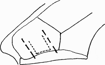
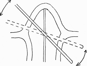
After vasoconstriction of the nose, the septum was infiltrated with a solution containing 2 per cent lignocaine and 1 in 80 000 adrenaline. A Killian's incision was made on the side opposite the sinus lesion and a mucoperichondrial flap raised. The septal cartilage was released from its lower attachment to the maxillary crest. Two releasing vertical incisions through the cartilage enabled a segment of the cartilage to be turned upwards but still retain its attachment to the rest of the septal cartilage superiorly (Figure 1). This turned-up segment of the cartilage also helped to keep the mucoperichondrial flap away and the access open. Instruments were passed through the cartilage window thus created (Figure 2). The mucosal incision on the side of the sinus lesion was made posterior to the posterior limit of the cartilage window. At the end of the procedure, the cartilage segment was turned down to its original position, and quilting stitches were used to hold the mucoperichondrial flaps together.
Diagram showing the septal incisions: long, interrupted lines represent the mucoperichondrial incisions on either side of the septum, while short, interrupted lines indicate the cartilage incision.

Cross-sectional view of nasal cavity showing the passage of instruments and the degree of mobility possible.

Patient one
A 49-year-old woman presented with nasal congestion and left middle meatal polyps.
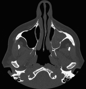
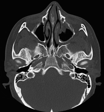
The patient underwent endoscopic nasal polypectomy. Histological examination confirmed an inverted papilloma. Computed tomography (CT) scanning showed the disease filling the left maxillary sinus completely, with extension into the nasal cavity (Figures 3 and 4).
Coronal computed tomography scan of sinuses, showing unilateral left maxillary sinus lesion.

Axial computed tomography scan of the lesion shown in Figure 3, showing involvement of the left anterior maxillary sinus.

An endoscopic medial maxillectomy was performed and the lesion completely removed using the septotomy approach.
Post-operatively, the patient developed an adhesion between the septum and the lateral wall of the nose. This was divided in the clinic, with no further problems.
At six months post-operative, the patient remained well. There was no sign of residual or recurrent disease, either clinically or on a magnetic resonance imaging scan. At the time of writing, she was under regular follow up.
Patient two
A 44-year-old woman presented with complaints of pain, nasal discharge, and altered sensation in her nose and headaches. Three years prior to this presentation, she had sustained a fracture of her nasal bone, septum and left zygoma complex in a road traffic accident. At that time, she had undergone manipulation of the nasal bones, septoplasty and inferior turbinectomy by another surgeon. Her clinical findings confirmed recurrent sinusitis affecting the left maxillary sinus.
A CT scan demonstrated polypoidal changes in the left maxillary sinus involving the anterior wall, along with osteomeatal complex obstruction (Figure 5).
Axial computed tomography scan showing a lesion involving the anterior wall of the left maxillary sinus.

We performed a left middle meatal antrostomy and, using the septotomy approach, removed all disease from the left maxillary sinus. No vasculitis, dysplasia or neoplasia was found on histological examination.
The patient's symptoms resolved well. At the time of writing, she remained under follow up.
Discussion
The trans-septal approach has traditionally been used to treat midline lesions such as those involving the pituitary gland.Reference Kern and Laws1 This approach involves gaining access in between the mucoperichondrial flaps of the septum. In contrast, septotomy involves going through the mucoperichondrial flaps sideways from the opposite nostril. Unlike the Draf type III procedure, permanent septal perforation is not created.
Septotomy enables access to a unilateral maxillary lesion from both sides of the septum. Whilst the endoscope is used from one side, the instruments can be passed from the opposite side through the septotomy. There are several advantages to this approach. Firstly, it leaves much more space to use the endoscope and instruments. Secondly, the instruments can be introduced at a much better angle to reach the anterior maxillary wall, as the septum is no longer a limiting factor. Thirdly, an assistant can access the operating side through the septotomy, helping either with suction to give a clear field and/or with retraction to assist dissection. This two-surgeon approach has been reported to work well.Reference Robinson, Patel and Wormald2, Reference Stamm, Vellutini, Harvey, Nogeira and Herman3 For midline lesions such as those involving the nasopharynx, the second surgeon can gain access through the opposite nasal passage. When the disease is more unilateral, the septotomy approach facilitates this process. It is also possible to pass both the endoscope and the instruments through the septotomy.
The septotomy approach adds little to the overall morbidity of the procedure. Septoplasty is often performed during endoscopic sinus surgery to gain access beyond a deviated septum. Septotomy adds only approximately 10 minutes to the operating time, and the resultant benefits are well worth it. The septotomy approach avoids any facial scar. It also reduces the risk of infraorbital anaesthesia, nasal vestibular stenosis and dental injury, associated with open approaches to the anterior maxilla (such as lateral rhinotomy, Caldwel–Luc and midfacial degloving).
We have used septotomy to treat maxillary sinus disease. It is also possible to extend its utility to deal with more posteriorly placed lesions, such as those involving the pterygopalatine fossa.Reference Robinson, Patel and Wormald2
In our patients, we did not encounter any complication due to septotomy, other than a minor adhesion which was treated in the clinic. The potential complications of the septotomy procedure should be similar to those of any septal surgery. Septal haematoma can be avoided by careful placement of quilting stitches. As the procedure does not involve removal of cartilage, one would not expect significant change in the shape of the nose. Septal perforation is avoided by taking care to place the mucosal incisions away from each other, and by keeping intact the cartilage deep to the incisions.