Introduction
Ectopic thyroid gland is a rare congenital anomaly which occurs as a result of failure of the thyroglossal duct to migrate from the foramen caecum to its final, prelaryngeal position during fetal development. An ectopic thyroid gland may be located anywhere from the base of the tongue to the prelaryngeal area.
Thyroid hemiagenesis is also a rare congenital anomaly in which one of the thyroid lobes fails to develop. Most cases remain undiscovered unless they become symptomatic. The diagnosis is usually made incidentally during evaluation of other thyroid disease. The exact prevalence of thyroid hemiagenesis is difficult to evaluate and remains unknown. However, the condition is estimated to occur in 0.2 per cent of normal children, being more common in females and on the left side of the thyroid gland.Reference Shabana, Delange, Freson, Osteaux and De Schepper1, Reference Piera, Garriga, Calabuig and Bargallo2
We present a case of recently diagnosed thyroid hemiagenesis which was accompanied by a contralateral goitre and an ectopic lingual thyroid; we also review the relevant literature.
Case report
A 26-year-old woman presented with a history of a long-standing, painless, palpable mass on the right side of the anterior neck. The neck mass had been gradually increasing in size, which caused mild compressive symptoms and cosmetic problems for the patient. The patient's medical and family histories were negative for thyroid disease or any other congenital disease.
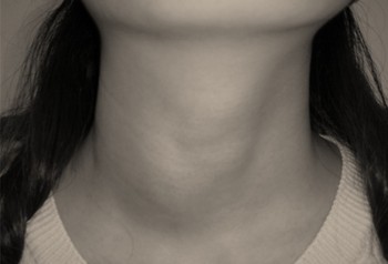
Clinical examination revealed a 2.5 × 3.0 cm thyroid mass type lesion on the right side of the anterior neck (Figure 1). The mass was soft, non-tender, mobile and moved upward during swallowing. There was no evidence of ulceration or colour change of the overlying skin. The rest of the examination, including fibre-optic nasopharyngoscopy and indirect laryngoscopy, was unremarkable.
Clinical photograph showing a swelling on the right side of the neck and a relative flattening on the left side, in the region of the thyroid gland.

Diagnostic investigation was performed, including thyroid function test, fine needle aspiration biopsy and computed tomography (CT) of the neck. Results for blood investigations and biochemical analysis were within the normal range, including analysis for thyroid-stimulating hormone (TSH), tri-iodothyronine and thyroxine. Fine needle aspiration cytology of the thyroid mass lesion revealed nodular adenomatous hypertrophy.
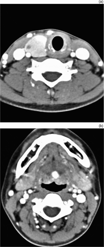
A CT scan of the neck at the level of the thyroid gland showed a 2.7 × 2.9 cm, well enhancing mass in the right lobe of the thyroid gland, without visualisation of the left lobe or isthmus of the thyroid gland (Figure 2a). An incidental CT image of the neck taken at the level of the submandibular gland showed a 0.8 × 0.9 cm, round, contrast-enhancing mass located in the base of the tongue, which was suspected to be a lingual thyroid (Figure 2b). Digital palpation and rigid endoscopy were performed in an attempt to locate this tongue base mass. However, clinical examination and endoscopy showed a normal appearance and normal mucosa. The patient denied experiencing any foreign body sensations or haemorrhage at the base of the tongue.
(a) Axial computed tomography (CT) scan of the neck at the level of the thyroid gland, showing a 2.7 × 2.9 cm, well enhancing mass in the right lobe of the thyroid gland, without visualisation of the left thyroid lobe or isthmus. (b) Axial CT scan of the neck at the level of the submandibular gland, showing a 0.8 × 0.9 cm, round, contrast-enhancing mass located in the tongue base, suspected to be a lingual thyroid.

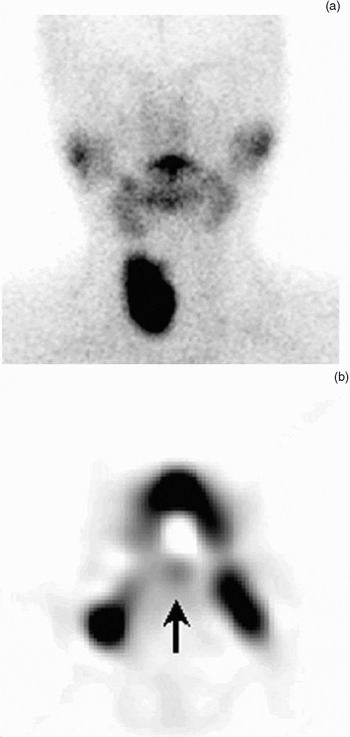
A thyroid scan was used to evaluate the thyroid gland function and the tongue base mass. Diffuse uptake of 99 mTC was noted in the right side of the thyroid gland, with no uptake in the left side (Figure 3a). However, the tongue base mass was difficult to find, even after rescanning following rinsing out saliva from the oral cavity. A SPECT (single photon emission computed tomography) scan with 99mTC scan was used to evaluate the function of the tongue base mass; weak uptake was seen into a lesion in the centre of the tongue at the level of the submandibular glands (Figure 3b).
(a) Thyroid scan with 99 mTC showing diffuse uptake in the right side of the thyroid gland, with no uptake in the left side. (b) SPECT (single photon emission computed tomography) scan with 99mTC scan showing weak uptake into a lesion (arrow) in the centre of the tongue at the level of the submandibular gland.

Based on these findings, a diagnosis of thyroid hemiagenesis and ectopic lingual thyroid with goitre was made.
The patient was commenced on a maintenance dose of thyroxine (0.05 mg daily). No treatment was given for the asymptomatic lingual thyroid.
Discussion
The embryological development of the thyroid gland begins during the fourth week of fetal gestation, with a median endodermal diverticulum on the floor of the pharynx at the foramen caecum. This primordium of the thyroid gland elongates and normally forms a bilobed diverticulum which descends in the neck to a final position anterior to the trachea in the seventh week of gestation.Reference McHenry, Walfis, Rosen, Lawrence and Paloyan3
Thyroid hemiagenesis was first reported by Handfield-Jones in 1866, followed by Luschka in 1876 and Ehlers in 1886.Reference Mikosch, Gallowitsch, Kresnik, Molnar, Gomez and Lind4 About 80 per cent of hemiagenesis cases are found in the left lobe and 20 per cent in the right lobe; agenesis of the isthmus is associated with 50 per cent of hemiagenesis cases.Reference McHenry, Walfis, Rosen, Lawrence and Paloyan3 Our patient had right lobe agenesis with isthmus agenesis. Thyroid hemiagenesis is more prevalent in females, with a 3:1 female:male ratio. However, this predominance seems to be based mainly on bias, due to a female preponderance in the populations investigated.Reference Mikosch, Gallowitsch, Kresnik, Molnar, Gomez and Lind4 As thyroid hemiagenesis does not cause clinical symptoms by itself, only a combination of thyroid hemiagenesis with other thyroid disease causing clinical symptoms leads to investigation and detection of the congenital anomaly. Such diseases included Graves' disease, Hashimoto's thyroiditis, subacute thyroiditis, nodular goitre, hyperfunctioning adenoma, and primary or metastatic carcinoma. In cases of thyroid hemiagenesis, hyperthyroidism or hypothyroidism may occur, and the thyroid function status mainly depends on iodine supply. Iodine deficiency is more commonly associated with hypothyroidism and iodine sufficiency with hyperthyroidism.Reference Mikosch, Gallowitsch, Kresnik, Molnar, Gomez and Lind4 In our case, the patient presented in a euthyroid state.
Ectopic thyroid is an uncommon embryological aberration characterised by the presence of thyroid tissue in a site other than its usual, pretracheal location. Arrest in descent can occur just below the foramen caecum (resulting in lingual thyroid), between the geniohyoid and mylohyoid muscles (causing sublingual thyroid), or just above or below the level of the hyoid bone (causing prelaryngeal thyroid). Although most ectopic thyroids are asymptomatic, lingual thyroid may be associated with a foreign body sensation, hoarseness, dysphagia, dyspnoea and haemorrhage. About 10 per cent of such patients present with hypothyroidism in spite of hyperplasia of the gland. The development of malignant disease in ectopic thyroid tissue is rare but well recognised. About 80 per cent of such tumours are papillary carcinomas.Reference Wong, Cunningham and Curtin5 Our patient had a lingual ectopic thyroid which was incidentally detected during evaluation of a thyroid mass. We could not see or palpate the mass because of its small size and its location deep within the submucosal and muscular layers of the base of the tongue. The patient had no symptoms of an ectopic lingual thyroid, such as foreign body sensation or haemorrhage.
Many cases of thyroid hemiagenesis associated with other diseases have been reported in the literature.Reference Mikosch, Gallowitsch, Kresnik, Molnar, Gomez and Lind4, Reference Melnick and Stemkowski6, Reference Karabay, Comlekci, Canda, Bayraktar and Degirmenci7 To the best of our knowledge, the present case represents the first report of thyroid hemiagenesis with ectopic lingual thyroid.
The thyroid scintiscan is an important diagnostic tool with which to evaluate the size of thyroid lesions and the functional status of the gland, and to locate normal thyroid tissue.Reference Ulug, Ulubil and Alagol8 The disadvantage of this technique is that it shows uptake in the salivary tissue and saliva within the oral cavity; it is also difficult to distinguish masses smaller than 1 cm. In our case, we were able to diagnose the ectopic thyroid tissue of the tongue base using SPECT (single photon emission computed tomography) scan with 99mTC; this lesion was difficult to distinguish using 99 mTC thyroid scanning.
Computed tomography scanning shows aberrant thyroid tissue as a high density mass, distinguishable from other soft tissue due to a high attenuation related to the high iodide content.Reference Baik, Choi and Lee9 Our patient's CT showed a contrast-enhancing, round mass located in the tongue base at the level of the submandibular glands.
The management of patients with thyroid hemiagenesis and ectopic thyroid is dependent on several factors, including the size of the lesion, presence of local symptoms, patient's age, thyroid gland status and presence of complicating factors (such as ulceration, haemorrhage, cystic degeneration or malignancy).Reference Baik, Choi and Lee9 Thyroid hormonal therapy will typically suppress the production of TSH, a useful treatment in cases of glandular enlargement. Our patient was prescribed thyroxine to reduce glandular enlargement.
Conclusion
Thyroid hemiagenesis and ectopic lingual thyroid are both known to be uncommon, and it is extremely rare to find them coexisting. The thyroid scintiscan is known to be the most accurate method of diagnosing an ectopic thyroid gland. SPECT (single photon emission computed tomography) scan with 99mTC is thought to be a more effective method of detecting small ectopic thyroid glands and distinguishing these from salivary tissue.