INTRODUCTION
Migraine is a common neurological disorder characterized by the episodic activation of trigeminovascular system (Waeber and Moskowitz, Reference Waeber and Moskowitz2005; Sanchez-Del-Rio et al., Reference Sanchez-Del-Rio, Reuter and Moskowitz2006). Under appropriate conditions of activation, the primary afferents of trigeminal ganglia that innerve extra- and intra-cranial vasculature, release several neuro-mediators, including calcitonin gene-related peptide (CGRP) and substance P. In particular, a robust evidence indicates that CGRP is the main mediator of trigeminal signaling (Edvinsson, Reference Edvinsson2001; Villalón and Olesen, Reference Villalón and Olesen2009). Indeed, increased levels of CGRP are found in the jugular vein during migraine attacks (Goadsby et al., Reference Goadsby, Edvinsson and Ekman1990; Goadsby and Edvinsson, Reference Goadsby and Edvinsson1993), and an increase in CGRP release occurs during peripheral sensitization of meningeal nociceptors, which is responsible for hyperalgesia and allodynia often associated with migraine (Strassman and Levy, Reference Strassman and Levy2006). Furthermore, it has been recently demonstrated that neurons can release CGRP from cell bodies within the ganglia (Zhang et al., Reference Zhang, Winborn, Marquez de Prado and Russo2007c; Li et al., Reference Li, Vause and Durham2008), and locally released CGRP is able to modulate neuronal transmission of pain signals (Thalakoti et al., Reference Thalakoti, Patil, Damodaram, Vause, Langford and Freeman2007). Within the trigeminal ganglion, neurons are surrounded by glial cells, referred to as satellite glial cells (SGCs), constituting morphological and functional units (Hanani, Reference Hanani2005; Dublin and Hanani, Reference Dublin and Hanani2007). It has been shown that SGCs express the CGRP receptor components CLR and RAMP1 (Eftekhari et al., Reference Eftekhari, Salvatore, Calamari, Kane, Tajti and Edvinsson2010), and we recently demonstrated that SGCs represent a direct target of CGRP action within the ganglia (De Corato et al., Reference De Corato, Lisi, Capuano, Tringali, Tramutola and Navarra2011). SGCs appear to respond to neuronal stimulation by secreting factors that can in turn modulate neuronal activity (Heblich et al., Reference Heblich, England and Docherty2001; Dublin and Hanani, Reference Dublin and Hanani2007). The existence of a cross-talk between neurons and glial cells within the trigeminal ganglion is now well established. In fact, SGCs modulate neuronal response when the trigeminal system is activated (Capuano et al., Reference Capuano, De Corato, Lisi, Tringali, Navarra and Dello Russo2009; Takeda et al., Reference Takeda, Takahashi and Matsumoto2009; Ceruti et al., Reference Ceruti, Villa, Fumagalli, Colombo, Magni and Zanardelli2011); such modulation could explain some features of migraine pain and other painful conditions related to trigeminal activation, such as pain chronicization and the lack of efficacy of pharmacological treatments.
Primary cultures of rat trigeminal neurons and SGCs are suitable in vitro models to investigate trigeminal responses. Moreover, we have recently developed a model to investigate the interactions between trigeminal neurons and SGCs (Capuano et al., Reference Capuano, De Corato, Lisi, Tringali, Navarra and Dello Russo2009). We characterized the pro-inflammatory activation of trigeminal SGCs upon different stimuli; we showed that SGCs can be activated by the exposure to interleukin-1β (IL-1β) or nitric oxide, releasing soluble pro-inflammatory and sensitizing mediators such as prostaglandin E2 (PGE2). Moreover, we demonstrated that the medium collected from activated SGCs cultures (conditioned medium (CM)) was able to modulate (‘sensitize’) neuronal response when applied to trigeminal neuron cultures.
OBJECTIVE
In the present paper, we further characterized the modulation of CGRP release from rat trigeminal neurons caused by the exposure of such neurons to CM collected from activated SGCs. The effects of CM were tested on neuronal cultures in different conditions of activation and at different time points. Moreover, we also studied the effects of a clinically relevant anti-migraine drug, sumatriptan, on the modulation of CGRP release. SGCs have been shown to induce, when activated, neuronal sensitization (Capuano et al., Reference Capuano, De Corato, Lisi, Tringali, Navarra and Dello Russo2009). The effects of sumatriptan were investigated under basal conditions and in different paradigms of glial-induced neuronal sensitization.
METHODS
Drugs
Diethylenetriamine/nitric oxide (DETA/NO), capsaicin and sumatriptan (as succinate salt) were purchased from Sigma-Aldrich (St. Louis, MO). DETA/NO and sumatriptan were dissolved in distilled water, capsaicin was dissolved in ethanol. Aliquots (10 mM) were made for each drug. Bradykinin was purchased by Vinci-Biochem (Florence, Italy) and dissolved in a 5% acetic acid solution at 100 mM. All drug aliquots were stored at −35°C. Human recombinant IL-1β (Endogen, Pierce Biotechnology, Rockford, IL) was dissolved in distilled water at the concentration of 40 µg/ml and stored in aliquots at −80°C. All working dilutions were made using cell culture medium. None of the tested compounds interfered with CGRP RIA.
Cell cultures
Rat trigeminal neuronal cultures and mixed neuronal/SGCs cultures from trigeminal ganglia were prepared as previously described (Capuano et al., Reference Capuano, De Corato, Lisi, Tringali, Navarra and Dello Russo2009), with some modifications. Six- to 7-day-old Wistar rats were used. The use of animals for this experimental work was previously approved by the Italian Ministry of Health (licensed authorization to P. Navarra). Briefly, both trigeminal ganglia were aseptically removed. Tissues were collected in a Petri dish containing 3–5 ml of ice-cold phosphate buffer saline without Ca2+ and Mg2+ (PBS w/o; Sigma), supplemented with antibiotics (100 IU/ml penicillin and 100 µg/ml streptomycin; Sigma) and d-glucose (6 g/l). Tissues were then digested in 5 mg/ml collagenase (Biochrom AG, Berlin, Germany) for 20 min at 37°C, followed by 10 min incubation with 0.125% trypsin (Biochrom), including DNAse I (Sigma) treatment in the last 5 min of incubation. At the end of the incubation time, the enzyme solution was replaced with 5 ml Ham's F12 medium (Biochrom), containing 10% heat-inactivated endotoxin-free fetal calf serum (FCS; GIBCO, Invitrogen Corporation, Paisley, Scotland) and antibiotics (complete culture medium). Cells were mechanically dissociated using a Pasteur pipette, plated on a 25 cm2 flask, and incubated at 37°C in a humidified atmosphere containing 5% CO2 for 2–3 h (pre-plating). This step allowed us to separate SGCs from neurons using their differential adhesion properties. At the end of the pre-plating time, neurons were floating in the incubation media and could be harvested by collecting the media. Trigeminal neurons were plated at a density of 100,000 cells/well in 48-well tissue culture plates, previously coated with poly-d-lysine (40 µg/ml; MW 70,000–130,000, Sigma). The incubation volume was 500 µl/well of complete culture medium (see above), enriched with 50 ng/ml of 2.5 S murine nerve growth factor (Alexis Biochemicals). The culture medium was changed within 24 h from seeding, and 20 µM cytosine arabinoside was added to further reduce non-neuronal cell growth (Capuano et al., Reference Capuano, De Corato, Lisi, Tringali, Navarra and Dello Russo2009). Experiments were carried out 4–5 days after dissection.
SGCs remained attached to the flask, in which 5 ml of fresh complete culture medium were added. Cells were incubated for a week, during which the medium was replaced twice, until reaching almost complete confluence. At this time, cells were detached from the flask by a 5-min 0.05% trypsin-EDTA (Biochrom) treatment at 37°C, resuspended in fresh complete culture medium, and plated at a density of 150,000 cells/ml using 48-well plates (500 µl/well) to establish co-cultures and 24-well plates (1 ml/well) for the preparation of CM. Cells were grown until reaching almost complete confluence (4–5 days).
Neuronal/SGCs co-cultures were established in two phases. Firstly, SGCs were grown in 48-well plates for 4–5 days. Then, neuronal cells obtained from a new dissection (see above) were plated over almost confluent satellite cells. Experiments on neuronal/satellite cell co-culture were carried out 48 h after neuronal cell plating.
Experimental procedures
In preliminary experiments, we characterized the effects of CM obtained from activated SGCs on neuronal CGRP release. CM from activated SGCs was generated according to our previously published protocol (Capuano et al., Reference Capuano, De Corato, Lisi, Tringali, Navarra and Dello Russo2009). Briefly, SGCs were incubated for 4 h with 10 ng/ml IL-1β or 6 h with 100 µM DETA/NO, then washed and incubated in fresh culture medium for another 24 h. At the end of this conditioning period, the medium (‘CM’) was collected and stored at −80°C until the experiments on neurons were performed. The CM were applied to trigeminal neurons at a 1:1 dilution. To characterize the effects of CM on trigeminal neuronal cultures, cells were incubated for various time with diluted CM and then further incubated for 30 min with different pro-inflammatory stimuli. In the same experimental paradigm, the effects of sumatriptan were tested, adding the drug (1–10 µM) together with the stimuli.
In some experiments carried out on neuronal/satellite cell co-culture, SGCs were previously treated with 10 ng/ml IL-1β (activated satellite cells) or with complete culture medium (resting satellite cells) for 24 h (see Fig. 4B). At the end of the incubation time, media were removed and cells were washed with fresh culture medium. Immediately after, neuronal cells were plated over satellite cells, previously activated or not with IL-1β. After 48 h in co-culture, experiments were carried out.
For all experiments on neuronal cells, Ham's F12 medium supplemented with 1 µM bacitracin, 10 mg/ml aprotinin and without Nerve Growth Factor, was used (plain medium). The incubation volume was 200 µl and experiments were performed at 37°C and 5% CO2. At the end of all the experiments, the medium was collected and stored at −35°C until the assessment of CGRP by a specific radioimmunoassay (RIA).
CGRP RIA
CGRP release was measured by a specific RIA technique validated in our laboratory and previously described in detail (Capuano et al., Reference Capuano, De Corato, Lisi, Tringali, Navarra and Dello Russo2009). Briefly, a buffer containing 10 mM sodium phosphate, 154 mM NaCl, 25 mM ethylene diamine tetraacetic acid (EDTA), 0.01% thimerosal, 0.5% BSA and 0.03% Tween-20, pH 7.2 was used. The RIA was performed as follows: 100 µl of sample or standard solution was diluted 3-fold into RIA buffer containing anti-hα-CGRP (kindly provided by Professor D. Currò) at a final dilution of 1:120,000. After 24 h at 4°C, 100 µl of [125I]-hα-CGRP (6000 cpm/tube) was added and the incubation continued at 4°C for 48 h. Separation of free from bound α-CGRP was achieved by adding anti-rabbit goat serum (at final dilution 1:200) and 500 µl of 6.6% polyethylene glycol solution in 5 mM phosphate buffer, pH 7.4. After 2 h at 4°C, the tubes were centrifuged at 3000 g for 30 min at 4°C, and the pellet counted in a γ-counter. The standard curve ranged from 1.95 to 1000 pg/tube of rα-CGRP. Each sample and standard were assayed in duplicate. The mean IC50 of the standard curve was 50.8 ± 2.0 pg/tube (n = 10) and non-specific binding of the labeled ligand was not different from the background of the γ-counter. The intra-assay (n = 6) and inter-assay (n = 10) coefficients of variation were ±1.27% and ±0.95% at the lowest (1.95 pg/tube) and ±13.7% and ±12.6% at the highest (1000 pg/tube) levels of standard, respectively. The detection limit was 19.5 pg/ml.
Statistical analysis
All data are reported as means ± SEM, unless otherwise stated. Data were analyzed by one-way or two-way analysis of variance (ANOVA), followed by Bonferroni post hoc test. All data were analyzed using a PrismTM computer program (GraphPad, San Diego CA, USA).
RESULTS
The effects of conditioned media on trigeminal neuronal cultures
SHORT-TERM EFFECTS OF CM ON NEURONAL CGRP RELEASE
As previously shown (Capuano et al., Reference Capuano, De Corato, Lisi, Tringali, Navarra and Dello Russo2009), SGCs can be stimulated by IL-1β or nitric oxide to release soluble pro-inflammatory and sensitizing mediators such as PGE2. Moreover, we demonstrated that the medium collected from activated SGCs cultures (CM) is able to modulate (‘sensitize’) neuronal response when applied to trigeminal neuronal cultures.
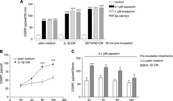
Figure 1A shows that a 30-min pre-exposure of trigeminal neurons to CM (1:1 dilution) further enhanced the increase in neuronal CGRP release evoked by different depolarizing agents (0.1 µM capsaicin, 1 µM bradykinin and 56 mM KCl, respectively) compared to the increase observed after pre-exposure to plain medium. The extent of this potentiation was similar regardless of the depolarizing stimulus used. Likewise, the effects of DETA-NO- and IL-1β-CM were also similar; therefore, only the latter was used in subsequent experiments.
Short- and long-term effects of conditioned media on neuronal CGRP release. (A) Trigeminal neurons were incubated for 30 min (short-term incubation) with plain medium or with CM obtained from IL-1β- and DETA/NO-activated SGCs. Neuronal cells were then stimulated with 0.1 µM capsaicin, 1 µM bradykinin or 56 mM KCl, and CGRP release was measured after 30 min incubation. Data are expressed as pg of CGRP/well/30 min. ***, P < 0.001 versus plain medium control; °°°, P < 0.001 versus capsaicin, bradykinin and 56 mM KCl, respectively, given in the presence of plain medium. (B,C) Trigeminal neurons were incubated for the indicated times (long-term incubations) with plain medium (controls) or with CM obtained from IL-1β-activated SGCs. At the end of the incubation times, media were collected, and CGRP released was measured. Data are expressed as pg of CGRP/well (B). At the end of experiments shown in (B), each experimental group was treated for 30 min with capsaicin, and CGRP released was also measured. Data are expressed as pg of CGRP/well/30 min (C); the amount of CGRP released during the previous part of the experiment was considered as a baseline level in each group, and therefore subtracted. * and ***, P < 0.05 and P < 0.001 versus the respective controls. All data are means ± SEM of n = 6–8 replicates for each experimental group.

LONG-TERM EFFECTS OF CM ON NEURONAL CGRP RELEASE
Time-course experiments were performed on trigeminal neuronal cultures to evaluate the effects of long-term exposures to CM. Neuronal cells were pre-incubated for 2, 4, 8 and 24 h with plain medium (controls) or with CM obtained from IL-1β-activated SGCs, and CGRP released in the incubation media was measured. We found that the long-term exposure of neuronal cells to CM (Fig. 1B) induced directly a time-dependent increase in CGRP release from 4 h onward, reaching statistical significance after 8 and 24 h of incubation. At the end of pre-incubation, each experimental group was exposed to 0.1 µM capsaicin for 30 min. Figure 1C shows that CGRP release was further increased in the groups pre-treated with CM for 2, 4 and 8 h compared to their respective controls, although the extent of such increase tended to decline with the time of pre-exposure, and was no longer significant in the group pre-exposed for 24 h.
The effects of sumatriptan on neuronal CGRP release
SUMATRIPTAN REDUCES CGRP RELEASE EVOKED BY DEPOLARIZING STIMULI
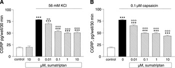
Trigeminal neuronal cultures were treated for 30 min with plain medium (controls) or medium containing 56 mM KCl or 0.1 µM capsaicin (Fig. 2A,B). Sumatriptan, tested in the range of 0.01–10 µM along with the stimuli, significantly inhibited the stimulatory effect of both triggers at each concentration tested, reaching the maximal effect from 0.1 µM onwards. At the higher concentration (10 µM), the drug did not affect the basal CGRP release (Fig. 2A).
The effects of sumatriptan on neuronal evoked CGRP release. Trigeminal neuronal cultures were incubated for 30 min with medium containing KCl (A) or capsaicin (B) alone or together with graded doses of sumatriptan. Data are expressed as pg of CGRP/well/30 min, the means ± SEM of n = 6 replicates for each experimental group. ***, P < 0.001 versus controls; °° and °°°, P < 0.01 and P < 0.001 versus each stimulus given alone.

SUMATRIPTAN DIFFERENTIALLY REGULATES THE EVOKED CGRP RELEASE AFTER SHORT- OR LONG-TERM APPLICATION OF CM
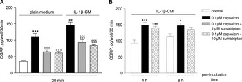
Trigeminal neuron cultures were pre-incubated for 30 min (short-term incubation) with plain medium or with IL-1β-CM. As shown in Fig. 3A, CM confirmed its ability to potentiate capsaicin-evoked CGRP release. In this experimental paradigm, 1 and 10 µM sumatriptan were able to significantly inhibit CGRP release evoked by 0.1 µM capsaicin in both plain medium and CM pre-treated neurons, with similar inhibitory potencies. The percent of inhibition of 10 µM sumatriptan on capsaicin-evoked CGRP release was 42.41% ± 3.05 (n = 6) in the group pre-treated with the plain medium, and 40.69% ± 2.34 (n = 6) in the group pre-treated with IL-1β-CM. Data are expressed as means ± SEM (n) per each group, and values are in the same order of magnitude of the inhibitory potencies displayed by sumatriptan over the challenges with depolarizing stimuli in the absence of pre-treatment. In fact, the percents of inhibition were 35.35% ± 2.12 (n = 6) and 43.78% ± 3.36 (n = 6) on the KCl and capsaicin challenge, respectively (Fig. 2A, B).
The effects of sumatriptan on the evoked CGRP release after short- or long-lasting application of conditioned media. (A) Trigeminal neurons were incubated for 30 min (short incubation) with plain medium or with CM obtained from IL-1β-activated SGCs. After the preincubation time, cells were stimulated with capsaicin alone or together with sumatriptan. ***, P < 0.001 versus controls (plain medium group); °°°, P < 0.001 versus capsaicin alone (plain medium group); ##, P < 0.01 versus capsaicin given in the absence of pre-incubation (IL-1β-CM group); §§§, P < 0.001 versus capsaicin alone (IL-1β-CM group). (B) Neuronal cells were pre-incubated for 4 and 8 h (long-lasting incubations) with IL-1β-CM, and then activated with capsaicin alone or together with sumatriptan. * and ***, P < 0.05 and P < 0.001 versus controls. All data are expressed as pg of CGRP/well/30 min, the means ± SEM of n = 6–8 replicates for each experimental group.

In a further series of experiments, the effects of sumatriptan were tested after long-lasting exposure to CM. In these experiments, 4 and 8 h pre-incubations were used, and sumatriptan was tested at the 10 µM concentration. As shown in Fig. 3B, cultured neurons were incubated with IL-1β-CM for 4 or 8 h. At the end of the incubation time, media were replaced with the plain medium (controls) or medium containing capsaicin alone or in the presence of 10 µM sumatriptan. In this experimental paradigm, capsaicin produced – as expected – a significant increase in CGRP release, but sumatriptan failed to counteract the stimulatory effect of capsaicin (Fig. 3B).
The effects of sumatriptan on neuronal-SGC co-cultures
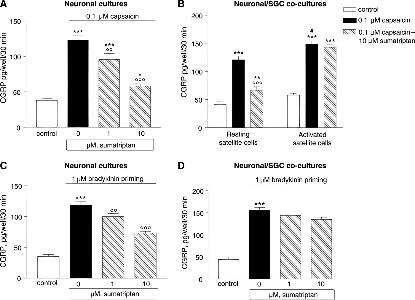
To further explore the influence of SGCs activation on sumatriptan effects, we used a different experimental paradigm. Sumatriptan was tested on both neuronal cultures (Fig. 4A,C) and neuronal/SGCs co-cultures (Fig. 4B,D) (see Methods). Figure 4A shows sumatriptan (1 and 10 µM) inhibition on capsaicin-evoked CGRP release from trigeminal neuronal cultures. In the co-culture paradigm, SGCs were previously treated with 10 ng/ml IL-1β (activated satellite cells) or with complete culture medium (resting satellite cells) for 24 h. At the end of the incubation time, neuronal cells were plated over satellite cells, previously activated or not with IL-1β and after 48 h in co-culture, experiments were carried out. In this experimental setting, 10 µM sumatriptan maintained its capability to counteract the increase in CGRP release stimulated by capsaicin in a 30 min challenge, but failed to inhibit capsaicin if SGCs were previously activated with IL-1β (Fig. 4B).
The effects of sumatriptan on the evoked CGRP release from neuronal cells alone or co-cultured with SGCs. (A) Inhibitory effect of sumatriptan on capsaicin-evoked CGRP release from cultured trigeminal neurons. * and ***, P < 0.05 and P < 0.001 versus controls; °° and °°°, P < 0.01 and P < 0.001 versus capsaicin given alone. (B) Effects of sumatriptan on neuronal/SGC co-culture: SGCs were previously activated (activated satellite cells) or not (resting satellite cells) for 24 h with 10 ng/ml IL-1β; at the end of the incubation time, neuronal calls were plated over SGCs and, after 48 h, the co-culture was treated or not with capsaicin alone or together with sumatriptan. ** and ***, P < 0.01 and P < 0.001 versus controls; °°°, P < 0.001 versus capsaicin alone; #, P < 0.05 versus the effects of capsaicin in resting satellite cells. (C,D) Differential effects of sumatriptan on neuronal (C) or neuronal/SGCs co-culture (D) in the bradykinin priming paradigm. Both neuronal and neuronal/SGCs co-culture were treated for 15 min with bradykinin (bradykinin priming). At the end of the incubation time, medium was replaced with bradykinin alone or in combination with sumatriptan. ***, P < 0.001 versus controls; °° and °°°, P < 0.01 and P < 0.001 versus bradykinin alone, respectively. All data are expressed as pg of CGRP/well/30 min, the means ± SEM of n = 6–8 replicates for each experimental group.
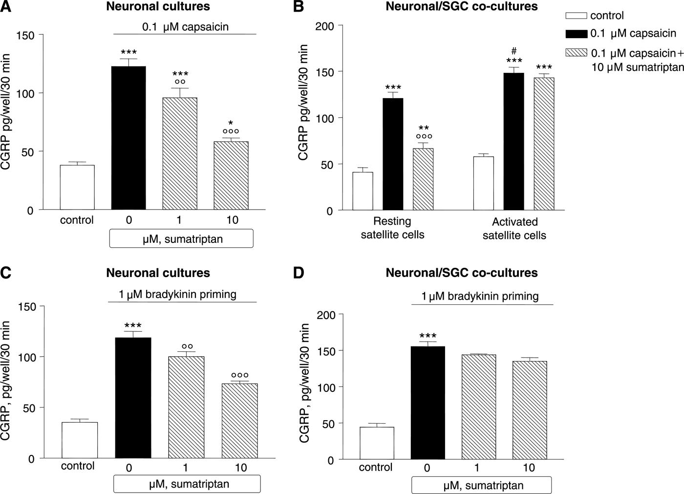
In the same experimental setting, bradykinin was used as a neuronal depolarizing agent. Both neuronal cultures and neuronal/SGCs co-cultures were pre-exposed to 1 µM bradykinin for 15 min (bradykinin priming). Subsequently, the medium was replaced with a medium containing 1 µM bradykinin alone or in combination with sumatriptan and CGRP was measured after 30 min of incubation. Interestingly, the bradykinin priming effect was greater in the neuronal/SGC co-culture than in the neuronal culture, as demonstrated by the different amount of CGRP measured in the incubation medium (Fig. 4C,D, black bars). As shown in Fig. 4C, both doses of sumatriptan were able to inhibit neuronal CGRP release induced by the previous bradykinin exposure in neuronal cultures, reaching maximal significance at the higher dose tested. On the contrary, when sumatriptan was tested on neuronal/SGCs co-cultures, it failed to inhibit the CGRP release evoked by bradykinin priming (Fig. 4D).
CONCLUSIONS
• Long-term exposures of trigeminal neurons to SGC-CM increased directly neuronal CGRP release and potentiated the stimulatory effect of capsaicin on neuronal CGRP release;
• sumatriptan was effective in inhibiting the evoked CGRP release from naïve neuronal cultures as well as from neurons pre-exposed for 30 min to CM, while it failed when tested after long-term pre-exposures;
• sumatriptan inhibited the evoked CGRP release from neuronal cells co-cultured with resting SGCs, whereas it failed when neurons were co-cultured with activated SGCs;
• when tested in the bradykinin priming paradigm, sumatriptan reduced CGRP release in neuronal cultures, whereas it was unable to exert any inhibitory effect in the co-culture model.
DISCUSSION
SGCs surround neuronal ganglion cells and form a tight envelope around them, which results in the formation of discrete units consisting of a neuronal cell body and the satellite cells surrounding it (Hanani, Reference Hanani2005). Apart from providing a mechanical and metabolic support to neurons, SGCs can directly modulate their functions and excitability (Hanani, Reference Hanani2005). Neuron–glial interactions have been shown to be involved in all stages of inflammation and pain associated with several diseases of the central nervous system (CNS) (Watkins and Maier, Reference Watkins and Maier2002; Wieseler-Frank et al., Reference Wieseler-Frank, Maier and Watkins2004). In a previous work (Capuano et al., Reference Capuano, De Corato, Lisi, Tringali, Navarra and Dello Russo2009), we demonstrated that SGCs can be activated by autocrine/paracrine stimuli such as IL-1β and NO, thus creating the potential for a positive feedback loop that may amplify and sustain inflammatory processes within the ganglia. Indeed, pre-exposure of trigeminal neurons for 30 min to CM significantly increased the release of neuronal CGRP evoked by stimulation with capsaicin (neuronal sensitization) (Capuano et al., Reference Capuano, De Corato, Lisi, Tringali, Navarra and Dello Russo2009). Once activated, these cells release glial factors, which in turn ‘sensitize’ neuronal cells, amplifying and sustaining inflammatory processes within the ganglia. Here, we further confirmed such sensitization effect, showing that the phenomenon is observed regardless of the activator used. One possible mediator of neuronal sensitization is PGE2, as suggested by recent observations highlighting the role of cyclooxygenase 2 activation after exposure of SGCs to IL-1β (Neeb et al., Reference Neeb, Hellen, Boehnke, Hoffmann, Schuh-Hofer and Dirnagl2011). Moreover, it has been shown that prostaglandins do not interfere directly with neuronal neurotransmitter release, such as CGRP release, but can reduce the firing threshold of sensory neurons in response to other depolarizing agents (Nicolson et al., Reference Nicolson, Foster, Bevan and Richards2007; Zhang et al., Reference Zhang, Strassman, Burstein and Levy2007b). However, it remains beyond the aims of the present study to characterize the composition of the CM, which is instead used as a pharmacological tool to study the effects of pro-inflammatory activated SGCs on trigeminal neurons. The pro-inflammatory milieu generated by SGCs activation into trigeminal ganglion and the subsequent neuronal sensitization may represent a new pathophysiological model of migraine pain. In particular, several findings indicate that glial–neuronal communication and their paracrine interactions contribute to primary afferent excitability and peripheral sensitization (Takeda et al., Reference Takeda, Tanimoto, Kadoi, Nasu, Takahashi and Kitagawa2007; Zhang et al., Reference Zhang, Chen, Wang and Huang2007a), which is characterized by increased neuronal excitability and a lower threshold for activation. From a clinical point of view, the increase in spontaneous firing and the sensitization of primary afferent neurons are crucial events for initiation of subsequent central sensitization (Burstein, Reference Burstein2001), characterized by enhanced or abnormal inputs into the CNS, thereby contributing to acute and chronic pain conditions. Here, we clearly showed that the long-term exposure of trigeminal neurons to CM augmented the evoked CGRP release, thus suggesting that the prolonged action of glial factors may facilitate the development of peripheral as well as central sensitization. Moreover, since CGRP co-localizes with glutamate in pre-synaptic central afferents (Merighi et al., Reference Merighi, Polak and Theodosis1991; Xiao et al., Reference Xiao, Richter and Hurley2008), an increased release of CGRP elicited by a depolarizing agent, such as capsaicin, consequently leads to central sensitization via a well-known glutamate-mediated mechanism (Pietrobon and Striessnig, Reference Pietrobon and Striessnig2003; Pietrobon Reference Pietrobon2005).
The influence of SGCs on neuronal excitability may affect the response to drugs used for migraine management. Indeed, one of the most intriguing and still not well-understood issues of migraine management is the lack of efficacy of current clinically available abortive treatments for migraine pain in chronic conditions. This point has been explored in animal as well as in human models of migraine, and sensitization of primary afferents seems to be one of the possible explanations (Strassman et al., Reference Strassman, Raymond and Burstein1996; Burstein and Jakubowski, Reference Burstein and Jakubowski2010). It has been hypothesized that the initial phase of migraine attack is mediated by the sensitization of peripheral trigeminovascular neurons innervating the meninges. If these neurons remain hyperactive, their ongoing input into the central trigeminovascular neurons eventually will render the latter sensitized as well, thus leading to the development of cutaneous allodynia later in the migraine attack. Triptans, 5HT1B/1D agonists, are the best studied and most commonly used family of anti-migraine drugs. Administration of triptans during the early phase of a migraine attack, while pain is still mild and allodynia is not fully developed, increases the success rate of rendering the patient pain free (Burstein et al., Reference Burstein, Collins and Jakubowski2004). Burstein and Jakubowski (Reference Burstein and Jakubowski2004) have shown that early treatments with sumatriptan provide a powerful means of preventing the initiation of central sensitization in a rat model of cutaneous allodynia. On the other hand, delayed treatments were not effective in reversing the increased spontaneous firing of the same sensitized trigeminovascular neurons nor the cutaneous allodynia, suggesting that sumatriptan cannot counteract an already established central sensitization in vivo.
The regulation of CGRP release from cultured trigeminal neurons by the anti-migraine drug sumatriptan was first shown by Durham and Russo (Reference Durham and Russo1999). We confirm this finding in our experimental model, where sumatriptan directly counteracted the increase in CGRP secretion from neuronal cultures in response to depolarizing agents. To further understand the putative role of SGCs in modulating the responses to sumatriptan, we adopted two different experimental models of trigeminal neuron sensitization, i.e. the CM and co-culture paradigms. Results obtained from these challenges highlight the importance of trigeminal SGCs activation in the development and maintenance of peripheral sensitization. In fact, while sumatriptan was able to inhibit the evoked CGRP release after 30-min exposure to CM, it failed to exert any inhibitory effect when neurons were exposed for longer times (4 and 8 h) to glial-derived factors. This finding clearly suggests that sumatriptan is unable to provide enough inhibition to significantly counteract an established and prolonged sensitization in trigeminal neurons. Concerning the co-culture experiments, sumatriptan maintained its capability to counteract the increase in CGRP release stimulated by capsaicin, but failed to inhibit capsaicin if SGCs were previously activated with IL-1β, further confirming our data. A possible explanation could be that the activation of 5HT1B/1D receptor by sumatriptan cannot generate functional inhibition when neurons are sensitized. Moreover, to further confirm our findings, sumatriptan was tested in the bradykinin priming paradigm, both on neuronal cultures and on co-cultures. We used bradykinin, a known activator of sensory neurons with potent inflammatory activity (Calixto et al., Reference Calixto, Cabrini, Ferreira and Campos2000), as a tool to stimulate CGRP release from neurons. Bradykinin priming (i.e. 15 min pre-exposure of cultures to 1 µM bradykinin, see subsection ‘The effects of sumatriptan on neuronal-SGC co-cultures’) is a well-established model to induce sensitization, as its acute application was shown to sensitize μ-opioid receptor signaling in trigeminal cultures (Berg et al., Reference Berg, Patwardhan, Sanchez, Silva, Hargreaves and Clarke2007) and to enhance the amplitude of purinoreceptor-mediated calcium response in trigeminal neurons (Ceruti et al., Reference Ceruti, Fumagalli, Villa, Verderio and Abbracchio2008). In our experimental model, sumatriptan exerted different actions on neuronal cultures and on co-cultures, after a brief pre-exposure to bradykinin. Indeed, the drug inhibited CGRP secretion from trigeminal neurons, while it was ineffective in the co-culture model. Indirect actions of bradykinin on SGCs activation have been postulated (Heblich et al., Reference Heblich, England and Docherty2001). Moreover, as shown in a recent work (Ceruti et al., Reference Ceruti, Villa, Fumagalli, Colombo, Magni and Zanardelli2011), it could be postulated that the CGRP release evoked by bradykinin application cause the activation of CGRP receptors on neighboring SGCs (co-culture) leading to subsequent release of soluble mediators, thus reproducing the pro-inflammatory milieu responsible for the inefficacy of sumatriptan. Even though we did not test the effects of bradykinin on SGC cultures, the priming was more effective in the co-culture system and our data confirm that SGCs are involved in sensitization of neuronal response also in the bradykinin paradigm since sumatriptan failed to reduce CGRP release in co-culture. Although it is not possible to compare our in vitro findings with those obtained in in vivo models, our data are consistent with the evidence that triptans can prevent initiation of central sensitization, which depends on incoming impulses from meningeal nociceptors, but cannot abort ongoing central sensitization once it becomes self-sustained (Burstein, Reference Burstein2001; Burstein and Jakubowski, Reference Burstein and Jakubowski2004).
In the last few years, novel findings on neuronal soma–SGC interactions in sensory ganglia (Thalakoti et al., Reference Thalakoti, Patil, Damodaram, Vause, Langford and Freeman2007; Takeda et al., Reference Takeda, Takahashi and Matsumoto2009; Ceruti et al., Reference Ceruti, Villa, Fumagalli, Colombo, Magni and Zanardelli2011) have led investigators to consider the interdependence between neuronal cell bodies and SGCs in regulating peripheral signaling. These interactions, and in particular those occurring within the trigeminal ganglion, represent a new focus of investigation in migraine pathophysiology. Our data demonstrate that trigeminal SGC activation leads to the generation of a pro-inflammatory milieu that could influence neuronal excitability, and that this event may affect the response of neurons to triptan treatment. This phenomenon provides the neurobiological basis to several features of migraine pain yet to be fully clarified, including the lack of efficacy of currently available abortive drugs such as triptans. Finally, this study confirms that both the CM and the co-culture model are suitable in vitro paradigms to study neuronal–glial interactions.
Statement of interest
None.