Increasing interest in recent years has been focusing on the use of donkeys’ milk for human consumption (D'Alessandro et al. Reference D'Alessandro, Martemucci, Jirillo and De Leo2011; Martemucci & D'Alessandro, Reference Martemucci and D'Alessandro2012) as a hypoallergenic substitute for children affected by cows’ milk protein allergy or multiple food intolerance (Tesse et al. Reference Tesse, D'Alessandro, Paglialunga, Rizzi, Bozza, Martemucci and Armenio2008) and as a promising nutraceutical for aged people due its effects of up-regulation of the immune system (Tafaro et al. Reference Tafaro, Magrone, Jirillo, Martemucci, D'Alessandro, Amati and Jirillo2007; Amati et al. Reference Amati, Marzulli, Martelli, Tafaro, Jirillo, Pugliese, Martemucci, D'Alessandro and Jirillo2010).
Machine milking may be considered of crucial importance in improving milk production owing to its effects on productive aspects (Thomas et al. Reference Thomas, Force, Bremel and Strasser1991; Ambord & Bruckmaier, Reference Ambord and Bruckmaier2010), the health of the animals’ udders (Zecconi et al. Reference Zecconi, Hamann, Bronco and Ruffo1992) and the work productivity of the animals (Ambord & Bruckmaier, Reference Ambord and Bruckmaier2010; Caria et al. Reference Caria, Murgia and Pazzona2011).
The conformation of the udders and teats of dairy animals are considered fundamental traits for milk production and important for efficiency of milking (Tilki et al. Reference Tilki, Inal, Çolak and Garip2005). The storage of milk between milkings and its yield at the time of milking is affected by the anatomy and morphology of the udder (Labussière, Reference Labussière1988) and varies among species. The spontaneously removed milk fraction can be easily collected by mechanical extraction to overcome the closing forces exerted by the teat sphincter, without the occurrence of milk ejection (McKusick et al. Reference McKusick, Thomas, Berger and Marnet2002). The distribution of milk in the udder, also affects the milk composition (De Bie et al. Reference De Bie, Berger and Thomas2000).
Equids have a different structure and a low storage capability (2 l in mare) of their udder compared with ruminants (Le Du, Reference Le Du1986). Moreover, unlike small ruminants in which most milk is cisternal (up to 75% in dairy breeds; Marnet & McKusick, Reference Marnet and McKusick2001; Salama et al. Reference Salama, Caja, Such, Peris, Sorensen and Knight2004), in equids 70–85% of secreted milk is alveolar (Le Du, Reference Le Du1986) and it can be drawn only if milk ejection occurs. However, in donkeys no information is available on the morphological characteristics of the udder, cistern size and milk storage, nor on the effect of machine milking characteristics in terms of the milking efficiency. Ultrasonography has been widely used to study the mammary gland in terms of its structure, to diagnose abnormalities and to evaluate the relationship between machine milking and changes in teat measurements (Bruckmaier & Blum, Reference Bruckmaier and Blum1992; Ambord & Bruckmaier, Reference Ambord and Bruckmaier2010).
The aims of this study were (i) to assess the morphological and anatomical characteristics of the udders and teats by measurements and ultrasonography; and (ii) to evaluate the effects of milking pulsation rates on milk yield, the degree of udder emptying in response to machine milking and chemical composition of spontaneously, and in response to i.m. oxytocin treatment, removed milk fractions.
Materials and methods
The animal handling procedures followed the recommendations of the European Union directive 86/609/EEC and Italian law 116/92 regarding animal care.
Animals, location and milking
Three studies were carried out in southern Italy on a total of 41 healthy adult jennies (6–10 years) of the Martina Franca breed, with an average weight of 320±20 kg and body condition score of 3·0–3·5 (scale 0–5; The Donkey Sanctuary, 2007). The experimental jennies, farmed in a semi-intensive system based on natural grazing and oats integration (2·0–2·5 kg/d per head), were in the third month of lactation. They were routinely milked in the presence of the foal (D'Alessandro & Martemucci, Reference D'Alessandro and Martemucci2012) twice daily at 11·00 and 14·00 with foals previously separated from dams for 3 h. In all three studies milking machine vacuum was set to 42 kPa with a pulsation ratio of 50 : 50; pulsation rate was set according to the experimental designs of individual studies. All the experiments started after 1 week of adaptation of the jennies to the milking pulsation rate corresponding to the experimental design. Individual milk yield per milking was measured with graduated cylinders.
Study 1: Evaluation of morphological and anatomical characteristics of the jennies’ udders by measurements and ultrasonographic scanning
The study was performed on sixteen jennies milked by milking machine set at the pulsation rate of 120 cycles/min (D'Alessandro & Martemucci, Reference D'Alessandro and Martemucci2012). After the period of adaptation to the milking conditions, the experiment lasted 3 weeks. During the first week of the trial, the measurements were performed before and after the milking at 14·00 because, on the basis of the data collected over the time preceding the trial, the amount of milk produced was greater at this milking time. The measurements were repeated twice within a 1-week lag by the same operator. For the udders, the measurements considered were: udder length, as the distance between the most cranial and caudal points of udder attachment at the intramammary groove; udder width, as the distance between the widest lateral points of the udder; and udder depth, as the distance between the abdominal wall at the base of the udder and the base of the teat. For the teats, the measurements consisted of: teat length, as the distance between the tip of the teat and its site of attachment to the udder; the diameter at the base and at the apex, the distances between the right and left teat at the base and at the apex. Moreover, the udder shapes were classified as bowl or globular, and the teat shapes were classified as conical or cylindrical, on the base of visual appraisal according to a study of udder morphology in buffaloes (Prasad et al. Reference Prasad, Sudhakar, Raghava Rao, Ramesh Gupta and Mahender2010).
In 5 jennies, additionally to previous measurements, the internal structures of the udder and cisternal cavities (gland and teat) were observed by ultrasonography with a 7·5-MHz linear array linear probe (Agroscan ALR 575; Foschi, Italy). The ultrasound images were obtained from the right half-udder moving the probe laterally, following the longitudinal direction of the teat cistern, until the cross-section with the largest cavity area was found. In each jenny, images of the glands and teats were recorded before and after milking. Then, closely after completion of the ultrasound scanning after milking, 40 IU of oxytocin (40 IU Pitocina 10 IU/ml; Intervet Productions, Aprilia, LT, Italy) was administered i.m. and ultrasound recording was repeated to monitor the changes during milk ejection.
Study 2: effect of milking pulsation rate on milk yield
Fifteen jennies were subdivided into three homogeneous groups (n=5) corresponding to the milking pulsation rate of 90, 120, and 150 cycles/min. The groups were balanced with respect to milk production (385±21·0 ml per milking) before the adaptation period to tested pulsation rates. The experiment lasted 3 weeks and the individual milk yield was measured for each of the two daily milkings.
Study 3: partitioning of milk fractions in mammary gland and their composition
The efficiency of machine milking and partitioning of milk in mammary gland was studied considering the pulsation rates at 90 and 120 cycles/min, on the basis of the results obtained in the study 2 where 120 cycles/min gave the higher milk production and no differences were found between 90 and 150 cylces/min. Therefore, the pulsation rate of 120 cycles/min represented the higher production level of milk while 90 cylces/min represented the lower level of milk.
Ten jennies were subdivided into two homogeneous groups (n=5) corresponding to the milking pulsation rates of 90 and 120 cycles/min. The groups were balanced with respect to milk production before the adaptation period to the milking pulsation rate. The experiment lasted 5 d and was performed in correspondence with the second daily milking (at 14·0), similarly to study 2. The trial consisted in evaluating the milk stored in the udder as spontaneously removed milk fraction or as residual milk; spontaneously removed milk fraction was defined as the amount of milk drawn by the machine milking, whereas residual milk was considered the amount of milk collected by an additional milking performed 5 min after an i.m. injection of 40 IU oxytocin (Pitocina 10 IU/ml; Intervet Productions, Aprilia, LT, Italy). The efficiency of milking in relation of the pulsation rates of 90 and 120 cycles/min was calculated as the percentage of spontaneously removed milk yield from total milk yield (milk from milking + residual milk amounts).
In all animals of both groups, the evaluation of the chemical composition of the spontaneously removed and residual milk fraction was performed. Individual milk samples were collected before and after oxytocin treatment over the 5 d of the trial. Milk samples were split into different aliquots (50 ml) and preserved (−20 °C) until the analysis of chemical composition and for the determination of somatic cell count (SCC).
The content of fat, protein, and lactose was analysed using an infrared milk analyser (Milkoscan 6000) previously calibrated for donkeys’ milk according to FIL-IDF 141C (2000). SCC was determined using an automatic cell counter (Fossomatic 360). No significant differences were found for the milk components between the groups and therefore the data of both groups were evaluated together.
Statistical analysis
All data were analysed using the GLM procedure for repeated measures (SAS, 2000) and for all the studies the animal entered the model as repeated factor. Data of study 1 were analysed by one-way analyses of variance where the fixed effect was the milking time (2 levels: before and after milking), with residual error being individual within milking time. Data of study 2 were analysed by one-way analyses of variance considering as fixed effect the milking pulsation rate (3 levels: 90, 120 and 150 cycles/min), with residual error being individual within a given milking pulsation rate. Differences were tested for significance using Bonferroni's t test. For study 3, the data on the yield of spontaneously removed milk, residual and total milk, and efficiency of milking were analysed considering as the fixed effect the milking pulsation rate (2 levels: 90 and 120 cycles/min), with residual error being individual within milking pulsation rate; for the data on milk composition and SCC content the fixed effect was the milk fraction (2 levels: spontaneously removed and residual milk), with residual error being individual within milk fraction. To obtain normal distribution, the data of SCC were log transformed to base 10 before the analysis. The means were compared using the t test. P values <0·05 were considered statistically significant.
Results
Study 1
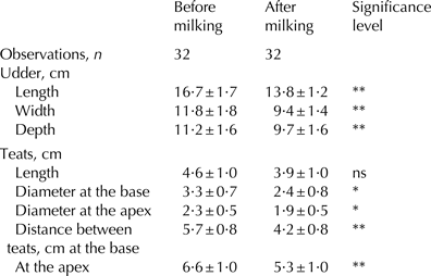
In Table 1 are reported the data on the measurements of the udders and teats before and after milking. Among the animals, the udder and teat morphologies were rather similar. Udder shape was classified as ‘bowl’ in 89% of the jennies and ‘globular’ in 11% of jennies; the teats were classified as ‘conical’ in 78% of the jennies and ‘cylindrical’ in 22% of the animals. As expected, all the measurements taken on the udder decreased after milking within a range from −11·6% (udder depth) to −25·7% (diameter of teat at the base). The teats appeared to have a direction slightly divergent towards the apex in respect to their base. After milking, the teats showed a significant decrease in the diameters at both the base and the apex (P<0·05), and in the distances between the teats measured at the base and at the apex (P<0·01).
Measurements of udder and teat characteristics before and after machine milking (means±sd)

ns, not significant
**P<0·01; *P<0·05
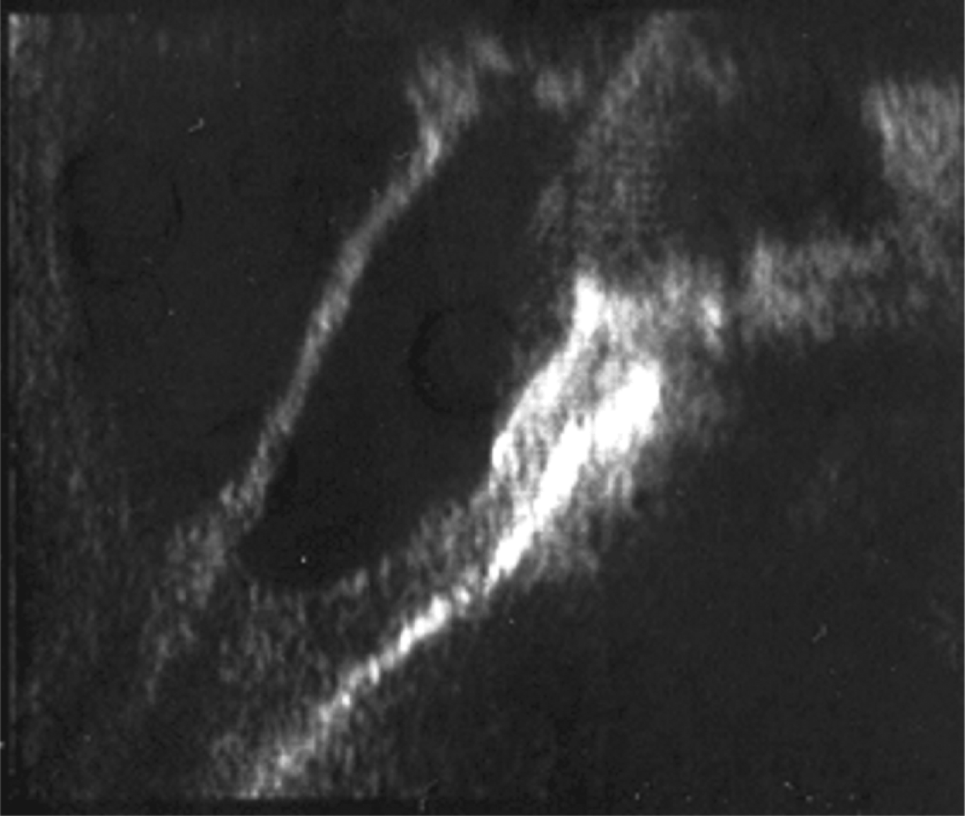
The ultrasound images of the mammary gland showed pocket-like areas of ducts empting into the teat instead of a single cisternal cavity (Fig. 1).
Ultrasound cross section of the udder of a jenny showing the pockets of ducts emptying directly in the teat.
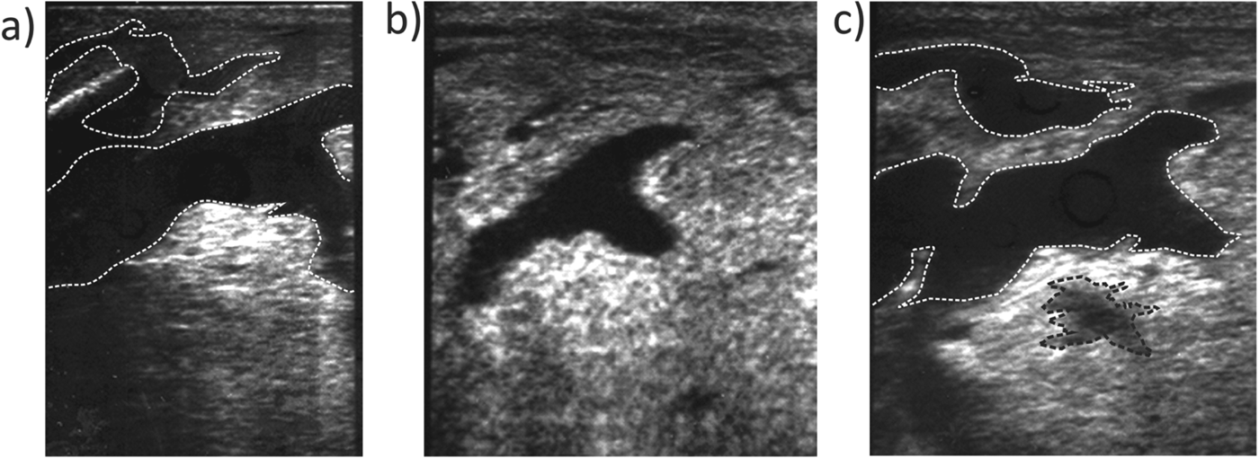
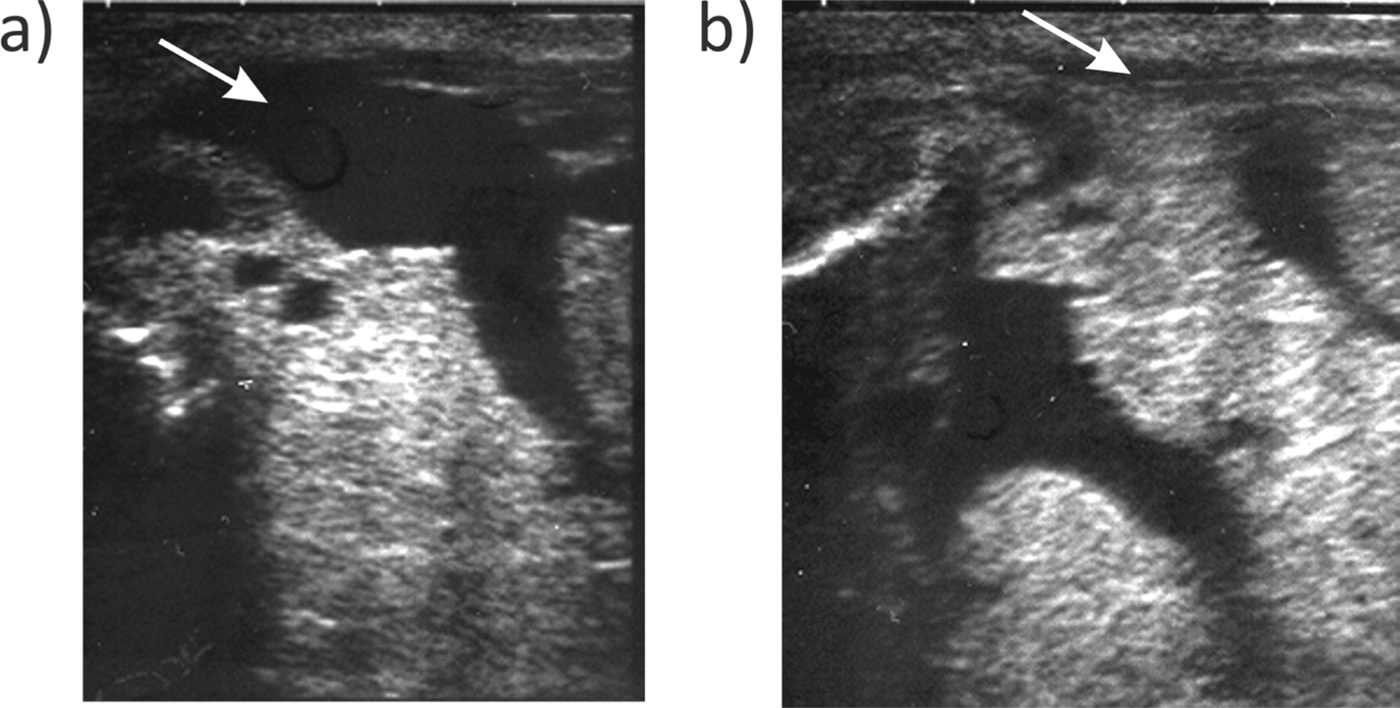
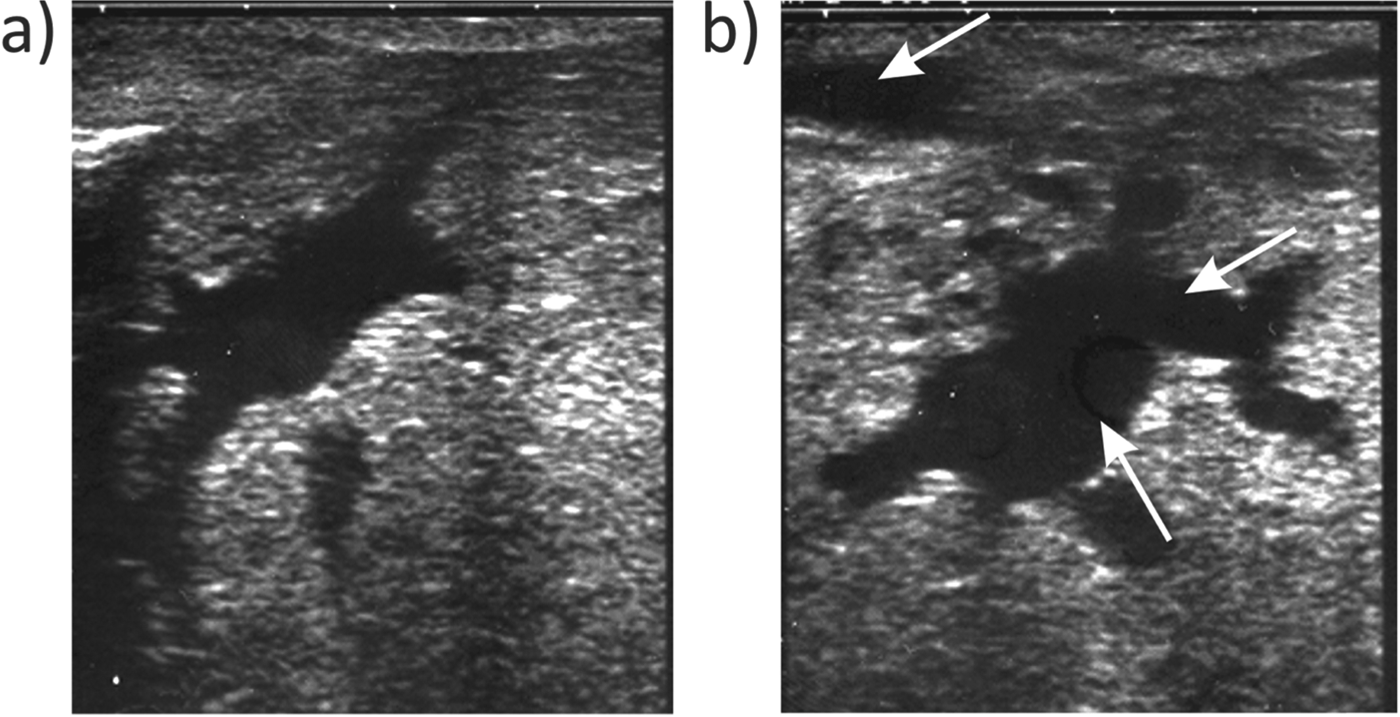
The cisternal areas in the gland observed before milking, after milking and after the oxytocin administration showed changes in their sizes (Fig. 2). After milking, the cisternal areas decreased in comparison with those recorded before milking (Figs. 2a, b & 3a, b). After the administration of oxytocin, a pronounced enlargement was then observed in the cisternal ducts (Figs. 2b, c & 4a, b).
Cisternal cavities in the udder of a jenny detected by ultrasonography before milking (a), after milking (b) and after the oxytocin administration (c). In the pictures (a) and (c), some cisternal areas are bordered by a white or black dotted line to show the variations of their sizes in comparison to those of the picture (b).
Cisternal cavities in the udder of a jenny detected by ultrasonography before milking (a) and after milking (b). In the pictures, the narrows indicate the same cisternal area showing the shrinkage that occurred after the milking.
Cisternal cavities in the udder of a jenny detected by ultrasonography after milking (a) and after the oxytocin administration (b). In the picture (b), the arrows indicate the enlargement of cisternal areas that occurred after the oxytocin administration.
Study 2
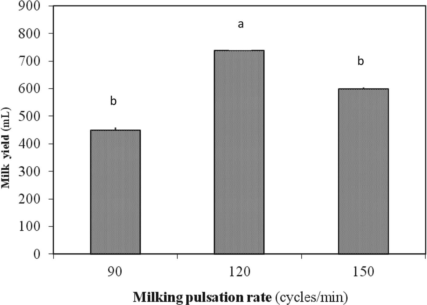
Data on the daily milk collected from the two milkings per day in relation to the milking pulsation rates are reported in Fig. 5.
Machine daily milk yield in jennies (n=5) in relation to the pulsation rate. Values are mean and sd. Different letters (a, b) show statistically significant differences (P<0·05).
The pulsation rate of 120 cycles/min resulted in a higher (P<0·05) daily milk yield (739·6 ml) in comparison with the pulsation rates of 90 and 150 cycles/min. No differences (P>0·05) in the milk recovered amounts were found between the pulsation rates of 90 and 150 cycles/min.
Study 3
The milking pulsation rate was a significant (P<0·05) source of variation for milk yield as spontaneously removed and residual milk fractions (Table 2). The milking pulsation rate of 120 cycles/min showed (P<0·05) the higher spontaneously removed milk fraction and the lower residual milk amounts compared with the pulsation rate of 90 cycles/min. Therefore, the pulsation rate of 120 cycles/min resulted in a higher efficiency of milking as regards the total milk production, consisting of the spontaneously removed milk plus the residual milk, compared with the pulsation rate of 90 cycles/min (73·6 vs. 55·8%; P<0·05).
Partitioning of spontaneously removed milk and residual milk fractions and efficiency of machine milking in relation to the pulsation rate of 90 and 120 cycles/min (means±sd)
ns, not significant
*P<0·05
† Milk from machine milking
‡ Milk from machine milking after oxytocin administration (40 IU i.m.)
§ Percentage of spontaneously removed milk yield from total milk yield
In Table 3 are given the data on chemical analysis of the spontaneously removed and residual milk fractions. Fat and SCC contents of residual milk were higher (P<0·05) than spontaneously removed milk fraction, while no differences were observed in relation to the lactose and protein contents.
Gross composition and somatic cell count (SCC) of spontaneously removed milk and residual milk fractions (means±sd)
ns, not significant
*P<0·05
† Milk from machine milking
‡ Milk from machine milking after oxytocin administration (40 IU IM)
Discussion
Donkeys’ mammary glands are located between the hind legs in the inguinal region. They are constituted by two half-udders, each including one teat with two orifices, separated by and supported by the medial suspensory ligament. In our experimental animals (Martina Franca breed) the udder is darkly pigmented and covered with hair, with the exception of the teats.
The dimensions of udder and teats obtained from this study provide a basis of knowledge on the morphological characteristics of the udder of donkeys, and may be useful to define the specific characteristics of the milking device for jennies and also for genetic selection. The ultrasound images collected in this study showed that the cistern cavity of the mammary gland of donkeys is characterised by the presence of several pockets that empty directly into the teat, instead of a single cisternal cavity. Administration of oxytocin induced visible and marked changes in the ultrasound images owing to the sudden increase of milk flow from the alveolar compartment, which increases the intramammary pressure (Bruckmaier et al. Reference Bruckmaier, Mayer and Schams1991) and enlarges the cisternal ducts. The dramatic enlargement in the cisternal area after milk ejection as induced by oxytocin administration has also been observed in cows, sheep and goats (Bruckmaier & Blum, Reference Bruckmaier and Blum1992; Caja et al. Reference Caja, Ayadi and Knight2004).
Taken together, the findings of this study relative to the udder anatomy and morphology, and the ability of the udder to stretch between milking (approximately 16% for the udder measurements and 18% for the udder and teat measurements as the overall mean values, respectively) may provide the basis of knowledge for the evaluation of udder ‘compliance’, defined as the capacity of an organ that receives, manages, and expels fluids, to accept dilatation. In cows, it has been proposed that compliance may also function in terms of the storage of milk (Davis et al. Reference Davis, Farr, Copeman, Carruthers, Knight and Stelwagen1998).
It is known that the equid udder has a lower storage capability compared with ruminants (Le Du, Reference Le Du1986) and is more adapted to frequent milk removal (2 or 3 h after separation from the foal, in mare; Doreau, Reference Doreau1991). However, our previous results in donkeys (D'Alessandro & Martemucci, Reference D'Alessandro and Martemucci2012) indicated an increase in the milk yield per milking induced by the increasing of milking interval from 3–8 h owing to the ability of the cistern to dilate, leading to a compensatory effect with respect to the decrease in milk synthesis. This is confirmed by the present findings about the capability of mammary gland to dilate and to permit a milking interval of 8 h.
The results of this study showed a marked increase in the amount of milk collected using the milking pulsation rate of 120 cycles/min in comparison with the pulsation rate of 90 or 150 cycles/min. In cows, the improvements in the quantity of collected milk related to the pulsation rate were found in some studies (Clough et al. Reference Clough, Dodd and Hughes1953; Stewart & Schultz, Reference Stewart and Schultz1958; Jorgensen & Coruolo, Reference Jorgensen and Coruolo1963) but not in others (Moore, Reference Moore1971; Rosen et al. Reference Rosen, Coruolo, Mochrie and Dickey1983; Thomas et al. Reference Thomas, Force, Bremel and Strasser1991). These discrepancies could be attributed to the different pulsation ratios adopted (Ricordeau & Labussiere, Reference Ricordeau and Labussiere1970) or to other experimental conditions such as lactation stage, lactation number and age of the animal (Bruckmaier et al. Reference Bruckmaier, Rothenanger and Blum1994; Bruckmaier & Hilger, Reference Bruckmaier and Hilger2001). In the present study, the pulsation rate of 120 cycles/min resulted in a higher efficiency of milking in terms of the milk spontaneously removed by milking (approximately 74% of the total milk) compared with the pulsation rate of 90 cycles/min. Whereas, from a physiological point of view, the residual milk fractions in the jennies udders corresponded to 26 and 44% of the total milk yield for pulsation rate of 120 and 90 cycles/min, respectively. In mares, the residual milk amount has been estimated to be 43% of the total (Dzidic et al. Reference Dzidic, Knopf and Bruckmaier2002); in cows, it can range from 10 to 30% (Bruckmaier & Blum, Reference Bruckmaier and Blum1998).
The higher fat content detected in the residual milk compared with the cisternal milk is in agreement with the results reported in dairy cows (Waldmann et al. Reference Waldmann, Ropstad, Landsverk, Sorenson, Solverod and Dahl1999) and in dairy ewes (McKusick et al. Reference McKusick, Thomas, Berger and Marnet2002) whereas the milk protein and lactose contents in the two milk fractions show slight differences (McKusick et al. Reference McKusick, Thomas, Berger and Marnet2002), as in this study. In a previous study (D'Alessandro & Martemucci, Reference D'Alessandro and Martemucci2012) a greater fat content in jennies’ milk was found at the 3-h milking interval in comparison with 5- and 8-h intervals. In dairy ewes, McKusick et al. (Reference McKusick, Thomas, Berger and Marnet2002) reported that the transfer of fat from the alveoli to the cistern may occur during early udder filling after which fat accumulation occurs in the alveolar compartment, and further draining requires the stimulation of myoepithelial contraction by oxytocin.
The similar contents of milk protein and lactose in the spontaneously removed milk fraction and residual milk fraction could show a different mechanism of draining in comparison with the fat transfer, presumably owing to the free transfer of the soluble constituents of milk (protein and lactose) from the alveolar to the cisternal udder compartment, as reported by McKusick et al. (Reference McKusick, Thomas, Berger and Marnet2002).
Our results for the SCC content in milk confirm findings obtained in other species (McKusick et al. Reference McKusick, Thomas, Berger and Marnet2002; Bruckmaier & Wellnitz, Reference Bruckmaier and Wellnitz2008). It is known that SCC, besides inflammation, is influenced by several physiological and management factors (Harmon, Reference Harmon1994; Ontsouka et al. Reference Ontsouka, Bruckmaier and Blum2003; Bruckmaier et al. Reference Bruckmaier, Ontsouka and Blum2004). In a study on cows that underwent longer milking intervals, the higher SCC content in the residual milk was ascribed to the high intramammary pressure which opens small junctions between cells, leading to the consequent influx of SCC (white blood cells and epithelial cells) into the mammary gland (Stelwagen et al. Reference Stelwagen, Farr, McFadden, Prosser and Davis1997).
In conclusion, the more frequent shapes were bowl udders and conical teats. After milking, measurements of the udders and teats showed a decrease (from −11·6 to −25·7%). Ultrasound scanning of the udders revealed several pockets of numerous ducts that empty directly into the teat, and a marked reduction of the cisternal areas in the gland after milking.
Daily milk yield was significantly higher with the machine milking performed at 120 cycles/min in comparison with the pulsation rate of 90 or 150 cycles/min. Use of 120 cycles/min reduced the residual milk fraction, with respect to the total milk yield, and improved milking efficiency compared with 90 cycles/min. Residual milk had a higher fat content and SCC in comparison with the spontaneously removed milk fraction.
These studies were supported by the University of Bari. The authors wish to thank Dr Mariella Masi for her support during the experimental work and for providing the experimental animals.