Introduction
Lymphatic filariasis (LF), a vector-borne parasitic disease of humans, is caused by the pathogenic nematodes Wuchereria bancrofti, Brugia malayi and Brugia timori. Of the total disease burden, W. bancrofti alone accounts for 90%, and this form of LF is known as Bancroftian filariasis (Taylor et al., Reference Taylor, Hoerauf and Bockarie2010). Currently the disease is known to occur in 55 countries, with an estimated total population of 120 million (WHO, 2016). It is the world's second leading cause of long-term disability (WHO, 2016), and is a major health concern in the tropics and subtropics of Asia, Africa, the Western Pacific and part of the Americas. Although the disease is global in its distribution, approximately one-third of the people infected live in India, one-third in Africa, and most of the remaining third in South-east Asia and the Pacific Islands. LF is currently classified as a “Neglected Tropical Disease” by the World Health Organization (WHO) due to its prevalence amongst the underprivileged. Major clinical manifestations of this disease, such as lymphedema, lymphangitis and elephantiasis, are caused mainly by the inflammatory response of the host to parasite infection (Allen and Loke, Reference Allen and Loke2001). In this context, filarial surface molecules mostly take part in the host–filarid interactions that decide the fate of the infection in a host (Araujo and Macedo-Soares, Reference Araujo, Macedo-Soares and Pereira2012). The proinflammatory responses resulting in immunopathology of LF are caused by the activation of immune cells (macrophage, dendritic cells, etc.) (Babu and Nutman, Reference Babu and Nutman2014; Mukherjee et al., Reference Mukherjee, Karmakar and Babu2016). Recently, we reported the presence of a microfilarial protein (MfP), a novel ligand of Toll-like receptor 4 (TLR4), from the sheath of microfilariae (larval form) of human filarid W. bancrofti that induces a proinflammatory response in macrophages through activation of the TLR4-NF-κB pathway (Mukherjee et al., Reference Mukherjee2017a). Surface proteins of the adult-stage bovine parasite Setaria cervi also induce inflammation by the same pathway when exposed to rodent or bovine macrophages (Mukherjee et al., Reference Mukherjee2017b). Thus, an obvious question arises about the identity of the factor from an adult-stage filarid that induces a proinflammatory response in the host. In this study, we report the presence of MfP in the adult-stage bovine filarial parasite Setaria cervi.
Materials and methods
Collection of parasites
We exploited the adult S. cervi as a model parasite to simulate the adult Bancroftian parasite of humans, W. bancrofti, which is rarely available. Setaria cervi was chosen due to its resemblance to W. bancrofti in its nocturnal periodicity, antigenic patterns, and metabolism (Kaushal et al., Reference Kaushal, Kaushal and Ghatak1987). It is recommended by the WHO as a model filarid and is widely used in filarial research (Mukherjee et al., Reference Mukherjee2014, Reference Mukherjee2017b, Reference Mukherjee2018). Adult S. cervi were collected from the peritoneal cavity of freshly slaughtered cattle at local abattoirs, washed several times with Kreb's Ringer Bicarbonate buffer (Sigma), and kept in Ringer's solution at 37°C until further processing.
Isolation of surface proteins from the adult-stage filarial parasite Setaria cervi
Surface proteins from adult-stage S. cervi were prepared following Mukherjee et al. (Reference Mukherjee2017b). In brief, parasitic cuticles (outermost layers) were carefully collected by dissecting adult females under a light microscope. Isolated cuticles were chopped into small pieces and somatic materials were removed by treating the cuticle samples through ten subsequent rounds of freezing/thawing, following Bisoffi and Betschart (Reference Bisoffi and Betschart1996). The cuticle fragments were treated with ethylenediaminetetra-acetic acid (EDTA; 1 mm), phenylmethyl sulphonyl fluoride (PMSF; 1 mm), sodium dodecyl sulphate (SDS; 1% v/v), and 2-mercaptoethanol (2-ME; 5% v/v) in phosphate buffered saline (PBS; 50 mm, pH 7.4) for 4 hours at 4°C with constant stirring.
Cuticles were washed with sterile PBS (50 mm and pH 7.4), treated with an antibiotic cocktail (100 U/ml penicillin, 100 μg/ml streptomycin and 0.25 μg/ml amphotericin B) and finally incubated with polymyxin B (10 μg/ml) for 6 hours. Cuticles were homogenized, subjected to ultrasonication (10 cycles each of 30 s at 20 KHz) in endotoxin-free PBS (100 mg total cuticle in 1 ml PBS; 0.1 N and pH 7.0) containing protease inhibitor (Fermentus) for 5 minutes. They were then centrifuged at 15,000 × g for 30 minutes at 4°C and the supernatant was removed as crude extract containing surface proteins (SPs).
Purification of Setaria cervi MfP
MfP was purified from S. cervi cuticle lysate following Mukherjee et al. (Reference Mukherjee2017a). In brief, cuticle lysate was prepared in PBS (50 mm and pH 7.4) and subjected to ultrafiltration employing a 50 kDa molecular weight cut-off (MWCO) membrane (Millipore, USA). The fraction containing proteins of > 50 kDa molecular weight was further loaded onto a p-amino-phenyl phosphorylcholine conjugated agarose column (Sigma, USA) following Mukherjee et al. (Reference Mukherjee2017a). The purity of the isolated protein was determined by SDS-PAGE.
Limulus amebocyte lysate (LAL) assay
Setaria cervi SPs and/or MfP were passed through a polymyxin B-coated agarose column (Sigma-Aldrich, USA) following the manufacturer's guidelines, and thereafter tested for endotoxin contamination by LAL assay kit (Hycult Biotech, Netherlands). We checked the possible involvement of lipopolysaccharide (LPS) in MfP-induced inflammation of macrophages. Our investigation revealed that the endotoxin (LPS) content in S. cervi-MfP was 0.006 EU/ml, which is far below the amount required for TLR4 activation (Rallabhandi et al., Reference Rallabhandi2006).
Immunodetection and immunolocalization of MfP in adult-stage filarial parasite Setaria cervi immunoblotting
Immunodetection of MfP in S. cervi cuticle was performed by immunoblot and enzyme-linked immunosorbent assay (ELISA) with an anti-MfP antibody developed in mice. For immunoblotting, S. cervi cuticle lysate was resolved in 12.5% SDS-PAGE, electro-transferred onto polyvinylidene difluoride (PVDF) membrane and blocked with 10% skimmed milk prepared in 1% tris buffered saline with tween 20 (TBST) for 2 hours. Blocked membranes were subjected to overnight incubation with anti-MfP antibody at a dilution of 1 : 1000 in blocking buffer (10% skimmed milk in 1% TBST). Membranes were washed twice for 15 minutes with 1% TBST and then incubated with polyclonal alkaline phosphatase (ALP)-conjugated IgG (goat-anti-rabbit IgG, Sigma Aldrich, MO, USA) (1 : 2000 dilution in blocking buffer) for 6 hours. Membranes were washed with TBST, developed with 5-Bromo-4-chloro-3-indolyl phosphate-Nitroblue tetrazolium (BCIP-NBT), analysed and photographed in a Gel-Doc (Bio-Rad, USA).
ELISA
ELISA was carried out in 96-well microplates following Mukherjee et al. (Reference Mukherjee2017a, Reference Mukherjeeb). ELISA plates were coated with 5 μg/well of the total protein (SP) from cuticle lysate overnight at 4°C with gentle stirring. After completion of incubation, each well was washed thrice with 1% PBST for 15 minutes and blocked with 1% bovine serum albumin (BSA) in PBST for 1 hour. Plates were further incubated with an anti-MfP antibody prepared in 1% BSA with various dilutions ranging from 1 : 50 to 1 : 2000 for 2 hours at 37°C. Wells were again washed with PBST twice for 15 minutes and incubated with ALP-conjugated rabbit IgG (1 : 5000) for 1 hour at 37°C. Upon completion of incubation, plates were washed with 1% PBST and developed with BCIP-NBT. Optical density was measured at 650 nm using a microplate reader (Bio-Rad, USA).
Immunohistochemistry
Adult S. cervi (female) were washed several times with PBS and fixed in 4% paraformaldehyde in cold PBS overnight, embedded in paraffin and cut into 3-μm thick sections following Haegeman et al. (Reference Haegeman2009) and Joardar et al. (Reference Joardar, Mukherjee and Sinha Babu2018). Tissue sections mounted on a glass slide were deparaffinized by treating with absolute xylene, and blocked with 1% BSA in PBST for 2 hours. Anti-MfP antibody was prepared in 1% BSA at 1 : 1000 dilution and tissue sections were incubated overnight at 4°C in a humidified chamber with gentle shaking. Slides were washed twice with 1% PBST for 15 minutes each and further incubated with fluorescein isothiocyanate (FITC) labelled IgG (1 : 500) (goat-anti-rabbit IgG, Sigma Aldrich, MO, USA) for 6 hours in the dark. Sections were washed twice with 1% PBST for 15 minutes and counterstained with Hoechst (1 μg/ml) for 1 minute in the dark. Stained sections were visualized and micrographed using an inverted fluorescence microscope (Carl Zeiss).
Blood sampling
The human serum samples were collected from microfilaraemic (circulating microfilariae in the blood) individuals with high W. bancrofti microfilariae count (> 50/20 μl nocturnal blood) residing in filaria endemic villages reported in our previous studies (Gayen et al., Reference Gayen2010, Reference Gayen2013; Mukherjee et al., Reference Mukherjee2014). In addition, serum from the endemic normal individuals was also collected.
Cross-reaction between the Wuchereria bancrofti-infected human serum and Setaria cervi MfP
In order to investigate the immunological relevance of S. cervi MfP, we performed immunoblotting of S. cervi MfP with W. bancrofti-infected serum following Mukherjee et al. (Reference Mukherjee2017a).
Comparative assessment on the immunoreactive potential of Wuchereria bancrofti MfP and Setaria cervi MfP
In order to investigate the immunoreactivity of S. cervi in comparison to W. bancrofti MfP, we incubated RAW 264.7 macrophages (1 × 10⁶/ml) separately with the MfPs (50 μg/ml) isolated from the two different sources, following Mukherjee et al. (Reference Mukherjee2017a). After completion of treatment, spent culture supernatants were collected and levels of the proinflammatory cytokines were determined by ELISA.
Moreover, we also investigated the effect of MfP immunodepletion from S. cervi lysate by immunoprecipitating MfP from S. cervi lysate using anti-MfP antibody developed against W. bancrofti MfP (Mukherjee et al., Reference Mukherjee2017a).
Finally, we checked the effect of TLR4 inhibition in macrophages (RAW 264.7) by applying a TLR4 pathway inhibitor (CLI-095; Invivogen, France) and anti-TLR4 antibody (Santa Cruz Biotechnology, USA) on S. cervi MfP induced macrophage activation, following Mukherjee et al. (Reference Mukherjee2017a). For the TLR4 blocking experiment, CLI-095 (3 μm; prepared in dimethyl sulfoxide) was added to the cell culture media of RAW 264.7 macrophages 1 hour prior to the addition of the parasitic stimulant. On the other hand, RAW 264.7 macrophages were treated with the anti-TLR4 antibody (2 μg/ml; prepared in sterile PBS), washed with culture media (Dulbecco's modified eagle medium) and thereafter treated with the filarial proteins.
Heat-inactivated (HIN) MfP was prepared by incubating the protein sample at 56°C in a water bath for 2 hours following Mukherjee et al. (Reference Mukherjee2017b). HIN-MfP was also used for macrophage stimulation study, as depicted earlier.
In all macrophage stimulation experiments, RAW 264.7 macrophages were incubated separately with 50 μg of parasitic proteins (SPs or SC-MfP or WB-MfP) or LPS (100 ng/ml) for 6 hours at 37°C in a humidified incubator. After termination of incubation, cell-free supernatant from each experimental set-up was collected through centrifugation at 3000 × g at room temperature. The cell-free supernatant was diluted with sterile PBS at a ratio of 1 : 2 and used for the determination of the cytokines through ELISA. The levels of TNF-α, IL-6 and IL-1β in the cell-free supernatant were determined by the sandwich ELISA kit (R&D Systems, USA) following the manufacturer's guidelines.
Statistical analysis
All the experiments were conducted in triplicate and repeated at least five times. Data are presented as mean ± SD. All data were subjected to one-way ANOVA analysis followed by Tukey's multiple comparison tests. P < 0.05 was considered statistically significant.
Results
Setaria cervi cuticles possess a homolog of Wuchereria bancrofti MfP
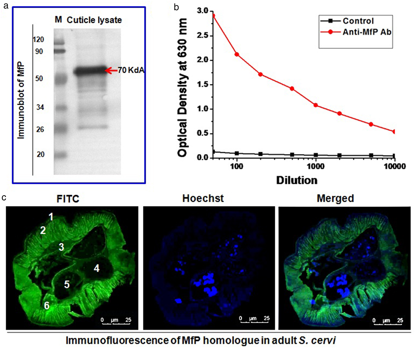
We detected a clear band corresponding to W. bancrofti MfP by immunoblot that clearly revealed the presence of a homolog of MfP in the cuticle lysate of adult S. cervi when immunoblotted with an anti-MfP antibody raised in mice (fig. 1a). Moreover, the position of the intense protein band (~70 kDa) was at the same position as the purified MfP of W. bancrofti was located (fig. 1a).
Immunodetection and immunolocalization of MfP in adult-stage filarial parasite Setaria cervi. (a) Immunoblot showing the presence of MfP homolog (indicated with red arrow) in the crude SPs of S. cervi cuticle. M: Molecular weight marker. Immunoblotting was performed using antibody developed against MfP isolated from Wuchereria bancrofti. (b) ELISA showing cross-reactivity between S. cervi SPs and anti-MfP antibody given at different dilutions. (c) Immunohistochemistry showing localization and distribution of MfP in the cross-section of adult-stage female S. cervi. Images were taken at 20× magnification. The marked body parts in the histological section of S. cervi are as follows: 1, cuticle; 2, hypodermis; 3, muscle layer; 4, ovary; 5, uterus; 6, intestine. Each experiment is a representative of three independent experiments repeated at least five times.
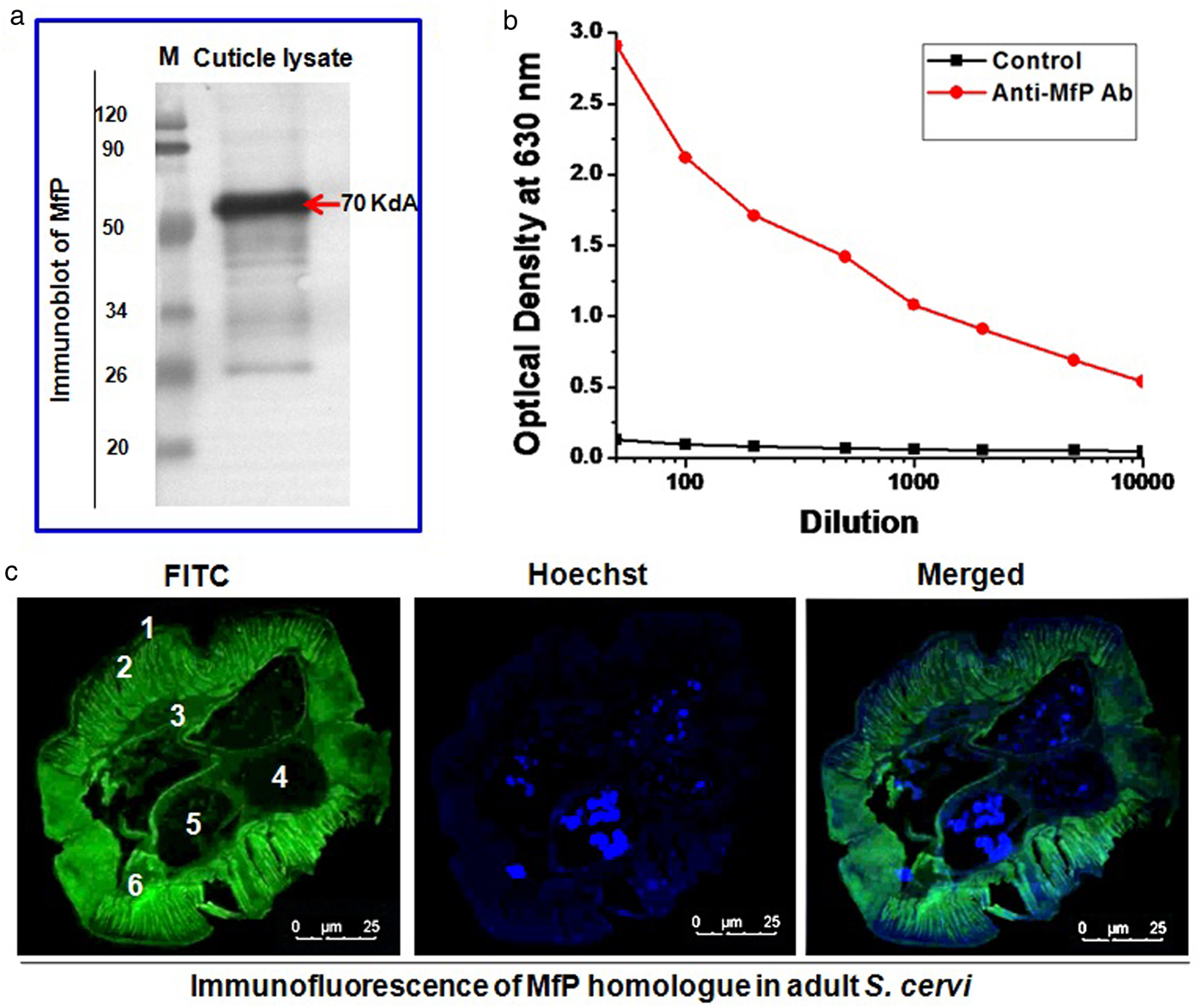
Similar to the immunoblot, we also detected a clear cross-reaction between S. cervi SPs and anti-MfP antibody as depicted in the ELISA analysis (fig. 1b). Furthermore, ELISA data revealed a high degree of sensitivity of the anti-MfP antibody (previously raised against W. bancrofti MfP) towards S. cervi MfP, as evident in fig. 1b. In this study, the anti-MfP antibody was found to cross-react with the MfP homolog in S. cervi SPs up to 1 : 20,000 dilution (fig. 1a).
MfP homolog in Setaria cervi is located on surface layers (cuticle) of the parasite
We conducted immunohistochemical staining of the parasite tissue with the anti-MfP antibody. Our in situ immunohistochemical observation further demonstrated the presence of the MfP homolog in adult-stage S. cervi as a surface protein distributed mostly in the cuticle and epicuticle of the parasite (fig. 1c). This finding corroborated our previous inference of MfP as a surface protein (Mukherjee et al., Reference Mukherjee2017a, Reference Mukherjeeb).
The immunological relevance of Setaria cervi MfP in Bancroftian filariasis research
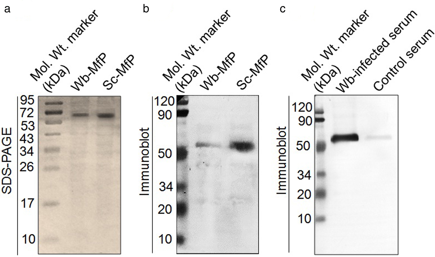
Our previous study revealed MfP as the major inflammation-inducing protein from the filarial parasite (Mukherjee et al., Reference Mukherjee2017a). In order to confirm the identity of the MfP homolog in S. cervi, we adopted similar purification strategies to those employed previously for purifying MfP from W. bancrofti (Mukherjee et al., Reference Mukherjee2017a). After purifying MfP from S. cervi, we compared S. cervi-MfP with W. bancrofti-MfP and both appeared as identical proteins in SDS-PAGE at c. 70 kDa band size with homogeneity (fig. 2a). In addition, we found clear cross-reaction between the anti-MfP antibody and purified S. cervi MfP, as shown in fig. 2b. The ability of anti-MfP antibody (developed against W. bancrofti) to bind S. cervi MfP was similar to its ability to bind W. bancrofti MfP (fig. 2b). Overall this suggests that S. cervi possesses MfP homolog.
The immunological relevance of Setaria cervi MfP. Comparative studies on the (a) SDS-PAGE profile and (b) immunoblot of MfP isolated from S. cervi and Wuchereria bancrofti. (c) Immunoblot showing cross-reactivity between S. cervi surface lysate and W. bancrofti-infected serum. The immunoblots are representative of experiments conducted in triplicate.
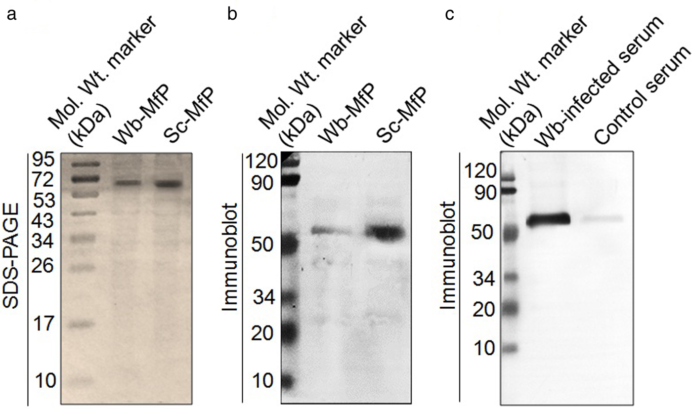
After confirming the presence of an MfP in S. cervi adult-stage parasites, we checked its immunological relevance in human filariasis research. The cross-reaction between the W. bancrofti-infected human serum and the S. cervi SPs was observed with a high sensitivity and specificity, as evident in the immunoblot data (fig. 2c). This clear cross-reactivity between the two signifies the immunological potential of S. cervi-MfP.
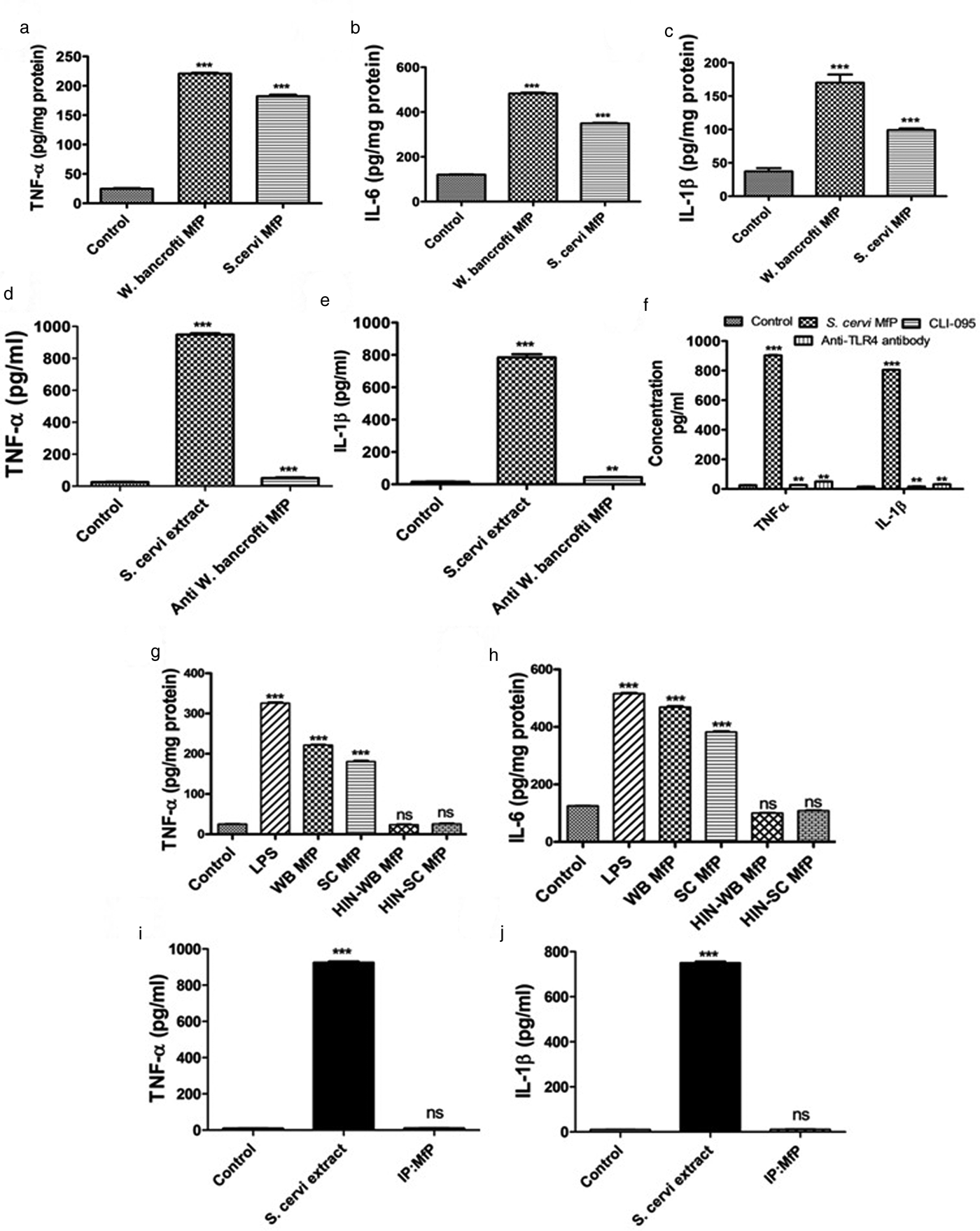
We also observed that the macrophage stimulation activity of S. cervi MfP is similar to that of W. bancrofti MfP (fig. 3). The levels of all the major proinflammatory cytokines, viz. IL-6, TNF-α and IL-1β, were significantly upregulated after treatment with S. cervi MfP (fig. 3a–c). Moreover, we also noticed that depletion of MfP from S. cervi lysate resulted in no sign of inflammation (fig. 3d–e), which suggests that the immunoreactive potential of S. cervi lysate is primarily dependent on MfP.
Comparative macrophage stimulatory activity of Setaria cervi MfP and Wuchereria bancrofti MfP. Level of (a) TNF-α, (b) IL-6 and (c) IL-1β in the spent culture supernatant of RAW 264.7 macrophages treated with MfP isolated from W. bancrofti and S. cervi. Effect of anti-W. bancrofti MfP antibody on S. cervi cuticle extract-induced secretion of (d) TNF-α, and (e) IL-1β from RAW 264.7 macrophages. (f) Effect of TLR4 inhibition by anti-TLR4 antibody and CLI-095 (3 μm) on S. cervi MfP-induced secretion of TNF-α and IL-1β in the spent culture media of the treated macrophages (RAW 264.7). (g & h) Effect of heat inactivation on S. cervi MfP induced macrophage (RAW 264.7) activation. (i & j) Effect of immunoprecipitation mediated immunodepletion of MfP from S. cervi extract on S. cervi cuticle extract-induced inflammation of RAW 264.7 macrophages. All the experiments were carried out in triplicate and repeated at least five times.

As W. bancrofti MfP was reported to exert its inflammatory activity through TLR4, we also found that inhibition of macrophage-TLR4 by TLR4 inhibitor (CLI-095) and anti-TLR4 antibody significantly diminished the inflammatory activity of S. cervi MfP (fig. 3f).
Our earlier observations on W. bancrofti-MfP and S. cervi SPs demonstrated that MfP exerts its immunostimulatory functions through its protein component (Mukherjee et al., Reference Mukherjee2017a, Reference Mukherjeeb). Therefore, we also checked the effect of heat denaturation in S. cervi-induced macrophage inflammation (fig. 3). The macrophage stimulatory action of S. cervi-MfP was diminished upon heat inactivation and this is at par with the characteristic of W. bancrofti MfP (fig. 3g–h). Therefore, S. cervi-MfP is not only homologous to W. bancrofti-MfP in its structure, but also in its functional attributes.
Discussion
Inflammatory consequences resulting from the host–parasite interaction are considered to be the major determinant in filarial immunopathogenesis. Particularly, the induction of inflammatory responses against the adult filarial parasite is a major contributor to the pathological transformation of the lymphatic system in filarial patients. The adult parasite usually survives for several years in the host's body and maintains chronic inflammation in the infected host, leading to immunopathological outcomes such as lymphedema, lymphangiogenesis and elephantiasis (Mukherjee et al., Reference Mukherjee2017a, Reference Mukherjeeb). However, the identity of the inflammation-inducing factor from the adult parasite is not yet clear.
In this study, we worked with the bovine filarial parasite S. cervi to study the presence of the inflammation-inducing antigen of W. bancrofti, as direct isolation of the antigen of interest, i.e. MfP from adult-stage W. bancrofti, is not possible. On the other hand, our previous study (Mukherjee et al., Reference Mukherjee2017a) demonstrated that MfP from W. bancrofti is the major proinflammatory antigen. As it is a surface/membrane protein, recombinant MfP is also difficult to prepare and its functionality could also be questionable. Therefore, taking all the facts into consideration, we aimed to search for a cost-effective native source of MfP.
To the best of our knowledge, this study is the first descriptive report on the presence of an inflammation-inducing protein homolog of W. bancrofti in a bovine filarial nematode. Thus, our study demonstrates the acceptability of S. cervi as a model parasite for Bancroftian filariasis research.
Inflammatory insults induced by the adult-stage parasite in LF patients are mostly associated with the pathological manifestations. Therefore, the therapeutic intervention of LF by targeting MfP could be an efficacious method. However, the physio-biochemical nature of MfP in S. cervi is not yet known and needs further investigation to develop S. cervi MfP for immunological and/or therapeutic research on Bancroftian filariasis in the near future. Particularly, inhibition of MfP could be very useful for filarial patients with pathological manifestations, and administration of purified MfP could potentially be useful in eliminating chronic filarial infection through the reestablishment of TH1 immunity. Therefore, an antibody raised against the whole MfP or particularly against the epitope could facilitate the therapeutic intervention of LF by targeting MfP and/or MfP as a vaccine candidate. Assessment of the efficacy of MfP in an animal model, and risk assessment at the subclinical level are currently underway.
Acknowledgements
Grateful thanks are due to Prof. Samir Bhattacharya for his generous help and support in isolating MfP from W. bancrofti and raising an antibody against MfP in mice. We thank the residents of Sian block, Birbhum, West Bengal, India for their help and active cooperation during blood sampling. Grateful thanks are due to Prof. Amrit Sen, Department of English & Other Modern European Languages, Visva-Bharati University and Dr. Biswanath Banerjee, Government Sanskrit College, Darbhanga, India for critically reviewing the language of the manuscript.
Author ORCIDs
S.P. Sinha Babu 0000-0002-7441-6426, S. Mukherjee 0000-0002-5709-9190 and N. Joardar 0000-0002-6460-0377.
Financial support
The authors acknowledge the Department of Science & Technology (DST), Govt. of India for providing funding for this research (SR/SO/AS-006/2014).
Conflict of interest
None.
Ethical standards
The authors declare that all procedures and protocols for this study comply with the ethical standards of the relevant guides on the care and use of laboratory animals and have been approved by the Institutional Animal Ethical Committee, Visva-Bharati, Santiniketan–731 235, West Bengal, India under the guidelines of Committee for the Purpose of Control and Supervision of Experiments of Animals (CPCSEA); Govt. of India (Registration No. 1819/GO/Ere/S/15/CPCSEA). The study was also approved by the sub-divisional hospital of Bolpur, Birbhum, West Bengal, India.